Clinically relevant phylogeny and embryology
The
middle cerebral artery (MCA) is the phylogenetically youngest cerebral
vessel. In “lower” species, primarily allocortical structures—such as
olfactory and hippocampal areas—are supplied by anterior cerebral artery
(ACA) and posterior cerebral artery (PCA)-like vessels, respectively,
while basal ganglia structures are served by “perforating” branches of
the proximal “cranial ramus” or ACA homologs. Expansion of the neocortex
in progressively “higher” species is accompanied by recruitment of
vessels from the existing lenticulostriate system to supply the
neocortical mantle. Thus, from an evolutionary perspective, the MCA is
both a branch of the ACA and a branch of lenticulostriate vessels, not
vice versa.1
Simply put, MCA or MCAs can arise from anywhere lenticulostriates do.
This perspective helps explain various accessory/duplicated MCA
configurations, fenestrations, as well as variations of lenticulostriate
anatomy.
Lenticulostriate vessels
The
lenticulostriate vessels are a contiguous family, with origins along
the traditional A2, A1, M1 and M2 segments. Subdividing them into
“medial” or “lateral” subgroups, or assigning special significance to
the artery of Heubner just because it is an eponym (it is simply the
most medial of lenticulostriates and frequently arises from the proximal
A2 segment2 3)
is helpful from a descriptive standpoint; however, it is a functionally
artificial concept that detracts from our appreciation of how the
family functions as a whole. As in any balanced system, a spectrum of
variation exists, leading to either common or separate origins of
individual perforator vessels from either the ACA or MCA and the
relative dominance of more medial versus more lateral trunks (figure 1).
A more dominant “Heubner” will pick up the classical “medial
lenticulostriate” territory of the proximal M1, while at the other end a
distinct “Heubner” may not be identifiable because vessels supplying
its territory happen to originate from the “medial lenticulostriate”
group of the MCA1 (figure 1).
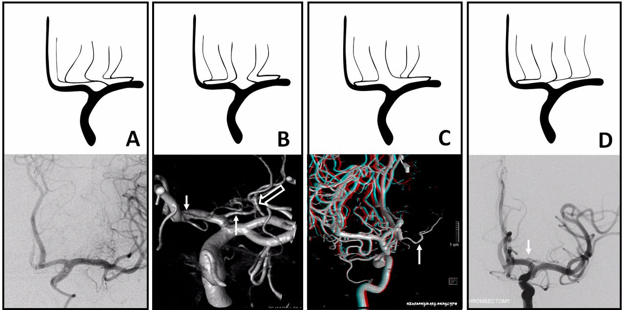
Figure 1 Schematic
and comparable in vivo variations of lenticulostriate anatomy. The row
of lenticulostriate perforators is a continuum of balance. Subdividing
them into medial and lateral lenticulostriates is anatomically
arbitrary. The Heubner (simply the most medial of perforators) may be
absent or small (A). A1 perforators can have separate origins (B,
arrows) or arise as a common trunk (C). In extreme, a highly dominant A1
system (C,D – white arrows) takes over the typical middle cerebral
artery (MCA) origin lenticulostriates. Classical dominance of the MCA
origin lenticulostriates is seen in (A). When short “M1” trunks are
present, more lateral perforators typically arise from the “M2” segment
(B, open arrow).
Accessory MCA/duplicated MCA
About
1% of the time, two MCA-like vessels are present. Distinction between
“accessory MCA” arising from the ACA, and “duplicated MCA” arising from
the distal ICA—with main classifications by Teal4 and Manelfe5—are
morphologically useful, but have no apparent basis in embryology. All
are expressions of the phylogenetic origin of the MCA from the
lenticulostriate system. The MCA is a hypertrophied lenticulostriate
vessel that captures cortical territory. A duplicated/accessory MCA
disposition simply reflects persistence of two separate vessels
responsible for cortical supply—conceptually similar to “duplicated”
anterior inferior or superior cerebellar arteries. Any place that “gives
rise” to lenticulostriate vessels can be a site of MCA-like vessel
origin. Figures 2 and 3
show the continuum between “duplicated” and “accessory” MCAs,
demonstrating that this distinction may simply be a consequence of
arbitrarily defining the location of the carotid terminus.6 (figures 2–4)
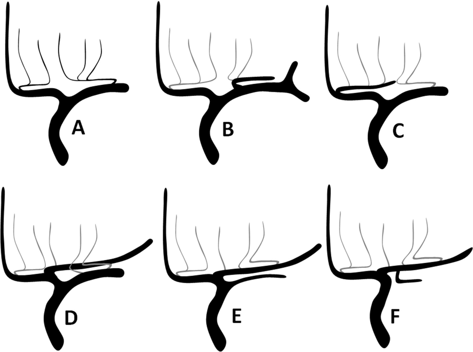
Figure 2 Schematic
of accessory/duplicated middle cerebral artery (MCA) variations,
underscoring a continuum based on the phylogenetic notion that MCA
represents a hypertrophied lenticulostriate origin vessel serving
neocortical territory. It can thus arise anywhere lenticulostriates come
from. (A) is classical disposition. (B) is a frequently unrecognized
variant origin of the usually frontal branch from the “M1” segment
lateral perforators, analogous to the more unusual origin of the same
branch from the A1 segment (C)—appropriately regarded as a hypertrophied
artery of Heubner, and commonly referred to as “accessory MCA”.
Criticism that this is not so because a separate Heubner-like artery may
be present (D) ignores the simple fact that lenticulostriate vessels
can have separate or common origins. Finally, (E) shows dominance of the
proximal “A1” origin MCA vessel, with the smaller caliber “duplicated”
MCA seemingly arising from the internal carotid artery (ICA). In fact,
this is simply based on arbitrarily deciding where the ICA ends and A1
begins. (F) shows the early origin of the anterior temporal branch, a
disposition that is contiguous with (E).
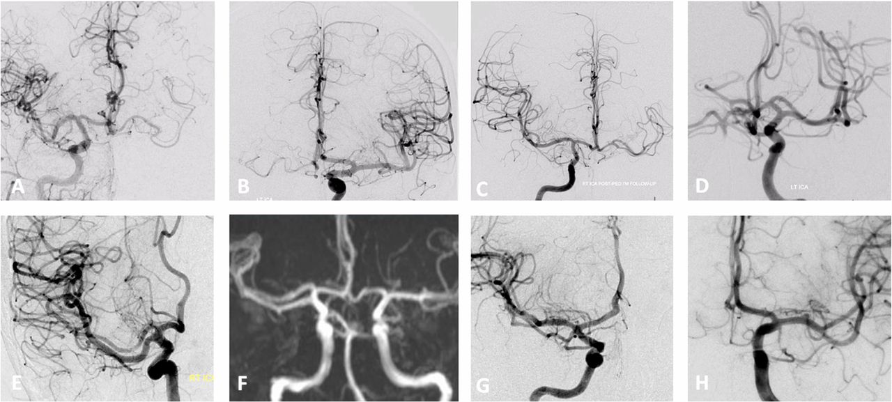
Figure 3 Spectrum
of duplicated/accessory middle cerebral artery (MCA) vessels, from ACOM
region Heubner-like variants (A,B) to the mid-A1 origin (C,D—same
patient, right and left internal carotid artery (ICA) injections), to
the more proximal “A1” site (E,F), to “ICA terminus”-like disposition
(G), to what is termed “duplicated” MCA (H). Which is “A1” origin and
which is “ICA” origin is really based on an arbitrary notion of what
represents the ICA terminus. In most cases, the “A1” origin MCA-like
vessels supply the frontal territory, whereas the smaller vessels
“arising” from the ICA (G,H) supply the anterior temporal lobe.
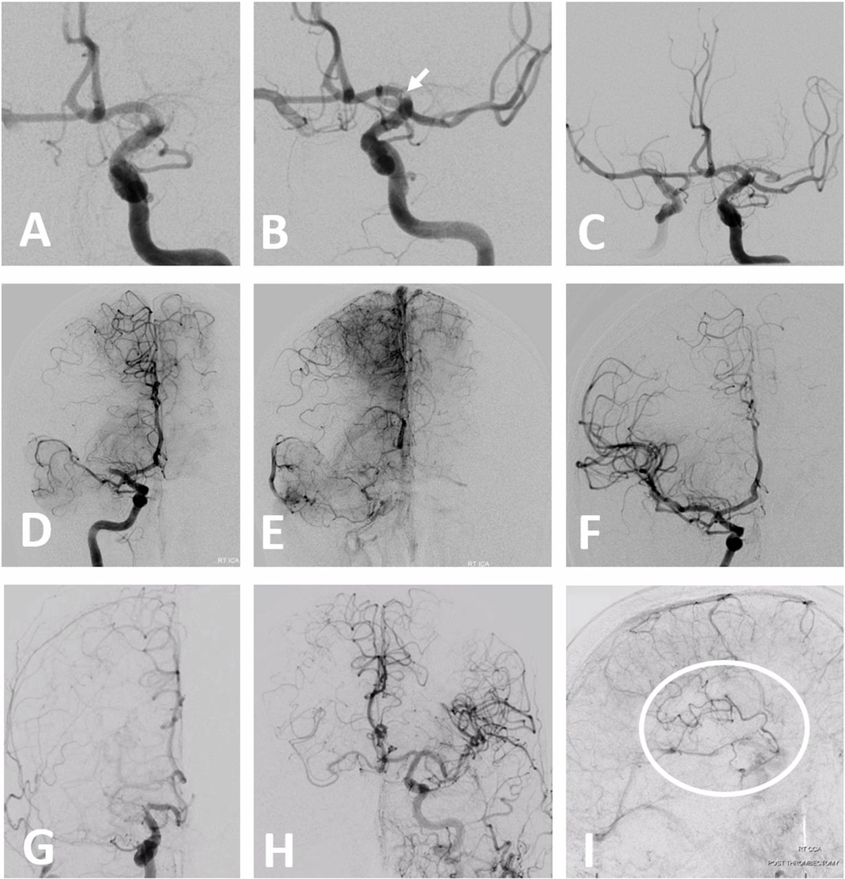
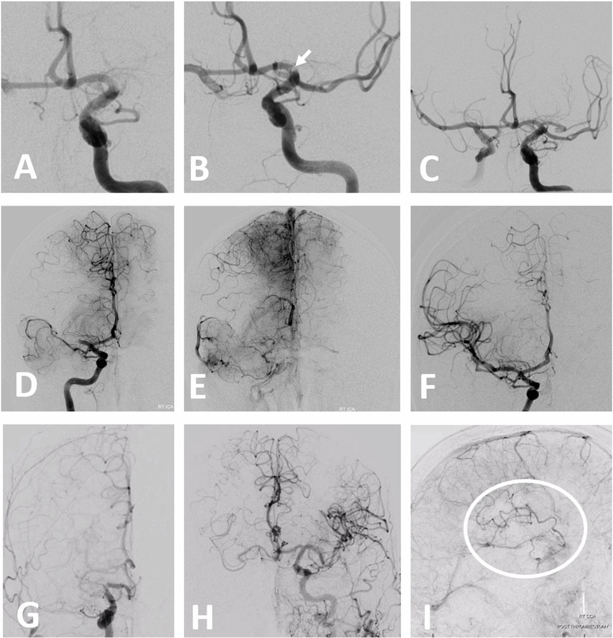
Figure 4 (A–F)
Examples of dual middle cerebral artery (MCA) thrombectomy. Case 1,
courtesy of Dr Daniel Sahlein (A,B,C): initial frontal view (A),
post-recanalization of dominant inferior division (B)—arrow points to
separate embolus in the still closed superior division—and final
recanalization (C). Case 2 (D,E,F): “duplicated” MCA supplying portions
of temporal lobe. Embolus lodges in the larger branch that follows the
vector of internal carotid artery (ICA) flow and supplies the bulk of
the MCA territory, with partial anterior cerebral artery (ACA)-MCA
leptomeningeal collateral support (E). Lenticulostriate arteries arise
proximal to the occlusion. Post-reperfusion (F). Case 3 (G,H,I): carotid
terminus occlusion. Frontal convexity (white oval) remains perfused in
an antegrade manner by the accessory MCA arising from the ACOM region
(H, I).
From
a practical perspective, when faced with two MCA-like vessels, key
questions are: what constitutes the territory of each trunk, and from
where do the lenticulostriates arise? It is not true that only one trunk
gives rise to lenticulostriates, particularly when a Heubner-like
accessory MCA is present. Typically, accessory MCAs with origins along
the ACA will tend to supply the superior division frontal territory,
whereas in classical “duplicated MCAs” the more proximal vessel usually
supplies the anterior temporal territory7 (figure 4).
The
very rarely seen proximal MCA fenestration might be relevant in
thrombectomy as either a point of occlusion or as a site of branch
origin—particularly the anterior temporal branch which seems to
originate frequently from one fenestration limb.8
MCA trunk and branching patterns
Here
is the classic story: M1 bifurcates into the superior and inferior
division M2 segments. One M2 is usually larger than the other (figures 5–10).
The superior division supplies the frontal lobe, the inferior division
supplies the temporal lobe. The parietal lobe is up for grabs—whichever
division supplies it is the “dominant” one.9 Typically, a small anterior temporal branch arises somewhere along the M1, before the “true” bifurcation.10 “Trifurcation”, with each branch dedicated to frontal, parietal, and temporal lobes, is seen in ~10%.9–11 It is important to realize that life frequently does not follow rules (figure 8), and be ready for anything when we cannot see what lies beyond the M1 stump.
Figure 5 Spectrum
of proximal middle cerebral artery (MCA) branching patterns. (A)
Duplicated/accessory MCA pattern is contiguous with a “short M1”
disposition (B), which is contiguous with a “classic” pattern (C) of M1
bifurcation into dominant and non-dominant M2s and any other conceivable
variation, including “trifurcation” (D), “pentafurcation” (E), or no
bifurcation at all (F), with vessels arising individually from a single
common trunk.
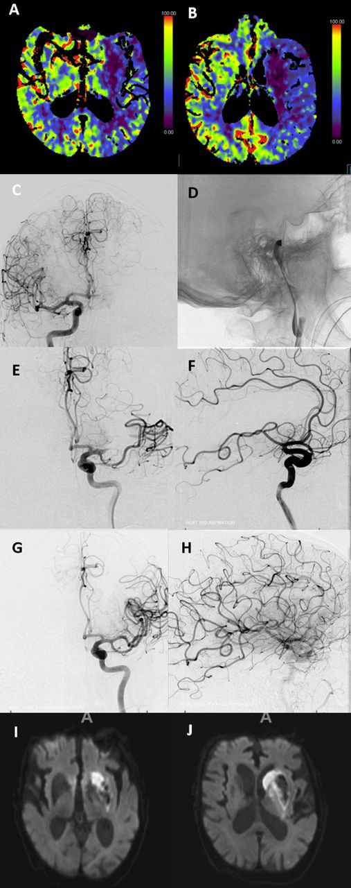
Figure 6 Analysis
of carotid terminus occlusion. Loss of anterior choroidal and
lenticulostriate territory is evident on perfusion imaging (A,B),
including caudate territory of the Heubner. From these two perfusion
images alone we can deduce that the embolus is likely located below the
choroidal artery, extends into the middle cerebral artery (MCA), and
that there is no A1 segment origin of the Heubner, with its territory
being served by occluded MCA lenticulostriates. Pre-thrombectomy (C,D),
after partial recanalization (E,F) with dominant inferior division
supplying frontal and parietal areas still closed, and full
recanalization (G,H). Delayed MRI (I,J) shows excellent correlation with
perfusion imaging in this case.
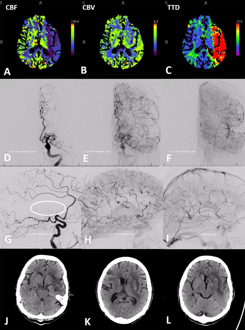
Figure 7 Classic
pattern of leptomeningeal collateral support in the proximal middle
cerebral artery (MCA) occlusion. Perfusion shows reduced CBF (A),
preserved CBV (B), and increased time to drain (TTD) (C) corresponding
with “penumbra” in the bulk of the MCA territory, with the exception of
the lateral basal ganglia and the external capsule region. “Fetal”
posterior cerebral artery (PCA) disposition allows for angiographic
visualization of both the anterior cerebral artery (ACA)-MCA and the
PCA-MCA leptomeningeal collaterals in sequential frontal (D,E,F) and
lateral (G,H,I) projections. Collateral response can be conceptualized
as a shift in watershed territory, with operculum and insula being most
vulnerable. (J,K,L) Day 2 post-TICI (thrombolysis in cerebral
infarction) 3 revascularization. CT reveals infarct of predicted lateral
basal ganglia with petechial hemorrhagic transformation, and external
capsule, as well as portions of the insula. Sparing of the caudate and
globus pallidus is related to the dominance of the A1 segment
lenticulostriate vessels (G, circle); unlike the patient in figure 6. the upper convexity was preserved. CBF, cerebral flood flow; CBV, cerebral blood volume.
Figure 8 Life
does not follow middle cerebral artery (MCA) branching pattern
classifications. Mean transit time (MTT) maps show posterior temporal,
parietal, and posterior frontal penumbra (A,B). (C,D) shows an MCA
“trifurcation”. The embolus (arrows) lodged in the largest diameter
straightest branch downstream of M1 segment flow vector. (E,F) Unlike
classic “trifurcation” theory of each branch picking up frontal,
parietal, and temporal lobes, this middle branch supplied both inferior
temporal and rolandic areas in addition to entire parietal
convexity—strategically producing a major set of clinical deficits
despite a relatively modest volume at risk. (G,H) Post-thrombectomy.
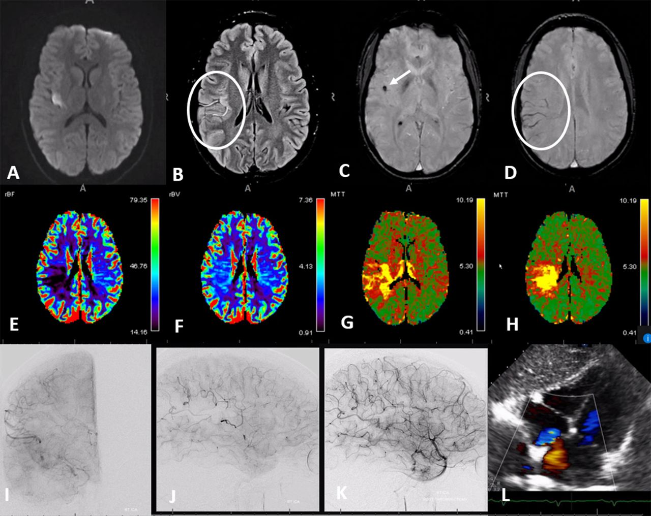
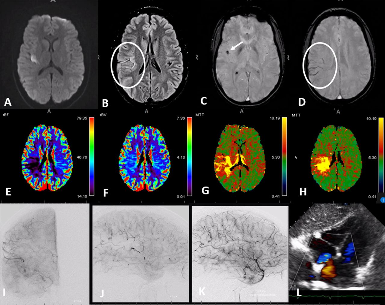
Figure 9 Advanced
MR imaging of acute ischemia in a young patient with delayed
presentation of the left upper extremity and facial weakness. Diffusion
(A) and FLAIR (fluid attenuation inversion recovery) (B) showing slow
flow in the distal fissure middle cerebral artery (MCA) branches.
Susceptibility weighted imaging (C,D) shows susceptibility in
hemosiderin-rich embolus (arrow) and increased hemosiderin content in
regional veins due to increased oxygen extraction (circle). MRI
perfusion (E–H) shows decreased flow (E), preserved volume (F),
increased mean transit time (MTT) (G), and increased delay (H).
Pre-thrombectomy angiography (I,J) shows occlusion of the parietal and
rolandic territory (arrow=embolus). Post-thrombectomy (K). Large patent
foramen ovale (PFO) (L).
Figure 10 Classification of middle cerebral artery cortical territories according to Gibo et al.9
The length of the main MCA trunk is as variable as everything else about the MCA (figure 5).
At one end of the spectrum there is no true MCA “bifurcation” or
“trifurcation”, with cortical vessels arising individually from the main
trunk. When bifurcation or trifurcation is identified, the consensus is
to call the main trunk “M1 segment”. Short M1 segments blend into the
“duplicated MCA” variant, underscoring limitations of defining what “M1”
really is (figure 5A,B).
In regard to thrombectomy, the major dilemma is deciding where M1 ends and M2 begins.12 13
This definition can impact trials and potentially lead to exclusion of
acute stroke patients from beneficial thrombectomy in less progressive
centers. As boundaries of thrombectomy are extended into “M3” and “M4”
segments, definitions become murkier still.
The surgical nomenclature of Gibo et al
9
of the M1 segment as extending horizontally along the sphenoid wing, M2
vertically along the operculum, M3 horizontally in the Sylvian fissure,
and M4 on the surface of convexity was not designed to suit
endovascular goals. Everything about MCA branching anatomy is summarized
by “variability”, hampering development of practical classifications
based on variable branching patterns.12 14
The majority of studies addressing “M2” thrombectomy do not
differentiate which side, division or territory was subject to
occlusion,14–16 raising the question of selection bias. This subject was investigated in a retrospective single-institution study by Seker et al,17
concluding that superior division occlusions, regardless of side, size
or dominance, were independently predictive of poorer outcome compared
with inferior division ones. Unsurprisingly, the National Institutes of
Health Stroke Scale (NIHSS) also carries predictive value in M2
occlusions.18 For more distal (M3, M4) occlusions, very little data are available to guide thrombectomy decisions19;
in all likelihood the bulk of “distal”—M3 and M4—interventions
currently involves patients ineligible for intravenous treatments (figure 9).
Endovascular
classifications should be based on supply territory rather than
non-vascular landmarks or sites of vessel origin. Thus, the only
thrombectomy classification that makes sense is one based on territory
at risk. Despite significant differences, ASPECTS,20 CTA ASPECTS,21 22 and CT perfusion23–25
methods are all rooted in the concept of vascular territories. While
the role of perfusion imaging in clinical decision-making remains a
source of endless controversy, its diagnostic value in identifying the
presence of a geographic hypoperfusion state and establishing the likely
location of an occlusion (particularly for more “distal” occlusions) is
invaluable (figures 6–9). A well-known naming system by Gibo et al
9 divides the MCA into 12 regions (figure 10). It is compatible with MCA variations and can be used in communicating both research and practice scenarios.
Hemodynamics and collateral circulation
Hemodynamic
factors are uniquely important from the perspective of thrombectomy.
Emboli of sizes comparable to branch vessel inner diameters tend to
lodge in the largest, straightest branches, with embolus lodging at a
bifurcation point that prevents further migration (figures 6, 8 and 9).26 27
The same path, for different reasons, is also likely to be taken by a
leading J-shaped microguidewire or by an aspiration catheter advanced by
itself (the so-called “snake technique”).28 Tortuous MCA configuration is associated with reduced stent-triever thrombectomy efficacy.29
Similarly, the more acute cranial angulation of the anterior and
mid-frontal superior division branches, combined with their relatively
smaller sizes, make for more challenging “distal” thrombectomy targets.
In contradistinction, vessels emerging more posteriorly from the Sylvian
fissure (such as the temporo-occipital or parietal) tend to be
straighter and usually somewhat larger in diameter,9 increasing the likelihood of both secondary embolization and successful distal thrombectomy (figures 8 and 9).
High quality data for “distal” or “medium” vessel thrombectomy is
currently lacking and significant challenges are anticipated in
collecting enough of it.19
Efficiency of collateral support is the single most
important factor determining brain tissue survival following acute
primary route occlusion (figure 7).30
Beyond the circle of Willis, the collateral potential of cortical MCA
territory (lenticulostriates have notoriously poor collateral support)
is related to the efficiency of leptomeningeal ACA-MCA-PCA anastomoses.
This efficiency is a complex product of genetic factors determining
intrinsic collateral potential and its modification by age, atheromatous
disease, pre-existing ischemic states favoring collateral enhancement,
and likely many other factors.31 32
Variation in individual collateral potential is simply astounding.
Equally intriguing is the degree to which leptomeningeal collateral
efficiency is enhanced by longstanding non-occlusive proximal flow
constraints (such as intracranial atheromatous stenosis) in some
patients, while similar lesions induce apparently little response in
others.
Aside from the double-edge sword of induced or
permissive hypertension, we have as of today no consistent way of
enhancing collateral response in the acute setting. It seems that even
highly effective leptomeningeal collaterals, producing initially low
NIHSS scores, ultimately fail, favoring early intervention.32
Non-embolic large vessel occlusive disease
Entities
such as large vessel atherosclerotic disease or intracranial
dissection—a minority of large vessel occlusion etiologies—likely
represent a substantial portion of modern thrombectomy “failures”.
Atherosclerosis is a far less common cause of large vessel occlusion compared with emboli, even in susceptible populations.33 34 SAMMPRIS35
(Stenting vs Aggressive Medical Management for Preventing Recurrent
Stroke in Intracranial Stenosis) does not apply in the hyperacute
setting. Prospective data on the safety and efficacy of acute
revascularization in atheromatous occlusion is non-existent.36
Plaques can affect side branches (commonly lenticulostriates) and/or
downstream territory. Only the latter is an appropriate present-day
reperfusion target, with untoward occlusion of side branches (snowplow
effect) being one of several major intervention hazards.35
Signs pointing to atheromatous (rather than embolic) disease include
the presence of other stenoses, tapered vessel morphology proximal to
occlusion, occlusion location not involving a major bifurcation and not
along the main vector of flow, and—importantly—failure of durable
reperfusion using well-established methods. Often, a small channel is
repeatedly created, followed by repeat re-occlusion. Induction of full
antiplatelet state and acute stenting are often required to maintain
vessel patency.34 MCA is a minority among this already small cohort, with more lesions involving basilar artery or carotid siphon.36
Intracranial dissection is a complex and controversial
entity; evolution spans acute and delayed ischemic and hemorrhagic
manifestations.37
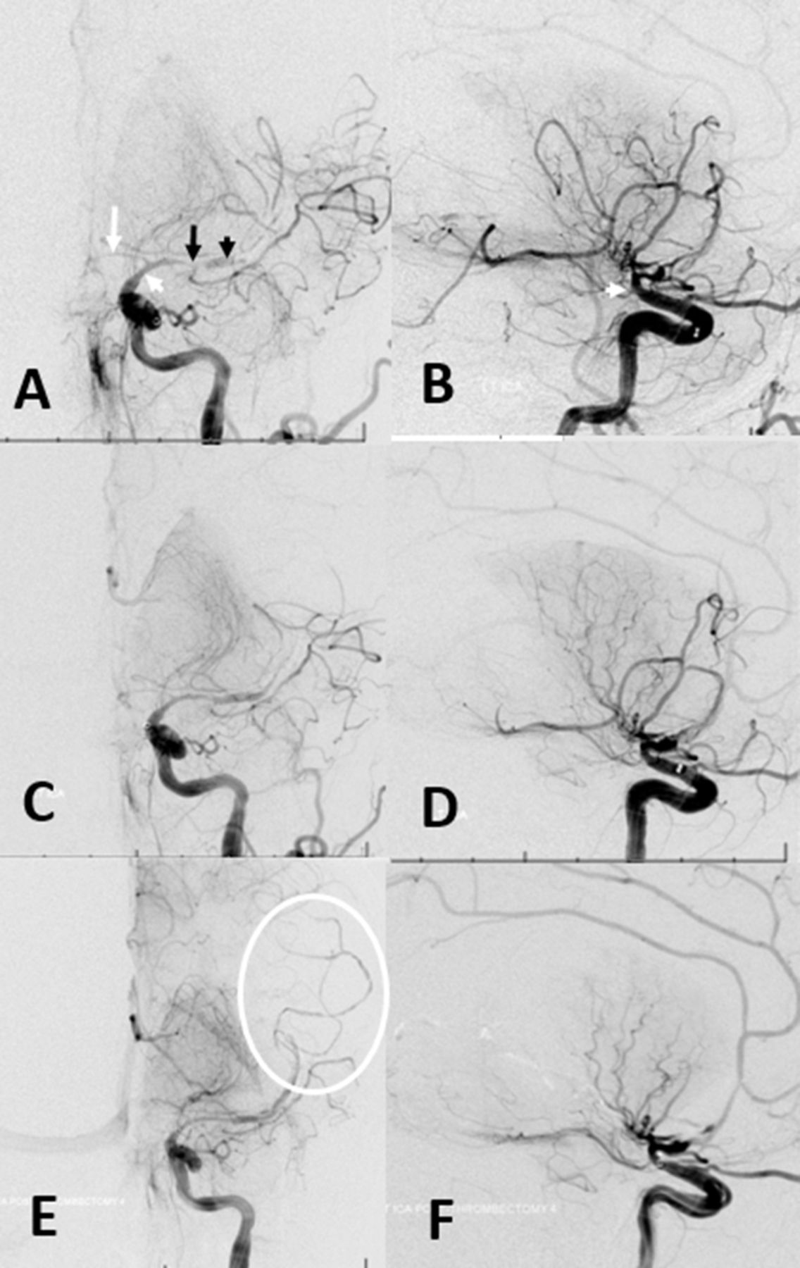
In our experience, acutely occlusive MCA-related dissections usually
present as a supraclinoid stenosis extending into M1 and A1 segments.
Inadvertent propagation of dissection during reperfusion attempts is a
distinct possibility. Unusual pruning of distal vessels, corkscrew
morphology, and rapid re-occlusion without apparent concentric stenosis
are some anatomical features (figure 11). Outcomes seem unpredictable, and more related to collateral efficiency rather than success of reperfusion.
Figure 11 Acute
intracranial dissection. (A,B) Initial images of supraclinoid stenosis
(dissection origin, white arrowhead) extending into A1 (white arrow) and
M1 (black arrow). A more “normal” caliber of the M2 segment (black
arrowhead) with no distal vessels reflects contrast in the false lumen.
(C,D) Following attempted thrombectomy, an apparent “improvement” in
proximal middle cerebral artery caliber reveals a corkscrew kind of
M1/M2 tapering with no overall improvement in perfusion. (E,F) Final
images show corkscrew appearance of dissection extending into the M2/M3
branches (E, oval).