Changing stroke rehab and research worldwide now.Time is Brain! trillions and trillions of neurons that DIE each day because there are NO effective hyperacute therapies besides tPA(only 12% effective). I have 523 posts on hyperacute therapy, enough for researchers to spend decades proving them out. These are my personal ideas and blog on stroke rehabilitation and stroke research. Do not attempt any of these without checking with your medical provider. Unless you join me in agitating, when you need these therapies they won't be there.

What this blog is for:

My blog is not to help survivors recover, it is to have the 10 million yearly stroke survivors light fires underneath their doctors, stroke hospitals and stroke researchers to get stroke solved. 100% recovery. The stroke medical world is completely failing at that goal, they don't even have it as a goal. Shortly after getting out of the hospital and getting NO information on the process or protocols of stroke rehabilitation and recovery I started searching on the internet and found that no other survivor received useful information. This is an attempt to cover all stroke rehabilitation information that should be readily available to survivors so they can talk with informed knowledge to their medical staff. It lays out what needs to be done to get stroke survivors closer to 100% recovery. It's quite disgusting that this information is not available from every stroke association and doctors group.

Showing posts with label contralateral. Show all posts

Monday, March 23, 2026

Stroke, here's how the brain's healthy hemisphere influences recovery: the revolutionary research

 Where is the EXACT PROTOCOL THAT DELIVERS RECOVERY? Oh NO, you fucking failed at your only job; creating stroke recovery protocols! You're fired! Your incompetent? doctor and hospital need to ensure human testing occurs.

Do you prefer your doctor, hospital and board of director's incompetence NOT KNOWING? OR NOT DOING? Your choice; let them be incompetent or demand action!

Stroke, here's how the brain's healthy hemisphere influences recovery: the revolutionary research

Rome, March 6. (Adnkronos Health) - After a stroke, attention focuses on the portion of the brain directly affected by the lesion, where neurons have been damaged. However, the functioning of the nervous system does not depend on isolated individual areas, but on networks of connections distributed between the two cerebral hemispheres. When one of the two is damaged, the other can also modify its activity, contributing decisively to recovery or hindering it. A study conducted by the Neuropharmacology Laboratory of Irccs Neuromed in Pozzilli (Isernia), in collaboration with Lund University in Sweden (Tadeusz Wieloch) and Washington University, St. Louis, USA (Adam Bouer), published in 'Stroke', focuses on this balance between the two cerebral hemispheres.


 The research - Neuromed informs - has identified a crucial node for the recovery of motor function precisely in the contralateral hemisphere, i.e., the one opposite to the lesion. In other words, after a stroke, the configuration of brain networks changes profoundly: the unaffected hemisphere can become excessively active, leading to a functional imbalance that hinders recovery.

 To identify possible pharmacological strategies capable of restoring the balance between the two hemispheres - a note explains - researchers focused on type 5 metabotropic glutamate receptors, or mGlu5, proteins that regulate communication between neurons and synaptic plasticity processes, i.e., the brain's ability to modify its connections. To precisely understand how mGlu5 receptors act in promoting or hindering post-stroke recovery, scientists used light-sensitive molecules, capable of being selectively activated or deactivated in specific brain regions. This approach, called photopharmacology, allows modulating the drug's effect in very circumscribed areas without genetically intervening on neurons.

 "Our research - explains Federica Mastroiacovo, from Neuromed's Neuropharmacology Laboratory, the study's first author - has shown that motor recovery after a stroke can be decisively influenced by the cerebral hemisphere not affected by the lesion. By selectively blocking mGlu5 receptors in the homotopic cerebral area contralateral to the lesion, we observed a significant improvement in function, while the same intervention in the lesioned area did not produce comparable effects." For the researchers, this is therefore an intervention aimed at functional recovery, regardless of the extent of ischemic damage and any previous vascular therapeutic strategies.

 "This study - comments Ferdinando Nicoletti, full professor of Pharmacology at Sapienza University of Rome and head of Neuromed's Neuropharmacology Laboratory - precisely identifies the cerebral site necessary for the blockade of mGlu5 receptors to promote recovery. The results indicate that the contralateral hemisphere is not merely a spectator of the damage, but an active participant in network reorganization processes. Understanding these mechanisms is essential for developing increasingly targeted interventions in the post-ischemic phase of stroke".

 The research - the note specifies - was conducted in animal models of stroke and represents an advancement in understanding the neurobiological mechanisms that regulate brain plasticity after ischemic damage. Further studies will be needed to verify if and to what extent these results can be translated into therapeutic applications in humans.

Adnkronos International: info@adnkronos.com

Thursday, March 19, 2026

Stroke, here's how the brain's healthy hemisphere influences recovery: the revolutionary research

 Since you didn't write AN EXACT PROTOCOL ON HOW TO USE THIS; COMPLETELY FUCKING USELESS! You're fired! And you knew how useless your research was by calling for further studies! That is world class incompetence!

Stroke, here's how the brain's healthy hemisphere influences recovery: the revolutionary research

Rome, March 6. (Adnkronos Health) - After a stroke, attention focuses on the portion of the brain directly affected by the lesion, where neurons have been damaged. However, the functioning of the nervous system does not depend on isolated individual areas, but on networks of connections distributed between the two cerebral hemispheres. When one of the two is damaged, the other can also modify its activity, contributing decisively to recovery or hindering it. A study conducted by the Neuropharmacology Laboratory of Irccs Neuromed in Pozzilli (Isernia), in collaboration with Lund University in Sweden (Tadeusz Wieloch) and Washington University, St. Louis, USA (Adam Bouer), published in 'Stroke', focuses on this balance between the two cerebral hemispheres.


 The research - Neuromed informs - has identified a crucial node for the recovery of motor function precisely in the contralateral hemisphere, i.e., the one opposite to the lesion. In other words, after a stroke, the configuration of brain networks changes profoundly: the unaffected hemisphere can become excessively active, leading to a functional imbalance that hinders recovery.

 To identify possible pharmacological strategies capable of restoring the balance between the two hemispheres - a note explains - researchers focused on type 5 metabotropic glutamate receptors, or mGlu5, proteins that regulate communication between neurons and synaptic plasticity processes, i.e., the brain's ability to modify its connections. To precisely understand how mGlu5 receptors act in promoting or hindering post-stroke recovery, scientists used light-sensitive molecules, capable of being selectively activated or deactivated in specific brain regions. This approach, called photopharmacology, allows modulating the drug's effect in very circumscribed areas without genetically intervening on neurons.

 "Our research - explains Federica Mastroiacovo, from Neuromed's Neuropharmacology Laboratory, the study's first author - has shown that motor recovery after a stroke can be decisively influenced by the cerebral hemisphere not affected by the lesion. By selectively blocking mGlu5 receptors in the homotopic cerebral area contralateral to the lesion, we observed a significant improvement in function, while the same intervention in the lesioned area did not produce comparable effects." For the researchers, this is therefore an intervention aimed at functional recovery, regardless of the extent of ischemic damage and any previous vascular therapeutic strategies.

 "This study - comments Ferdinando Nicoletti, full professor of Pharmacology at Sapienza University of Rome and head of Neuromed's Neuropharmacology Laboratory - precisely identifies the cerebral site necessary for the blockade of mGlu5 receptors to promote recovery. The results indicate that the contralateral hemisphere is not merely a spectator of the damage, but an active participant in network reorganization processes. Understanding these mechanisms is essential for developing increasingly targeted interventions in the post-ischemic phase of stroke".

 The research - the note specifies - was conducted in animal models of stroke and represents an advancement in understanding the neurobiological mechanisms that regulate brain plasticity after ischemic damage. Further studies will be needed to verify if and to what extent these results can be translated into therapeutic applications in humans.

Thursday, June 30, 2022

Next-gen stroke rehab: Robot at home

 Now the only question is: EXACTLY WHAT DAMAGE DIAGNOSIS will this be useful for? I lost my motor cortex and most of my pre-motor cortex so the only possibility for me would be if my contralateral side could be trained to control both hands.

Next-gen stroke rehab: Robot at home

Exoskeleton controlled by brain, developed at University of Houston, now in clinical trials

Grant and Award Announcement

University of Houston

University of Houston engineering professor Jose Luis Contreras-Vidal, an international pioneer in noninvasive brain-machine interfaces and robotic device inventions

image: University of Houston engineering professor Jose Luis Contreras-Vidal, an international pioneer in noninvasive brain-machine interfaces and robotic device inventions, has developed a portable robot for stroke rehabilitation. view more 

Credit: University of Houston

When 66-year-old Oswald Reedus had a stroke in 2014, he became one of 795,000 people in the United States who annually suffer the same fate. This year he also became the first stroke patient in the world to use a robotic arm controlled by his brainwaves - at home - to recover the use of a limb.  

Reedus was lucky to live in Houston and have access to this futuristic-looking, portable device - an invention of University of Houston engineering professor Jose Luis Contreras-Vidal, an international pioneer in noninvasive brain-machine interfaces and robotic device inventions. His team developed the portable brain-computer interface (BCI) exoskeleton to restore upper limb function. 

It’s the next generation of stroke rehabilitation, and now Reedus’ name will forever be associated with it. 

“If I can pass along anything to help a stroke person’s life, I will do it. For me it’s my purpose in life now,” said Reedus, whose determination sharpened after his mother and younger brother both died of strokes. 

Reedus realized he had lost the use of his left arm the night he had the stroke. His wife roused him from sleep, asking him to get up because he was mumbling, and she couldn’t understand his words. He tried but couldn’t use his left arm to help him rise.  

The stroke also caused Reedus to suffer aphasia, a difficulty with speech, barely noticeable now. 

“I don’t know why God spared me, but I want to leave here helping someone,” he said. 

Now he’s helping usher in a pivotal moment in stroke rehabilitation and medical science. Goal achieved. 

Using the robot 

Most neuro technologies are limited to the lab or clinic and are very expensive and hard to operate. This brain-controlled robotic arm requires no surgery and is accessible to robotically guide stroke rehabilitation both in clinic and at home. Reedus’ use of it in his Houston home follows clinical trials at TIRR Memorial Hermann. 

“The broader impact and commercial potential of this project is to advance national health by accelerating development, efficacy and use of brain-controlled robotic rehabilitation after stroke by capitalizing on the benefits of non-invasive brain interfaces that extract information about the patient’s motor intent and the real-time assessment of impairment and recovery of motor function," said Contreras-Vidal, Hugh Roy and Lillie Cranz Cullen Distinguished Professor of electrical and computer engineering at UH. “Brain-machine interfaces based on scalp electroencephalography (EEG) have the potential to promote cortical plasticity following stroke, which has been shown to improve motor recovery outcomes.” 

Neuroplasticity is the brain’s ability to modify, change, adapt and recover itself. Like a plastic material, which can be stretched and shaped to a desired design, there are certain properties in the brain that induce flexibility to recover even decades after a stroke or brain injury.

Friday, August 13, 2021

Neurohormones: A major new dimension in the problem of brain injury

 No clue.

Neurohormones: A major new dimension in the problem of brain injury

  1. Jonathan R Wolpaw  Is a corresponding author
  2. Jonathan S Carp
  1. National Center for Adaptive Neurotechnologies, Albany Stratton VA Medical Center, United States
  2. Albany Stratton VA Medical Center, United States
Insight

Cite
as: eLife 2021;10:e72048 doi: 10.7554/eLife.72048

Abstract

Evidence that neurohormones contribute to the contralateral effects of unilateral brain injury challenges a fundamental assumption of basic neuroscience and clinical neurology.

Main text

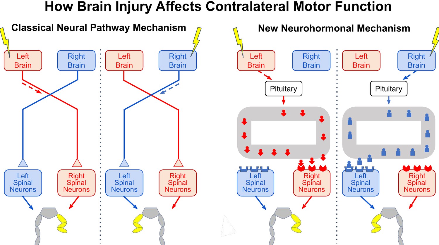
As Hippocrates knew, an injury to one side of the brain, such as a stroke, affects muscle control on the opposite side of the body (Clarke and O’Malley, 1996). For centuries, it has been assumed that this contralateral effect is due entirely to abnormal activity in neural pathways from the injured side of the brain that cross the midline to activate the spinal cord neurons that control muscles on the opposite side of the body (Figure 1, left side).

Figure 1

Now, in eLife, Georgy Bakalkin (Uppsala University), Jens Schouenborg (Lund University) and colleagues in Denmark, Portugal, Russia and Sweden – including Nikolay Lukoyanov, Hiroyuki Watanabe, Liliana Carvalho, Olga Nosova and Daniil Sarkisyan as joint first authors – report that neurohormones produced by the pituitary gland at the base of the brain may also contribute to the contralateral effect of brain injury (Lukoyanov et al., 2021). They build on previous studies showing that pituitary neurohormones can have side-specific effects in uninjured rats. If confirmed, these latest results are enormously significant, both scientifically and clinically.

In a straightforward set of experiments, the spinal cord of rats was completely transected in the mid-back, thereby cutting the neural pathways from the brain to the spinal neurons that control the hind-leg muscles on the opposite side. Then, the sensorimotor area of cerebral cortex was injured on one side of the brain. Surprisingly, the researchers observed an abnormal posture in the opposite hind leg similar to that seen when the sensorimotor cortex was injured in rats in which the spinal cord had not been transected.

To explore this further, the researchers removed the pituitary gland before transecting the spinal cord and injuring one side of the brain. With the pituitary removed and the spinal cord transected, the brain injury did not cause an abnormal posture in the opposite leg. Moreover, injecting healthy animals with specific pituitary hormones, or with an extract from the blood of brain-injured animals, produced a comparable abnormal posture on one side, thereby confirming a previously unrecognized contribution of neurohormones to the contralateral effect of a unilateral brain injury. This effect may result from the asymmetrical distribution of some neurohormonal receptors in the spinal cord (Figure 1, right side; Kononenko et al., 2017; Watanabe et al., 2021).

Perhaps the most remarkable aspect of these findings is that the phenomenon the researchers describe has gone largely unrecognized for so long. Certainly, the results require further studies, also involving other species. A thorough search of the clinical literature for reports about the effect of hemispheric stroke in people with complete spinal cord injuries would also be in order. But assuming that the results are confirmed and do occur in other species, in particular primates, the scientific questions they raise – and the clinical possibilities they introduce – are significant and exciting.

A variety of studies show that lateralized effects of neurohormones contribute to normal brain function (e.g., Deliagina et al., 2000; Marlin et al., 2015; Nation et al., 2018; Watanabe et al., 2015). It remains to be seen whether the lateralized effects of neurohormones following brain injury are limited to motor function or, more plausibly, whether they also affect other nervous system functions, such as vision or language. Language problems often occur with stroke on the left side of the brain, and neurohormones might play a role in these difficulties.

It is also unclear how lateralized neurohormonal effects interact with lateralized neural pathway effects. They clearly differ in mechanisms, and probably in other respects as well, such as time frames. Are their interactions synergistic, additive or opposing, or does this vary from one situation to another? Another important question is how the lateralized effects of neural pathways and neurohormones operate in normal life. How do they interact to ensure that normal function is maintained throughout life? For example, learning new motor skills is currently studied and understood entirely in terms of neuronal and synaptic plasticity in neural pathways (e.g., Dayan and Cohen, 2011). Do neurohormones contribute as well?

The work of Lukoyanov et al. adds a whole new dimension to the problem of brain injury and to the opportunities for new therapies that enhance recovery. They studied neurohormonal effects in the first few hours after injury; thus, the long-term effects are unknown. For example, neurohormones might contribute to the abnormal movement patterns that emerge after stroke (e.g., Senesh et al., 2020). Knowledge of their acute and chronic effects could help shape the design of new therapeutic regimens, both in general and for individual patients; appropriate treatments may well differ depending on which side of the brain is injured. Furthermore, new classes of therapeutic agents that mimic or oppose neurohormones, or affect their endogenous production, might enhance recovery of useful function beyond that achievable with present methods.

In summary, the truly groundbreaking research of Lukoyanov et al. opens a new research area and demands a host of further studies. If the results are replicated, the impact, excitement and activities they will generate are likely to continue growing well into the future.

The effects of brain injury on contralateral motor function.

Injury to one side of the brain (yellow lightning bolt) impairs motor function on the opposite side of the body, causing the abnormal flexed posture of the opposite hind leg (yellow). It now appears …


Wednesday, March 20, 2019

Health Beat: Stroke shoe retrains the brain

Interesting that this is put on the good foot, similar to this? This probably would do nothing for me, the spasticity turning my foot out and lack of a free swinging lower leg wouldn't be corrected  by this.

Exercising the good side to recover the 'bad' side.


Health Beat: Stroke shoe retrains the brain


"I used to walk three to five miles a day before my stroke. and it would be nice if I could just walk a half a mile," she said.
Hintz is making strides with a patented portable shoe. It's called the Moterum iStride device. It was invented at the University of South Florida in Tampa. Doctors have been working for years to get it just right, and they're almost to the finish line.
"It took a lot of math, a lot of engineering and quite a few different prototypes to get it to work just right," said Kyle Reed, an associate professor of mechanical engineering at USF.
Many stroke patients are left with a limp because of damage to their central nervous system. The shoe helps rewire the brain so they can correct their gait. Doctors said it's more effective and cheaper than the typical split belt treadmill treatment, and patients can even take it home.
"The iStride device causes one foot to move backwards while they're walking and this helps to exaggerate one of the feet so it becomes more asymmetric, especially when they take it off," Reed continued. "They have a corrected gait where it's more symmetric afterwards."
"Don't forget the patient is wearing the shoe on their good side," said Seok Hun Kim, an associate professor of physical therapy and rehabilitation sciences at USF.

Monday, December 4, 2017

Empowering the Mind to Heal the Brain for Stroke

The picture of Dr. Eric Leuthardt hovering over someones' head with a drill and drill bit does not make me want to try this although it might be the only way I'll ever recover my left hand function. That picture is inaccurate since no surgery is required for this.

Empowering the Mind to Heal the Brain for Stroke


MIT Technology Review picture of Dr Eric Leuthardt

The capability to “decode the brain” now makes thought-controlled machines a reality. Even more fundamentally theses brain computer interfaces empower the mind to now heal the physical substance of the brain after stroke. The Neurolution laboratory has created brain computer interfaces that enable a person with a chronically paralyzed had to recover function by using brain signals from the uninjured side of their brain. With continued practice, while the patient’s machine-empowered thoughts enable them to move their paralyzed hand, something extraordinary happens — the patients begin to recover function.

Dr. Eric Leuthardt is an Associate Professor of Neurological Surgery and Biomedical Engineering at Washington University School of Medicine, where he specializes in brain-mapping for patients with brain tumors and epilepsy; Director of Washington University’s Center for Innovation in Neuroscience and Technology, a collaborative collective for developing new technologies to improve neurosurgery; and a published author of the hit psychological thriller RedDevil_4.
He has a 17 minute TEDx video at the link.

Saturday, September 17, 2016

Contralaterally Controlled Functional Electrical Stimulation Improves Hand Dexterity in Chronic Hemiparesis: A Randomized Trial

So what if it does? Useless without a protocol being written for it. Does no one ever think of the survivors?
http://stroke.ahajournals.org/content/strokeaha/early/2016/09/08/STROKEAHA.116.013791.full.pdf
1
N
euromuscular electrical stimulation (NMES) of the paretic wrist and finger extensors is routinely used in
stroke rehabilitation to promote recovery of muscle strength and upper extremity function. A recent review of 31 randomized controlled trials concluded that there is strong evidence that NMES applied in the context of task practice improves upper extremity function in subacute and chronic stroke.
1
This is corroborated by a recent systematic review with meta-analysis that concluded that functional electrical stimulation improves activity compared with training alone.
2
Cyclic NMES (cNMES) is a commonly used and widely available method of administering NMES in stroke rehabilitation.
3
With cNMES, stimulation is delivered according to an on–off cycle, with the cycle timing, repetitions, and intensity of stimulation set by the therapist. Thus, cNMES requires no active participation from the patient, and because the patient does not control the timing or intensity of stimulation,
cNMES is not easily used to assist functional task practice (FTP). Nevertheless, several studies have shown that cNMES can reduce upper limb motor impairment compared with control groups
4,5
although the longevity of effect is inconsistent across studies.
6
Contralaterally controlled functional electrical stimulation (CCFES) is a new NMES modality that enables the patient to actively open their paretic hand and perform functional tasks. With CCFES, the patient controls the stimulation to their paretic hand in real-time by opening and closing their strong hand. An instrumented glove worn on the strong hand modulates the stimulation intensity to the paretic hand extensors so that both hands open synchronously (Figure I in the online-only Data Supplement
).
7
CCFES may be more effective than cNMES because the stimulation is intention driven; the patient
Background and Purpose
It is unknown whether one method of neuromuscular electrical stimulation for poststroke
upper limb rehabilitation is more effective than another. Our aim was to compare the effects of contralaterally controlled functional electrical stimulation (CCFES) with cyclic neuromuscular electrical stimulation (cNMES).
Methods
Stroke patients with chronic (>6 months) moderate to severe upper extremity hemiparesis (n=80) were randomized to receive 10 sessions/wk of CCFES- or cNMES-assisted hand opening exercise at home plus 20 sessions of functional task practice in the laboratory for 12 weeks. The task practice for the CCFES group was stimulation assisted. The primary outcome was change in Box and Block Test (BBT) score at 6 months post treatment. Upper extremity Fugl–Meyer and
Arm Motor Abilities Test were also measured.
Results
At 6 months post treatment, the CCFES group had greater improvement on the BBT, 4.6 (95% confidence interval
[CI], 2.2–7.0), than the cNMES group, 1.8 (95% CI, 0.6–3.0), between-group difference of 2.8 (95% CI, 0.1–5.5),
P
=0.045. No significant between-group difference was found for the upper extremity Fugl–Meyer (P
=0.888) or Arm Motor Abilities Test (P=0.096). Participants who had the largest improvements on BBT were <2 years post stroke with moderate (ie, not severe) hand impairment at baseline. Among these, the 6-month post-treatment BBT gains of the CCFES group, 9.6 (95% CI, 5.6–13.6), were greater than those of the cNMES group, 4.1 (95% CI, 1.7–6.5), between-group difference of 5.5 (95% CI, 0.8–10.2), P=0.023.
Conclusions
CCFES improved hand dexterity more than cNMES in chronic stroke survivors.
Clinical Trial Registration
URL:
http://www.clinicaltrials.gov
. Unique identifier: NCT00891319.
(
Stroke
. 2016;47:00-00. DOI: 10.1161/STROKEAHA.116.013791.)
Key

Friday, August 26, 2016

Combined Action Observation and Motor Imagery Neurofeedback Up-Regulates Contralateral Sensorimotor Activity

Whoa, three big words to confuse us peons; Up-regulates, Contralateral, Sensorimotor. I don't think researchers even want us to read research which is why they obsfucate their blatherings.
https://dalspace.library.dal.ca/handle/10222/72090
Author
Friesen, Christopher
 
Motor imagery (MI) and action observation have proven to be efficacious adjuncts to traditional physiotherapy, to enhance motor recovery from stroke. Recently, researchers have used a combined approach called imagined imitation (II), where an individual watches a motor task being performed, while simultaneously imagining they are performing the movement. While neurofeedback (NFB) has been used extensively with MI to improve patients’ ability to modulate sensorimotor activity and enhance motor recovery, the feasibility of using NFB with II is unknown. This project tested whether healthy controls could modulate sensorimotor lateralization during II-NFB of a unilateral handshake using electroencephalography, and whether this ability transferred to subsequent MI. Thirty-two subjects, receiving real or sham NFB attended four sessions where they engaged in II-NFB training and subsequent MI. Results showed the NFB group demonstrated more sensorimotor activity during sessions three and four, and that this NFB effect transferred to subsequent MI.
 

Tuesday, April 19, 2016

tDCS: "Current Distribution in the Brain From Surface Electrodes"

It seems that earlier research on tDCS had no clue what it was doing because they never knew what current actually got thru to the brain. I'm sure there was no measurement of thick skulls and attributing lack of success to that. Ask your doctor whether your skull is thin enough to successfully use tDCS, either regular or anodal. Ipsilateral or contralateral? And it has only been out there since 1968.

http://journals.lww.com/anesthesia-analgesia/Citation/1968/11000/Current_Distribution_in_the_Brain_From_Surface.16.aspx

RUSH, STANLEY Ph.D.; DRISCOLL, DANIEL A. M.E.E.

Anesthesia & Analgesia:
SCIENTIFIC ARTICLE: PDF Only


Thursday, October 2, 2014

Stroke victims recover use of weakened limbs by exercising unaffected limbs, research finds

This is only 2 years old, has any of this been implemented in your rehabilitation center? Or 120 years from the original discovery. Is that enough time for even the most ossified doctor to find and use?
http://blogs.vancouversun.com/2012/12/10/stroke-victims-recover-use-of-weakened-limbs-by-exercising-unaffected-limbs-research-finds/
Stroke victims can make astonishing gains in strength in weakened limbs by training the unaffected limbs on the other side of their body, according to new research by the University of Victoria.
Neuroscientist Paul Zehr and PhD candidate Katie Dragert designed “ridiculously simple” devices made of wooden boards and cloth straps that stroke victims used to strengthen the muscles in their legs and ankles. Patients completed a six-week high-intensity training regime — not with the limbs weakened by the stroke, but with the limbs that were less affected or unaffected.
What happened surprised even the researchers.
Patients gained as much strength in the weakened leg as they did in the leg that did the exercises. Patients achieved strength gains of about 30 per cent in both the trained and untrained legs, a far more dramatic effect than previous research on healthy people had achieved.
The finding promises to be a boon to patients whose limb strength is so impaired by stroke that they can’t lift or train the affected parts at all.
“Weakness is a big part of what happens after a stroke and if you can do something to increase people’s strength, you can help them get walking and all kinds of stuff,” said Zehr.
Patients in the study suffered their stroke on average about 80 months before training. That suggests patients can benefit from the program years after a debilitating event.
Study participant Barb Oliver suffered from weakness in her left leg after a stroke 10 years ago, but continues to make gains through UVic’s experimental programs.
“I couldn’t walk at all and they didn’t think I would ever walk again,” said Oliver. “Now, I get around with a cane.”
Zehr and Dragert employed a mostly forgotten 1894 discovery by Yale University researchers who found that when people train one arm, the other arm also gained strength.
“The arm that they trained got stronger, but the other arm got stronger, too, even though it wasn’t trained,” Zehr explained. “Over the years people have looked at cross-education of strength on different parts of the body, upper and lower limbs, and it pretty much shows up everywhere to a greater or lesser degree.”
Most of the research found that the untrained limb gains about half as much strength as the trained limb.
“A 30-per-cent gain on the trained side usually results in a 15-per-cent gain on the untrained side,” he said. “We thought that with all the damage caused by the stroke that we might see a five- or 10-per-cent gain in our patients’ untrained limbs.”
But the strength gains recorded in the UVic study of stroke victims were twice as high as the gains achieved by healthy people in past studies.
Much of the training gain in strength and skill that people achieve through exercise takes place in the brain and the nervous system rather than the muscles themselves, Zehr said.
The surprising strength of the cross-education effect suggest the training program may be tapping into communication pathways between the left and right sides of the brain and activating built-in — but little used — duplications in the neural wiring that controls movement, he said.
The study will be published in the journal Experimental Brain Research and has been published by that journal online.

Thursday, July 24, 2014

New Rehabilitation Institute of Chicago Brain Stimulation Study Reveals Breakthrough in Stroke Recovery

Ok, the RIC I guess can do good things also. Bad stuff here.
I like the fact that this was for chronic, a 5 year survivor.  This sounds like applying the earlier studies on contralateral recovery like these;
Cortical Reorganization After Stroke How Much and How Functional?

New model of how brain functions are organized may revolutionize stroke rehab

Magic for Stroke Patients: The One-Sided Workout

 So ask your therapists why this isn't available in their clinic and exactly what the protocol is.
http://www.wspa.com/story/25587087/new-rehabilitation-institute-of-chicago-brain-stimulation-study-reveals-breakthrough-in-stroke-recovery#.U8OcnUKrd_0.email
Doctors at the Rehabilitation Institute of Chicago (RIC) revealed the results of a study wherein 80 percent of stroke survivor participants regained arm and hand use – 30 percent more than possible with standard therapies. The breakthrough study combines for the first time a new, non-invasive brain stimulation technique with traditional occupational therapy (OT) to improve upper limb (arm and hand) movement recovery – and give patients back the independence they lost as a result of their strokes.
Richard L. Harvey, MD, medical director at the RIC Center for Stroke Rehabilitation and lead scientist of this study, researched and developed a new approach to modulate brain healing and improve lasting arm and hand use in stroke survivors. This type of brain stimulation is the only technology that has been shown to deliver greater functional improvements when accompanied with an OT session. Typically, only about 50 percent of post-stroke patients regain full upper limb use through OT alone.1 The results from RIC's study indicate this combined treatment could increase that success rate to 80 percent.
RIC worked with Nexstim Corporation to apply this novel type of non-invasive brain stimulation – navigated brain stimulation (NBS) – to standard stroke rehabilitation. After a stroke blocks blood to the brain, some regions of the affected brain hemisphere become less active, while some areas on the healthy side of the brain become more active. By externally and painlessly stimulating the overly active parts of the healthy brain with NBS magnetic waves, these targeted parts calm down. The RIC study indicates that when the overly active healthy brain areas quiet down, the stroke-damaged areas respond more fully and sustainably to occupational therapy. This allows the patient to have a better recovery and increased arm use.
"This study represents what could be a significant breakthrough in the treatment of strokes," said Harvey. "Our results indicate that targeting stimulation to the correct area can set up the brain to learn and retain more during traditional stroke rehabilitation. We know that stroke is a leading cause of serious disability and costs the U.S. more than $36 billion each year from lost work days and long-term health care. To these patients, regaining arm use and mobility means increasing their ability and, thus, their quality of life."
Darryl Holmes, one of the 30 stroke survivors with severe arm impairments in this pilot study, had a stroke in 2009 at age 57. After his stroke, his movement was limited. He had trouble using his left side, including his arm and hand, which prevented him from performing many of his regular activities, like driving and writing.
In 2013, Holmes participated in RIC's six-month stroke study and received treatment with NBS in combination with occupational therapy. This study used NBS to stimulate the brain with magnetic waves using a navigation system akin to a GPS map. Adding navigation to regular brain stimulation allows doctors to locate the exact area of the brain that should be inhibited, or calmed down, by this type of stimulation.
"Since the RIC study treatment, I've gone back to work," said Holmes. "I've gone into private practice. I drive a new car. Participating in this research has made me a healthier person, both physically and mentally. I have my life back, and it's good."
The Center for Stroke Rehabilitation at RIC integrates research directly into patient care and is the only federally-designated Rehabilitation Research and Training Center for Stroke, as recognized by the National Institute on Disability and Rehabilitation Research. RIC is the first hospital to run a trial with NBS as an aid to OT in stroke survivors. Now that the pilot study is complete, RIC has expanded the study and currently manages 12 other research centers in a nationwide study – the first large-scale trial of its kind to take a major step toward clinical availability of this treatment.
About The Rehabilitation Institute of Chicago
The Rehabilitation Institute of Chicago (RIC) is the nation's leading provider of comprehensive physical medicine and rehabilitation care to patients from around the world. Founded in 1954, RIC has been designated the "No. 1 Rehabilitation Hospital in America" by U.S. News & World Report every year since 1991. RIC sets the standard of care in the post-acute market through its innovative applied research and discovery programs, particularly in the areas of neuroscience, bionic medicine, musculoskeletal medicine and technology transfer. For more information, go to www.ric.org.

Tuesday, January 21, 2014

Cortical Reorganization After Stroke How Much and How Functional?

Your doctor will absolutely need to get this.
http://nro.sagepub.com/content/20/1/56.abstract?etoc
  1. Christian Grefkes1,2
  2. Nick S. Ward3,4
  1. 1Department of Neurology, Cologne University Hospital, Cologne, Germany
  2. 2Max Planck Institute for Neurological Research, Cologne, Germany
  3. 3Sobell Department of Motor Neuroscience, UCL Institute of Neurology, London, UK
  4. 4The National Hospital for Neurology and Neurosurgery, London, UK
  1. Christian Grefkes, Department of Neurology, Cologne University Hospital, Kerpener Strasse 62, Cologne 50624, Germany. Email: christian.grefkes@uk-koeln.de

Abstract

The brain has an intrinsic capacity to compensate for structural damage through reorganizing of surviving networks. These processes are fundamental for recovery of function after many forms of brain injury, including stroke. Functional neuroimaging techniques have allowed the investigation of these processes in vivo. Here, we review key advances over the past two decades that have shed light on the neural mechanisms enabling recovery of motor function after stroke. We first provide an overview on invasive stroke models in non-human primates that provided insights into lesion-induced changes in the cortical representations of the upper limb. We then present key findings from neuroimaging studies in human stroke patients, which suggest that the role of contralesional motor hemisphere in supporting recovered function depends on factors such as time since stroke, lesion location and anatomical region. More recently, research has been directed at understanding how surviving brain regions influence one another during movement. It appears that it is not only the corticospinal tract but also brainstem pathways and interhemispheric connections that affect cortical reorganization patterns and functional recovery. In summary, neuroimaging opens the way for greater understanding of the mechanisms of recovery and potentially improves our ability to deliver effective restorative therapy.

Friday, April 26, 2013

New model of how brain functions are organized may revolutionize stroke rehab

I'm sure this won't help us for at least 40 years but you could challenge your doctor to describe how this is different from your current stroke protocol.
http://news.psu.edu/story/273127/2013/04/15/research/new-model-how-brain-functions-are-organized-may-revolutionize
A new model of brain lateralization for movement could dramatically improve the future of rehabilitation for stroke patients, according to Penn State researcher Robert Sainburg, who proposed and confirmed the model through novel virtual reality and brain lesion experiments.
Since the 1860s, neuroscientists have known that the human brain is organized into two hemispheres, each of which is responsible for different functions. Known as neural lateralization, this functional division has significant implications for the control of movement and is familiar in the phenomenon of handedness.
Understanding the connections between neural lateralization and motor control is crucial to many applications, including the rehabilitation of stroke patients. While most people intuitively understand handedness, the neural foundations underlying motor asymmetry have until recently remained elusive, according to Sainburg, professor of kinesiology and neurology and participant in the neuroscience and physiology graduate programs at the University's Huck Institutes of the Life Sciences.
Research by Sainburg and his colleagues in the Center for Motor Control and published in the journal Brain has revealed a new model of motor lateralization that accounts for the neural foundations of handedness. The discovery could fundamentally change the way post-stroke rehabilitation is designed.
"Each hemisphere of the brain is specialized for different aspects of motor control, and thus each arm is 'dominant' for different features of movement," said Sainburg. "The dominant arm is used for applying specific force sequences -- such as when slicing a loaf of bread with a knife -- and the other arm is used for impeding forces to maintain stable posture, such as holding the loaf of bread. Together these specialized control mechanisms are seamlessly integrated into every day activities.
"Our research has shown that this integration breaks down in neural disorders such as stroke, which produces different motor deficits depending on whether the right or left hemisphere has been damaged," Sainburg continued. "Traditionally, physical rehabilitation professionals have used the same protocols to practice movements of the paretic arm, regardless of the hemisphere that has been damaged. Our research shows that each arm should be treated for different control deficits, and it also indicates that therapists should directly retrain patients in how to use the two arms together in order to recover function."
In preparing to test their model, Sainburg and his team selected study participants from the New Mexico Veterans Administration Hospital and Penn State Milton S. Hershey Medical Center based on specific criteria in order to accurately distinguish the motor control mechanisms specific to each brain hemisphere. Participants were then asked to perform a series of tasks on a virtual reality interface, programmed and designed by Sainburg, which allowed the researchers to record detailed 3D position and motion data. The data for all the participants' hand trajectories and final positions were then aggregated to compare the effects of left versus right hemisphere damage on different aspects of control.
"Our results indicated that while both groups of patients showed similar clinical impairment in the contralesional arm, this was produced by different motor control deficits," Sainburg said. "Right hemisphere damaged patients were able to make straight movements that were directed toward the targets, but were unable to stabilize their arms in the targets at the end of motion. In contrast, left hemisphere damaged patients were unable to make straight and efficient movements, but had no difficulty stabilizing their arms at the end of motion. These results confirmed that each hemisphere contributes unique control to its contralesional arm, verifying why our arms seem different when we use them for the same tasks."
Results mirror those of Sainburg's prior studies of motor deficits in unilateral stroke patients, focused on the ipsilesional arm, which formed the basis for his model of lateralization.
"Because both arms in stroke patients show motor deficits that are specific to the hemisphere that was damaged, we have concluded that the left arm is not simply controlled with the right hemisphere and vice versa," Sainburg said. "This is a revolutionarily new perspective on sensorimotor control: each hemisphere contributes different control mechanisms to the coordination of both arms, regardless of which arm is considered dominant."
Sainburg and his colleagues are currently designing follow-up studies that will aid the development of new rehabilitation protocols addressing the specific motor deficits associated with each hemisphere.
This work was supported by grants from the National Institutes of Health, National Institute for Child Health and Human Development, Department of Veterans Affairs Research and Development Medical Merit Review, and Rehabilitation Research and Development.
Read the unabridged article by Seth Palmer on the website of the Huck Institutes of the Life Sciences.

Thursday, December 20, 2012

Magic for Stroke Patients: The One-Sided Workout

Your therapists should be able to construct a stroke protocol from this. Ask them for it.
The columnist writing a article here:
Magic for Stroke Patients: The One-Sided Workout
The original research from the university here:
Brain to brawn: Training one leg strengthens both after stroke

To recover strength and ultimately perhaps the ability to walk, the best bet after a severe stroke might just be to forego working the weaker, more-affected side. It seems counter-intuitive, but high-intensity strength training on the less-affected side could have remarkable potential for helping recover mobility after a stroke, new UVic research indicates.
The notion of cross-education of strength—training one side of the body achieves strength gains in the corresponding muscles on the other side—has had considerable study since it was first proven at Yale University in 1894. Typically, the corresponding strength gain in the non-trained side is about half the improvement on the trained side.
While an interesting phenomenon, it’s not had serious clinical application, since most people or patients are looking to strengthen both sides of their body symmetrically.
“It just sat in the literature as one of those quirky observations,” says Dr. E. Paul Zehr, professor of neuroscience in the Division of Medical Sciences and School of Exercise Science, Physical and Health Education at UVic.
But Zehr, who studies how the arms and legs work together while we walk and how that information can be used to help recover walking when it’s been lost due to nervous system damage, thought cross-education might be useful for people with an existing asymmetry, such as those who’ve suffered a stroke.
“After a stroke, you’re weaker all over, but it’s more prominent on one side,” says Zehr. “The standard way to recover walking is to strength-train that weak side, particularly the leg.”
A couple years ago, Zehr started thinking about “tapping into” the weaker side indirectly by using the less-affected limb and decided the idea needed to be explored.
And his study showed remarkable results. Training on the less-affected side of the body in post-stroke patients achieved equivalent strength gains (about 30 per cent) on both sides. The researchers had predicted about a 5–10-per-cent strength gain.
“I never imagined that it would come out like this,” says Zehr. “The results exceeded any normal expectations.”
The results of the study, the first time the theory was tested for stroke sufferers, are to be published in the journal Experimental Brain Research and are already online.
Zehr notes that the results show huge potential for neurological rehabilitation because the strength gain was equal on both sides, but also because the training effect was shown well after a stroke event. Of 19 study participants, many were years after a stroke event and the average was 80 months after a stroke.
“It doesn’t matter how long it’s been after a stroke, strength can still be recovered,” says Zehr.
Study participants completed six weeks of training, involving three sessions a week—two at home and one in the lab, without aid of any specialized equipment.
The study focused on the legs with an eye toward walking recovery. Zehr says the next steps are to test cross-education of strength in the upper body “to come at the more affected leg from different angles” and work toward integrating the practice into a full walking retraining program.
“The big-picture view is looking at changing the way we do mobility rehabilitation,” Zehr says.

Monday, April 16, 2012

Chronic In Vivo Imaging Shows No Evidence of Dendritic Plasticity or Functional Remapping in the Contralesional Cortex after Stroke

In vivo means while live. So I don't know if this was because the scan  wasn't strong enough to find the changes or if they really don't occur. 4 weeks seems rather short.
http://cercor.oxfordjournals.org/content/early/2012/04/10/cercor.bhs092.abstract

Abstract

Most stroke survivors exhibit a partial recovery from their deficits. This presumably occurs because of remapping of lost capabilities to functionally related brain areas. Functional brain imaging studies suggest that remapping in the contralateral uninjured cortex might represent a transient stage of compensatory plasticity. Some postmortem studies have also shown that cortical lesions, including stroke, can trigger dendritic plasticity in the contralateral hemisphere, but the data are controversial. We used longitudinal in vivo two-photon microscopy in the contralateral homotopic cortex to record changes in dendritic spines of layer 5 pyramidal neurons in green fluorescent protein mice. We could not detect de novo growth of dendrites or changes in the density or turnover of spines for up to 4 weeks after stroke. We also used intrinsic optical signal imaging to investigate whether the forepaw (FP) sensory representation is remapped to the spared homotopic cortex after stroke. Stimulation of the contralateral FP reliably produced strong intrinsic signals in the spared hemisphere, but we could never detect a signal with ipsilateral FP stimulation after stroke. This lack of contralateral plasticity at the level of apical dendrites of layer 5 pyramidal neurons and FP sensory maps suggests that the contralesional cortex may not contribute to functional recovery after stroke and that, at least in mice, the peri-infarct cortex plays the dominant role in postischemic plasticity.