Changing stroke rehab and research worldwide now.Time is Brain! trillions and trillions of neurons that DIE each day because there are NO effective hyperacute therapies besides tPA(only 12% effective). I have 523 posts on hyperacute therapy, enough for researchers to spend decades proving them out. These are my personal ideas and blog on stroke rehabilitation and stroke research. Do not attempt any of these without checking with your medical provider. Unless you join me in agitating, when you need these therapies they won't be there.

What this blog is for:

My blog is not to help survivors recover, it is to have the 10 million yearly stroke survivors light fires underneath their doctors, stroke hospitals and stroke researchers to get stroke solved. 100% recovery. The stroke medical world is completely failing at that goal, they don't even have it as a goal. Shortly after getting out of the hospital and getting NO information on the process or protocols of stroke rehabilitation and recovery I started searching on the internet and found that no other survivor received useful information. This is an attempt to cover all stroke rehabilitation information that should be readily available to survivors so they can talk with informed knowledge to their medical staff. It lays out what needs to be done to get stroke survivors closer to 100% recovery. It's quite disgusting that this information is not available from every stroke association and doctors group.

Showing posts with label Normobaric oxygen. Show all posts

Sunday, October 13, 2024

Evaluating the Effectiveness of Neuroprotective Strategies in Enhancing Post-stroke Recovery: A Systematic Review of Meta-Analyses and Clinical Trials

 First of all, stop using the term, neuroprotection! Neuronal cascade of death is the correct term! It gives absolutely NO SENSE OF URGENCY! If you tell your patients, you know nothing about stopping  the 5 causes of the neuronal cascade of death in the first week thus letting die hundreds of millions to billions of neurons.  They might just get angry with your incompetence!

Send me hate mail on this: oc1dean@gmail.com. I'll print your complete statement with your name and my response in my blog. Or are you afraid to engage with my stroke-addled mind? I would like to know why you aren't solving stroke to 100% recovery, because this doesn't solve stroke recovery at all!

Evaluating the Effectiveness of Neuroprotective Strategies in Enhancing Post-stroke Recovery: A Systematic Review of Meta-Analyses and Clinical Trials 

Published: October 12, 2024

DOI: 10.7759/cureus.71343 

 Peer-Reviewed

Cite this article as: Hassan B, Dabas M M, Kanemitsu K, et al. (October 12, 2024) Evaluating the Effectiveness of Neuroprotective Strategies in Enhancing Post-stroke Recovery: A Systematic Review of Meta-Analyses and Clinical Trials. Cureus 16(10): e71343. doi:10.7759/cureus.71343

Abstract

This systematic review evaluates the effectiveness of various neuroprotective strategies in enhancing recovery following acute ischemic stroke, focusing on interventions such as normobaric oxygen (NBO), lithium, selective serotonin reuptake inhibitors (SSRIs), and Cerebrolysin. Drawing upon data from six primary studies, including randomized controlled trials (RCTs) and meta-analyses, we assessed these therapies' impact on functional outcomes, motor recovery, and neurological improvement. Normobaric oxygen, across 12 RCTs, demonstrated limited efficacy in improving recovery outcomes or reducing mortality. Lithium, supported by animal models but with inconclusive human data, showed potential in reducing stroke volume but did not significantly enhance functional recovery in clinical trials. SSRIs, particularly fluoxetine, showed moderate success in improving motor recovery, as evidenced by the FLAME (Fluoxetine for Motor Recovery after Acute Ischaemic Stroke) trial and meta-analyses. Cerebrolysin demonstrated consistent improvement in early neurological function and motor recovery, with a number-needed-to-treat (NNT) of 7.1 for early NIHSS (National Institutes of Health Stroke Scale) score improvements. Our Preferred Reporting Items for Systematic Reviews and Meta-Analyses (PRISMA)-guided search covered PubMed, Medline, Embase, and the Cochrane Library up to September 2024. These findings emphasize the mixed efficacy of these neuroprotective interventions and underscore the necessity for personalized treatment protocols and further large-scale, controlled trials to clarify their roles in clinical practice. This review contributes to the ongoing dialogue on optimizing post-stroke recovery and highlights the critical need for evidence-based neuroprotective strategies.

Introduction & Background

Stroke remains one of the leading causes of death and disability worldwide, with ischemic stroke accounting for the majority of cases. The pathophysiology of stroke involves a cascade of events, including excitotoxicity, oxidative stress, inflammation, and apoptosis, all contributing to brain injury and long-term neurological deficits [1]. Early intervention with neuroprotective therapies has the potential to limit such damage and enhance functional recovery [2]. Despite significant advancements in acute stroke care, such as thrombolysis and thrombectomy, there is still an unmet need for effective neuroprotective strategies that can be implemented during the subacute phase of stroke recovery [3].

We selected normobaric oxygen (NBO), lithium, selective serotonin reuptake inhibitors (SSRIs), and Cerebrolysin for review due to their unique mechanisms of action and their emerging roles in neuroprotection and post-stroke recovery. These therapies have shown varying degrees of promise in both preclinical studies and early-phase clinical trials. NBO has been explored for its ability to increase oxygen delivery to ischemic brain tissue, though meta-analyses suggest that it may not significantly improve functional outcomes [4,5]. Lithium, widely known for its mood-stabilizing effects, has demonstrated potential in reducing stroke volume and improving post-stroke function in animal models, but human data remain inconclusive. SSRIs, particularly fluoxetine, have garnered attention for their ability to enhance motor recovery post-stroke, independent of their antidepressant effects, as supported by the FLAME (Fluoxetine for Motor Recovery After Acute Ischemic Stroke) trial and subsequent meta-analyses [6]. Cerebrolysin, a neuropeptide preparation, has been the subject of multiple randomized controlled trials (RCTs) and meta-analyses, with studies suggesting its potential for early neurological improvement, though evidence of long-term benefits remains limited [7].

These four interventions were selected based on their clinical relevance, availability of substantial data from RCTs and meta-analyses, and their distinct neuroprotective mechanisms. Compared to other emerging interventions, such as hypothermia, neurotrophic factors, and stem cell therapies, NBO, lithium, SSRIs, and Cerebrolysin have garnered the most clinical attention and research, making them well-suited for a focused systematic review. The objective of this review is to evaluate the efficacy of these neuroprotective strategies in improving post-stroke recovery, particularly in terms of functional outcomes, motor recovery, and neurological improvement. By synthesizing data from clinical trials and meta-analyses, this review aims to provide a clearer understanding of the benefits and limitations of these therapies in stroke rehabilitation.

More at link.

Monday, August 5, 2024

Research progress on high-concentration oxygen therapy after cerebral hemorrhage

 So still NO PROTOCOL  on oxygen delivery. 

 I can't see any use for HBOT unless it's delivered in the first week and there are vastly easier options for delivering oxygen than that.


  • oxygen delivery (29 posts to January 2020) Many ideas in here, if your doctor isn't already using them to save neurons immediately post stroke; you don't have a functioning stroke doctor!

Research progress on high-concentration oxygen therapy after cerebral hemorrhage

  • 1Department of Neurology, Clinical Medical School of Jiujiang University, Jiujiang, Jiangxi, China
  • 2Jiujiang Clinical Precision Medicine Research Center, Jiujiang, Jiangxi, China
  • 3Department of Anorectal Surgery, Third Affiliated Hospital of Wenzhou Medical University, Zhejiang, China

Recently, the role of high-concentration oxygen therapy in cerebral hemorrhage has been extensively discussed. This review describes the research progress in high-concentration oxygen therapy after cerebral hemorrhage. High-concentration oxygen therapy can be classified into two treatment methods: hyperbaric and normobaric high-concentration oxygen therapy. Several studies have reported that high-concentration oxygen therapy uses the pathological mechanisms of secondary ischemia and hypoxia after cerebral hemorrhage as an entry point to improve cerebral oxygenation, metabolic rate, cerebral edema, intracranial pressure, and oxidative stress. We also elucidate the mechanisms by which molecules such as Hypoxia-inducible factor 1-alpha (HIF-1α), vascular endothelial growth factor, and erythropoietin (EPO) may play a role in oxygen therapy. Although people are concerned about the toxicity of hyperoxia, combined with relevant literature, the evidence discussed in this article suggests that as long as the duration, concentration, pressure, and treatment interval of patients with cerebral hemorrhage are properly understood and oxygen is administered within the treatment window, it can be effective to avoid hyperoxic oxygen toxicity. Combined with the latest research, we believe that high-concentration oxygen therapy plays an important positive role in injuries and outcomes after cerebral hemorrhage, and we recommend expanding the use of normal-pressure high-concentration oxygen therapy for cerebral hemorrhage.

1 Introduction

Spontaneous intracerebral hemorrhage (ICH) refers to the hemorrhage of brain parenchyma caused by vascular rupture caused by non-traumatic causes. With an increase in population age and widespread use of antithrombotic drugs, risk factors such as hypertension, diabetes, obesity, and alcohol abuse have increased, and the incidence of ICH is also increasing (1). Its high mortality and disability rates are closely related to neuronal damage caused by pathological reactions such as perifocal hypoxia after ICH. Surviving patients often experience permanent sequelae (2).

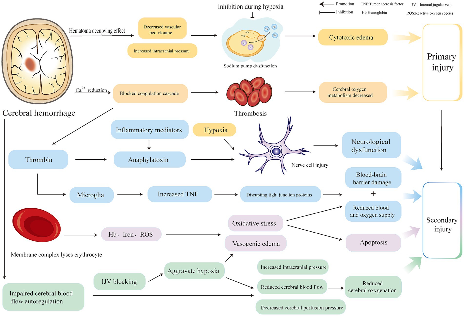
Brain injury after ICH can be classified into primary and secondary injuries. The primary injury is mechanical compression and expansion of the hematoma, which are key factors in determining the progression and outcome of ICH. They are generally caused by continued bleeding from ruptured blood vessels (3), usually occurring within 6 h after ICH, which can induce a space-occupying effect, compress blood vessels, reduce the volume of the vascular bed, increase intracranial pressure (ICP), decrease local perfusion, inhibit membrane ion pump activity (4), increase intracellular sodium ions, decrease intracellular crystal osmotic pressure, and form cytotoxic edema (5). The decrease of calcium ions in serum impairs the thrombin cascade reaction, and coagulation dysfunction (3) forms thrombosis, resulting in cerebral microcirculation obstruction and decreased oxygenation.

Hematoma components can induce secondary ICH. Thrombin is one of the components of hematoma. A high concentration of thrombin activated by the thrombin cascade reaction after ICH is closely associated with secondary ICH injury (4, 6). Thrombin can induce the production of inflammatory mediators, cause nerve cell damage (7), and influence neurological outcomes after ICH. Thrombin can also cleave the protease-activated receptors (PARs) receptor in microglia, which in turn phosphorylates the Src family kinase, thereby aggravating brain edema and the destruction of the blood–brain barrier. Stimulated microglia can also destroy tight junction proteins by up-regulating the expression of tumor necrosis factor (TNF) (6), increasing blood–brain barrier permeability. The complement cascade activated by thrombin may promote inflammation and edema after ICH through anaphylactoid toxins, or it may lyse red blood cells through membrane complexes to produce hemoglobin, iron, and oxygen free radicals, promote edema and oxidative stress and accelerate apoptosis after ICH, death, and blood–brain barrier disruption (4, 6). Red blood cells are also one of the components of hematoma. After intracerebral hemorrhage, the blood overflows from the ruptured blood vessels, and some red blood cells that are not completely phagocytized by microglia and infiltrating macrophages are directly released into the central system. Substances such as hemoglobin, iron, and carbonic anhydrase-1 are neurotoxic and are closely related to secondary brain injury after ICH (8). In addition, the automatic adjustment function of cerebral blood flow is based on a formula for cerebral blood flow, cerebral perfusion pressure (CPP), and cerebral vascular resistance (CVR). CVR can be adjusted to ensure the supply of cerebral blood flow (CBF) when ICP increases and CPP decreases. Expanded hematoma and high intracranial pressure after ICH cause decompensation of this regulation, further reducing CPP and aggravating cerebral ischemia (9). It is important to note that because hypertension is the most common cause of ICH (10), antihypertensive measures are often taken to alleviate the expansion of ICH hematoma; however, this is undoubtedly a challenge to the automatic regulation of cerebral blood flow. Therefore, attention should be paid to the tolerance of patients with hypertensive ICH to rapid blood pressure reduction; otherwise, insufficient cerebral perfusion will occur (11). Adnan et al. recommended that for intensive blood pressure reduction in ICH, systolic blood pressure should be controlled at 130–150 mmHg (12). Notably, cerebral venous outflow disorder also has a significant impact on the pathological mechanisms of ICH. Some researchers have found that cerebral venous outflow disorders are closely related to cerebrospinal fluid dynamics (13), especially in the internal jugular vein (IJV), which is the main route of brain drainage (14). Feng et al. (14) found that among patients with ICH, those with positive internal jugular venous reflux had larger perihematomal edema (PHE) volume than those with negative internal jugular venous reflux and demonstrated a positive correlation between jugular venous reflux (JVR) and PHE volume. When JVR worsens, it can further increase ICP, reduce CBF and CPP, aggravate vasogenic edema, and reduce cerebral oxygenation (14, 15). In general, secondary injuries can include vasogenic edema, neuroinflammatory reactions, blood–brain barrier damage, decompensation of cerebral blood flow autoregulation, excessive lowering of blood pressure, and cerebral venous return disorder, which are related. The pathological mechanisms are different, but to a certain extent, they can synergistically aggravate pathological reactions such as brain edema, neuroinflammatory reactions, high ICP, low CPP, oxidative stress, cell apoptosis, and destruction of the blood–brain barrier after ICH. From a comprehensive literature review, we believe that ischemia and hypoxia after ICH intersect and interact with these pathological reactions (16, 17). (The details are shown in Figure 1.)

Figure 1
www.frontiersin.org

Figure 1. Injury mechanism of cerebral hemorrhage.

A green stroke channel has been established clinically, and the slogan “time is brain” has been put forward because ICH is dangerous and progresses rapidly. Although there is currently no specific clinical treatment for ICH, the pathological basis of ICH can be explored to improve patient prognosis. Recently, high-concentration oxygen therapy has become increasingly active in the public eye. This emerging and effective intervention can alleviate ischemic hypoxic conditions after ICH, reduce intracranial pressure and cerebral edema, and improve neuroinflammatory reactions and other adverse effects. This study aimed to explore the progress in the efficacy of high-concentration oxygen therapy for ICH. Clinically, common high-concentration oxygen therapies used after ICH can be categorized into hyperbaric oxygen (HBO) and normobaric oxygen (NBO).

Sunday, April 18, 2021

Dynamics of cerebral perfusion and oxygenation parameters following endovascular treatment of acute ischemic stroke

But we still have NO PROTOCOL on how to deliver oxygen immediately post stroke to save neurons.

If your doctor is doing nothing about oxygen delivery to the brain s/he is letting more neurons die than should. ARE YOU OK WITH THAT?

I'm going to be demanding the following from my doctors but since I'm not medically trained you can't listen to me. But does your doctor have ANYTHING AT ALL? 

Possible solutions: Obviously not vetted coming from me. Don't do them.

You can look at the years these were reported on and tell how long your hospital has been incompetent.


How to Improve Your Brain Function with An Oxygen Concentrator April 2018 

Or is it more important to increase the loading ability of red blood cells to carry more oxygen? 

Like this?

University of Glasgow Study Demonstrates the Ability of Oxycyte® to Supply Oxygen to Critical Penumbral Tissue in Acute Ischemic Stroke  August 2012

Or like this?

chronic cannabis users have higher cerebral blood flow and extract more oxygen from brain blood flow than nonusers. August 2017   

Vinpocetine increases cerebral blood flow and oxygenation in stroke patients: a near infrared spectroscopy and transcranial Doppler study May 2015 

Or this? having red blood cells release more oxygen.

Methylene blue shows promise for improving short-term memory


HOW FUCKING LONG WILL YOU LET YOUR INCOMPETENT STROKE HOSPITAL STILL TREAT PATIENTS LIKE NOTHING NEW HAS OCCURRED IN THE PAST 50 YEARS?

The latest here:


Dynamics of cerebral perfusion and oxygenation parameters following endovascular treatment of acute ischemic stroke

  1. Gianluca Brugnara1,
  2. Christian Herweh1,
  3. Ulf Neuberger1,
  4. Mikkel Bo Hansen2,
  5. Christian Ulfert1,
  6. Mustafa Ahmed Mahmutoglu1,
  7. Martha Foltyn1,
  8. Simon Nagel3,
  9. Silvia Schönenberger3,
  10. Sabine Heiland1,
  11. Peter Arthur Ringleb3,
  12. Martin Bendszus1,
  13. Markus Möhlenbruch1,
  14. Johannes Alex Rolf Pfaff1,
  15. Philipp Vollmuth1
  1. Correspondence to Dr Philipp Vollmuth, Department of Neuroradiology, University Hospital Heidelberg, Heidelberg 69120, Baden-Württemberg, Germany; philipp.vollmuth@med.uni-heidelberg.de

Abstract

Background We studied the effects of endovascular treatment (EVT) and the impact of the extent of recanalization on cerebral perfusion and oxygenation parameters in patients with acute ischemic stroke (AIS) and large vessel occlusion (LVO).

Methods Forty-seven patients with anterior LVO underwent computed tomography perfusion (CTP) before and immediately after EVT. The entire ischemic region (Tmax >6 s) was segmented before intervention, and tissue perfusion (time-to-maximum (Tmax), time-to-peak (TTP), mean transit time (MTT), cerebral blood volume (CBV), cerebral blood flow (CBF)) and oxygenation (coefficientof variation (COV), capillary transit time heterogeneity (CTH), metabolic rate of oxygen (CMRO2), oxygen extraction fraction (OEF)) parameters were quantified from the segmented area at baseline and the corresponding area immediately after intervention, as well as within the ischemic core and penumbra. The impact of the extent of recanalization (modified Treatment in Cerebral Infarction (mTICI)) on CTP parameters was assessed with the Wilcoxon test and Pearson’s correlation coefficients.

Results The Tmax, MTT, OEF and CTH values immediately after EVT were lower in patients with complete (as compared with incomplete) recanalization, whereas CBF and COV values were higher (P<0.05) and no differences were found in other parameters. The ischemic penumbra immediately after EVT was lower in patients with complete recanalization as compared with those with incomplete recanalization (P=0.002), whereas no difference was found for the ischemic core (P=0.12). Specifically, higher mTICI scores were associated with a greater reduction of ischemic penumbra volumes (R²=−0.48 (95% CI –0.67 to –0.22), P=0.001) but not of ischemic core volumes (P=0.098).

Conclusions Our study demonstrates that the ischemic penumbra is the key target of successful EVT in patients with AIS and largely determines its efficacy on a tissue level. Furthermore, we confirm the validity of the mTICI score as a surrogate parameter of interventional success on a tissue perfusion level.

Monday, July 13, 2020

Anticoagulation Timing in Cardioembolic Stroke and Recurrent Event Risk

To me it seems vastly more important to get more oxygen to your brain to prevent more neurons dying. Like maybe these. I don't consider HBOT an option, too expensive and not easily obtained.  

Possible solutions: Obviously not vetted coming from me. Don't do them. 

Normobaric oxygen (10)

How to Improve Your Brain Function with An Oxygen Concentrator April 2018 

Or is it more important to increase the loading ability of red blood cells to carry more oxygen? 

Like this?

University of Glasgow Study Demonstrates the Ability of Oxycyte® to Supply Oxygen to Critical Penumbral Tissue in Acute Ischemic Stroke  August 2012

Or like this?

chronic cannabis users have higher cerebral blood flow and extract more oxygen from brain blood flow than nonusers. August 2017  

The latest here:

Anticoagulation Timing in Cardioembolic Stroke and Recurrent Event Risk


First published: 12 July 2020
This article has been accepted for publication and undergone full peer review but has not been through the copyediting, typesetting, pagination and proofreading process, which may lead to differences between this version and the Version of Record. Please cite this article as doi: 10.1002/ana.25844.

Abstract


Objectives

Guidelines recommend to initiate anticoagulation within 4‐14 days after cardioembolic stroke. Data supporting this did not account for key factors potentially affecting the decision to initiate anticoagulation such as infarct size, hemorrhagic transformation, or high risk features on echocardiography.

Methods

We pooled data from stroke registries of 8 comprehensive stroke centers across the United States. We included consecutive patients admitted with ischemic stroke and atrial fibrillation. The primary predictor was timing of initiating anticoagulation (0‐3 days, 4‐14 days, or >14 days) and outcomes were recurrent stroke/TIA/systemic embolism, symptomatic intracerebral hemorrhage (sICH), and major extracranial hemorrhage (ECH) within 90 days.

Results

Among 2084 patients, 1289 met the inclusion criteria. The combined endpoint occurred in 10.1% (n = 130) subjects (87 ischemic events, 20 sICH, and 29 ECH). Overall, there was no significant difference in the composite endpoint between the three groups: 0‐3 days [10.3% (64/617)], 4‐14 days [(9.7%) 52/535)], >14 days [10.2% (14/137), p=0.933]. In adjusted models, patients started on anticoagulation between 4‐14 days did not have a lower rate of sICH (vs. 0‐3 days) (OR 1.49 95% CI 0.50 – 4.43) neither did they have a lower rate of recurrent ischemic events (vs. > 14 days) (OR 0.76 95% CI 0.36 – 1.62, p = 0.482).

Interpretation

In this multicenter real world cohort, the recommended (4‐14 days) time frame to start oral anticoagulation was not associated with reduced ischemic and hemorrhagic outcomes. Randomized trials are required to determine the optimal timing of anticoagulation initiation.
This article is protected by copyright. All rights reserved.

Sunday, April 19, 2020

Oxygen Cost During Walking in Individuals With Stroke: Hemiparesis Versus Cerebellar Ataxia

Useless. You describe a problem, give NO SOLUTION.  Not even any mention of normobaric oxygen. Does anybody in stroke have any clue at all in what they are doing?
Possible solutions: Obviously not vetted coming from me. Don't do them.

How to Improve Your Brain Function with An Oxygen Concentrator April 2018 

Or is it more important to increase the loading ability of red blood cells to carry more oxygen? 

Like this?

University of Glasgow Study Demonstrates the Ability of Oxycyte® to Supply Oxygen to Critical Penumbral Tissue in Acute Ischemic Stroke  August 2012

Or like this?

chronic cannabis users have higher cerebral blood flow and extract more oxygen from brain blood flow than nonusers. August 2017


The latest here:

Oxygen Cost During Walking in Individuals With Stroke: Hemiparesis Versus Cerebellar Ataxia

First Published February 24, 2020 Research Article Find in PubMed


Background.
Understanding the factors that limit mobility in stroke patients is fundamental for proposing appropriate rehabilitation strategies. A high oxygen cost during walking (Cw) has a strong impact on the community ambulation of hemiparetic patients. The Cw in poststroke cerebellar ataxia is poorly evaluated, unlike hemiparetic gait.  
Objective.
To compare the oxygen cost/self-selected walking speed (S) relationship in stroke individuals with cerebellar ataxia or hemiparetic gait.  
Methods. Thirty-three subjects were included (14 cerebellar stroke, 19 hemispheric stroke), with stroke confirmed by brain imaging and able to walk without human assistance. We measured Cw using the Metamax3B. The relationship between Cw and self-selected walking speed was modelled by logistic regression and then compared between the cerebellar and hemispheric groups.  
Results.
No significant difference was found between the 2 groups for all characteristics of the population, except motor impairments, spasticity, and ataxia (P < .01). We identified 2 separate Cw/S relationships with different logistic regression equations for the 2 groups. Faster than 0.4 m s−1, Cw was 30.6% to 39.9% higher in patients with cerebellar stroke in comparison with hemispheric stroke individuals. The Cw was correlated with ataxia (r = 0.88; P < .001) in the cerebellar group, whereas there was a correlation with motor impairments (r = −0.61; P < .01), spasticity (r = 0.59; P < .01), and ataxia (r = 0.81; P < .01) in hemispheric stroke individuals.  
Conclusion.
The Cw in poststroke cerebellar ataxia is significantly higher compared with hemiparetic patients at an equivalent walking speed. The impact on community walking needs to be explored in stroke survivors with cerebellar stroke.

Walking is a meaningful activity in view of social participation and quality of life in individuals with poststroke sequelae.1 Unfortunately, at 6 months after stroke, less than 30% of patients have regained the ability to walk independently, and almost 50% believe they have restricted mobility in the community.2 The limitation of walking activity may be due to metabolic factors, because individuals with stroke suffer from significant cardiorespiratory deconditioning. The VO2 peak in these patients was found to be 26% to 87% lower than that of age- and sex-matched healthy control subjects.3 In addition, secondary to their neurological impairments, stroke individuals consume between 1.5 and 2 times more oxygen when walking than healthy subjects, which causes intense metabolic strain, leading to an increase in fatigability and consequently to limitation of activity.4 The walking efficiency of an individual can be quantified by measuring the oxygen cost of walking (Cw), which is the consumption of oxygen per meter expressed in mL kg−1 m−1.5 The Cw is of crucial importance in stroke individuals, because this parameter is associated with walking ability4 and social participation.6 Franceschini et al reported that stroke individuals with a Cw greater than 0.55 mL kg−1 m−1 have walking restrictions for community activities.6
Neurological impairments following stroke depend on the damaged neurological structure. Thus, after a hemispheric stroke, damage to the pyramidal pathway is classically responsible for spastic hemiparesis, associated or not with cognitive, sensory, and neuro-vegetative impairments, depending on the extent of the injury. These impairments lead to a stiff knee gait and circumduction of the impaired lower limb.7 After a cerebellar stroke, individuals typically have coordination and balance disorders, resulting in characteristic ataxic walking.8
Among these 2 types of clinical conditions, several deficiencies may induce an increase in Cw. The presence of co-contractions related to spasticity are associated with an increase in Cw.9,10 The increase in the variability of the trajectories of the limb segments due to the loss of selectivity of the central control11 is also associated with the increase in Cw.9,12
The gait patterns of cerebellar stroke and hemispheric stroke patients are very different. Cerebellar ataxic gait is typically characterized by an increased step width, variable foot placement, irregular foot trajectories, and a resulting unstable, stumbling walking path with very high movement variability and a high risk of falling. This type of gait is clinically recognizable and is easily differentiated from the gait pattern of hemiparetic individuals, who have a loss of symmetry, with a tendency for increased stance time on the unaffected limb. The affected lower limb appears stiff-legged, demonstrating a synergy pattern with extension, adduction, internal rotation of the hip, extension of the knee, and plantarflexion/inversion of the foot/ankle. This extended limb posture leads to an impairment of limb clearance during the swing phase, and compensatory maneuvers include hip hiking, lateral trunk sway, circumduction, and, less commonly, contralateral vaulting (Olney and Richards, 1996)13. In a previous study, we showed that the relationship between the Cw and self-selected walking speed (S) was strong in individuals with poststroke hemispheric sequelae (r = −0.94; R2 = 0.97, P < .001).14 We assumed from this work and our clinical experience that this strong relationship is explained by the fact that the walking pattern was similar from one hemiparetic individual to another. In addition, Zamparo et al, in a similar study, compared the Cw of 20 hemiparesis patients and 17 healthy subjects. The authors found different Cw/S relationships between the groups of subjects (P < .001 in the covariance analysis).15
The Cw/S relationship seems to be a characteristic of the walking efficiency of individuals that would be specific to the individual’s walking pattern. To support this hypothesis, it must be compared with a very different gait pattern, that of ataxic individuals with post–cerebellar stroke. To our knowledge, no studies have explored the Cw of cerebellar stroke individuals or the Cw/S relationship.
The objective of this study was to compare the relationship between Cw and S in individuals with cerebellar and hemispheric stroke. Our hypothesis is that individuals with cerebellar stroke sequelae do not have the same Cw/S relationship as hemiparetic stroke individuals, due to the differences in impairments and gait patterns between these 2 stroke types. If this hypothesis is confirmed, this would imply that a specific Cw/S relationship could be identified for patients, depending on the stroke location.

More at link. 

Friday, February 28, 2020

Study Shows Hyperbaric Oxygen Therapy (HBOT) Protocols Can Improve Cognitive Function in Stroke Patients

I don't trust this since it could easily be biased since the study backers own HBOT services. 

Normobaric oxygen would be much easier and cheaper to deliver.

There is this from March, 2005:

A Pilot Study of Normobaric Oxygen Therapy in Acute Ischemic Stroke

 And this in October, 2010

Normobaric oxygen therapy in acute ischemic stroke: A pilot study in Indian patients

And this from October 2012:

Normobaric Oxygen Therapy

But all this other research shows no help for HBOT.

HBOT hasn't had any earlier success in either TBI or stroke.

Oxygen therapy no better than placebo for treating concussion, study finds

Can Hyperbaric Oxygen Repair the Damaged Brain?

Mayo clinics take:

Agency Research for Healthcare and Quality:

hbot as stroke therapy - quackery?

Peter Levine talking about problems of HBOT here:Stroke and Hyperbaric Oxygen Therapy

Enormous Inferno Kills Man Who Tried Smoking a Cigarette in a Hyperbaric Chamber


 The latest on HBOT here:

Study Shows Hyperbaric Oxygen Therapy (HBOT) Protocols Can Improve Cognitive Function in Stroke Patients

TEL AVIV, Israel, Feb. 27, 2020 /PRNewswire/ -- The Sagol Center for Hyperbaric Medicine and Research at Shamir Medical Center, one of the largest hyperbaric medicine centers in the world, announced study results indicating its hyperbaric oxygen therapy (HBOT) protocols induce significant improvement in most cognitive domains of patients who suffered stroke – even months to years after the event.
The retrospective analysis study, published in Restorative Neurology and Neuroscience, demonstrated that through elevating oxygen concentration in the blood and injured brain tissue, the Sagol Center's HBOT protocols supply the energy needed for brain tissue recovery – regardless of the stroke location or cause.
"Despite nearly half of stroke survivors suffering from differing degrees of cognitive dysfunction, most rehabilitative efforts focus on restoring motor functions," said Shai Efrati, MD, Director of the Sagol Center for Hyperbaric Medicine and Research, Shamir Medical Center, and study co-author. "Our study shows that stroke-related cognitive deficits can be treated as brain wounds, regardless of the stroke's location or origin – ischemic or hemorrhagic. The rehabilitation potential lies in the metabolic characteristics of the chronic damage, or brain wound. By utilizing our HBOT protocols, the injured but viable brain tissue can be recovered even years after the acute insult."
Stroke is the second highest cause of mortality and the third leading cause of disability worldwide. When stroke occurs, whether ischemic or hemorrhagic, dysfunction typically presents in the affected brain region and impacts more than one cognitive domain such as memory or attention. Cognitive recovery after stroke mainly transpires within the first 30 days, with some patients gaining additional mild improvement up to 90 days after.
The study evaluated the sustained impact of Sagol's HBOT protocols on all aspects of cognitive function including memory, executive function, visuospatial skills, verbal function, attention, information processing and motor skills. Results demonstrated clinically significant improvement in cognitive function in most evaluated cognitive domains after HBOT.
The study found the following:
  • 86% of the stroke patients achieved clinically significant improvement in their cognitive function
  • The memory domain had the most prominent improvements
  • Baseline cognitive function was a significant predictor of rehabilitation potential
  • Hemorrhagic strokes had significantly higher improvement in information processing compared to ischemic strokes, though ischemic stroke patients still showed improvement
  • Left hemisphere strokes had a greater increase in the motor domain, compared to right hemisphere strokes
Of the 351 patients assessed for eligibility, a total of 162 patients met the inclusion criteria for the final analysis. Criteria comprised the receipt of treatment after more than three months post-injury, the completion of the Sagol Center HBOT protocols and at least two cognitive evaluations, conducted using a fully computerized cognitive testing battery (NeuroTrax test battery®). Patients were treated in a multi-place hyperbaric chamber at the Sagol Center.
"This is the largest cohort of stroke patients who have undergone strict cognitive, imaging and neurological assessments to evaluate the rehabilitation potential of HBOT treatment," said Amir Hadanny, MD, Chief Medical Research Officer at the Sagol Center for Hyperbaric Medicine and Research, Shamir Medical Center, and lead author of the study. "To identify the true cognitive and clinical impact of our protocols, all patients were evaluated with two validated alternate forms of the cognitive test battery prior to and post-HBOT. Significant improvements were demonstrated in all cognitive domains including memory, information processing speed, attention and executive functions. We are thrilled to bring a new hope to stroke patients worldwide."
About the Sagol Center for Hyperbaric Medicine and Research at Shamir Medical Center
The Sagol Center for Hyperbaric Medicine and Research at Shamir Medical Center (formerly Assaf Harofeh Medical Center), is a leader in advancing our understanding of the impact of hyperbaric medicine on cognitive and physical function. Serving as one of the largest Hyperbaric centers worldwide, the Sagol Center offers highly advanced large multi-place chambers, treating more than 200 patients daily. Research conducted at the Center has proven that brain rejuvenation is possible across a wide range of neurological pathologies and illnesses.
Media Contact:Nicole Grubner
Finn Partners for The Sagol Center for Hyperbaric Medicine and Research
+1-929-222-8011
nicole.grubner@finnpartners.com
SOURCE The Sagol Center for Hyperbaric Medicine and Research

Wednesday, February 19, 2020

Advances in Normobaric Hyperoxia Brain Protection in Experimental Stroke

Useless, NOTHING ON STROKE PROTOCOLS FOR USE. And even worse you ask for followup to cover up your failures in your research. 

Maybe these might help you talk to your doctor.


Advances in Normobaric Hyperoxia Brain Protection in Experimental Stroke

  • 1Department of Neurology, Xuanwu Hospital, Capital Medical University, Beijing, China
  • 2Department of Neurosurgery, School of Medicine, Wayne State University, Detroit, MI, United States
  • 3Department of Neurosurgery, Xuanwu Hospital, Capital Medical University, Beijing, China
  • 4Center of Stroke, Beijing Institute for Brain Disorders, Beijing, China
As we all know that stroke is still a leading cause of death and acquired disability. Etiological treatment and brain protection are equally important. This review aimed to summarize the advance of normobaric-hyperoxia (NBHO) on brain protection in the setting of experimental stroke and brain trauma. We analyzed the data from relevant studies published on PubMed Central (PMC) and EMBASE, about NBHO on brain protection in the setting of experimental ischemic and hemorrhagic strokes and brain trauma, which revealed that NBHO had important value on improving hypoxia and attenuating ischemia damage. The mechanisms of NBHO involved increasing the content of oxygen in brain tissues, restoring the function of mitochondria, enhancing the metabolism of neurons, alleviating blood-brain barrier (BBB) damage, weakening brain cell edema, reducing intracranial pressure, and improving cerebral blood flow, especially in the surrounding of injured area of the brain, to make the neurons in penumbral area alive. Compared to hyperbaric oxygen (HBO), NBHO is more safe and more easily to transform to clinical use, whereby, further studies about the safety and efficacy as well as the proper treatment protocol of NBHO on human may be still needed.

Thursday, January 9, 2020

Dark chocolate improves coronary vasomotion and reduces platelet reactivity

What is your doctor doing IMMEDIATELY post stroke to increase oxygen to your brain? Maybe this dark chocolate  by dilating your arteries?

Or your doctor will never suggest this one:

 The team that found chronic cannabis users have higher cerebral blood flow and extract more oxygen from brain blood flow than nonusers.

 Or maybe beet juice?

 The study showed that beet juice with high amounts of nitrate made the blood vessels relax and return to normal function

Or this?

Sesquiterpenes, a natural compound found in essential oils of Vetiver, Patchouli, Cedarwood, Sandalwood and Frankincense, can increase levels of oxygen in the brain by up to 28 percent 

There is this from March, 2005:

A Pilot Study of Normobaric Oxygen Therapy in Acute Ischemic Stroke

 And this in October, 2010

Normobaric oxygen therapy in acute ischemic stroke: A pilot study in Indian patients

And this from October 2012:

Normobaric Oxygen Therapy

If your doctor is doing nothing about oxygen delivery to the brain s/he is letting more neurons die than should. ARE YOU OK WITH THAT?

The latest here:

Dark chocolate improves coronary vasomotion and reduces platelet reactivity

Author information

1
Cardiovascular Center, Cardiology, University Hospital Zurich, Raemistr 100, CH-8091 Zurich, Switzerland.

Abstract

BACKGROUND:

Dark chocolate has potent antioxidant properties. Coronary atherosclerosis is promoted by impaired endothelial function and increased platelet activation. Traditional risk factors, high oxidative stress, and reduced antioxidant defenses play a crucial role in the pathogenesis of atherosclerosis, particularly in transplanted hearts. Thus, flavonoid-rich dark chocolate holds the potential to have a beneficial impact on graft atherosclerosis.

METHODS AND RESULTS:

We assessed the effect of flavonoid-rich dark chocolate compared with cocoa-free control chocolate on coronary vascular and platelet function in 22 heart transplant recipients in a double-blind, randomized study. Coronary vasomotion was assessed with quantitative coronary angiography and cold pressor testing before and 2 hours after ingestion of 40 g of dark (70% cocoa) chocolate or control chocolate, respectively. Two hours after ingestion of flavonoid-rich dark chocolate, coronary artery diameter was increased significantly (from 2.36+/-0.51 to 2.51+/-0.59 mm, P<0.01), whereas it remained unchanged after control chocolate. Endothelium-dependent coronary vasomotion improved significantly after dark chocolate (4.5+/-11.4% versus -4.3+/-11.7% in the placebo group, P=0.01). Platelet adhesion decreased from 4.9+/-1.1% to 3.8+/-0.8% (P=0.04) in the dark chocolate group but remained unchanged in the control group.

CONCLUSIONS:

Dark chocolate induces coronary vasodilation, improves coronary vascular function, and decreases platelet adhesion 2 hours after consumption. These immediate beneficial effects were paralleled by a significant reduction of serum oxidative stress and were positively correlated with changes in serum epicatechin concentration.

Comment in

PMID:
17984375
DOI:
10.1161/CIRCULATIONAHA.107.713867