Showing posts with label Neurodegeneration. Show all posts
Showing posts with label Neurodegeneration. Show all posts
Thursday, August 27, 2026


Disrupted brain-immune signaling may help drive neurodegeneration
Will your competent? doctor and hospital get followup research initiated that will create protocols that prevent neurodegeneration?
Do you prefer your doctor, hospital and board of director's incompetence NOT KNOWING? OR NOT DOING? Your choice; let them be incompetent or demand action!
Disrupted brain-immune signaling may help drive neurodegeneration
From gut-primed T cells to microglial signaling and persistent gene-regulatory states, researchers map an intricate immune network that connects the brain with the rest of the body.
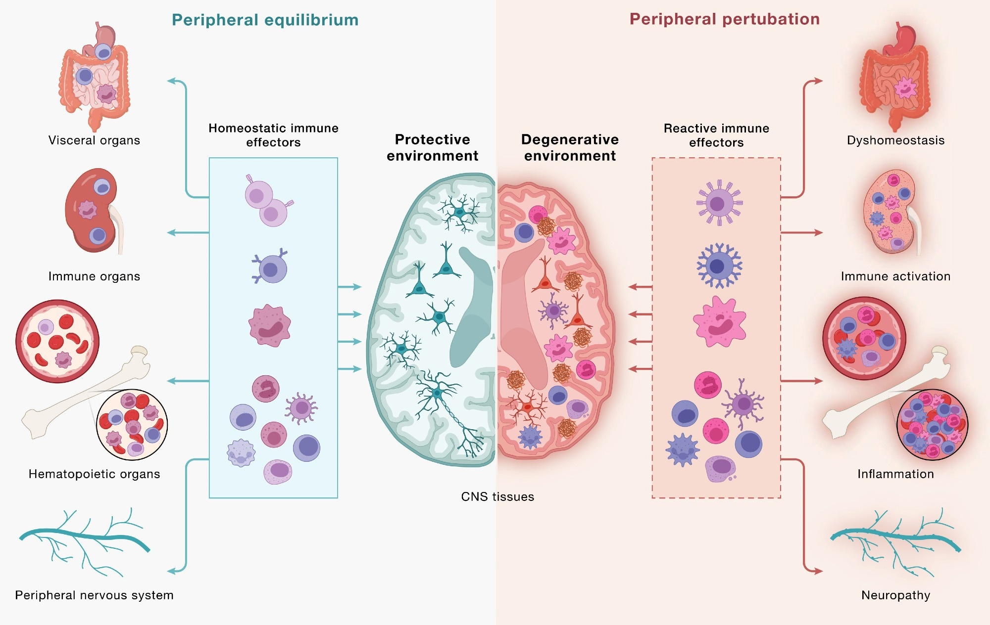
Immune
cells communicate across a continuous, bidirectional circuit between
the CNS and periphery. Immune cells from across the body engage CNS
tissues and, in turn, are shaped by signals returning from the brain.
(Left) Under peripheral equilibrium, immune effectors mobilized from
visceral organs, secondary lymphoid organs, hematopoietic tissues, and
the peripheral nervous system (PNS) - including CD4+ and CD8+
T cells, B cells, monocytes (top 3 cell types), NK cells, dendritic
cells, mast cells, neutrophils, and innate lymphoid cells (bottom
cluster of cells) - engage the CNS in a homeostatic dialogue that
supports neural cell health, plasticity, and repair, sustaining a
protective environment. (Right) Age-associated dysfunction across the
same peripheral compartments reshapes this circuit: altered immune
effectors enter the CNS and contribute to shifting the local environment
toward a degenerative state in which microglia adopt disease-associated
programs, astrocytes become reactive, and neurons accumulate pathology.
This model underscores how the same circuitry can yield protective or
pathogenic outcomes and that neurodegeneration is downstream of
dysfunctional brain-immune crosstalk. Study: Neurodegeneration as a dysregulation of neuroimmune crosstalk
A recent perspective published in the journal Cell synthesizes scientific evidence suggesting that neurodegeneration involves intricate crosstalk between neurons and immune cells, linking the brain to peripheral immunity through bidirectional exchange. Strategies that restore immune homeostasis or recalibrate neuroimmune signaling may potentially slow neurodegeneration and promote recovery.
Historically, immune dysregulation has often been considered a consequence of neurodegenerative disorders. Recent studies, however, are beginning to change this scientific mindset, suggesting that disordered communication between the brain and immune cells may also contribute to disease onset and progression. The authors describe immune dysfunction as a “concause” of neurodegeneration, meaning it may interact with neuronal and glial vulnerabilities without necessarily being the initial trigger. It is essential to advance understanding of the pathophysiology of neurodegenerative diseases to inform therapeutic development and the development of immune-based strategies.
In this perspective, researchers examined brain-immune interactions and their potential role in neurodegeneration. They organized emerging evidence into three frameworks: “outside-in” effects driven by peripheral immunity, “inside-out” signaling coordinated by brain-resident microglia, and “locked-in” gene regulatory programs that can stabilize maladaptive neuroimmune states.
The brain-immune communication network
The brain continuously communicates with peripheral immune networks. Components of the CNS, including the choroid plexus, meninges, and lymphatic and vascular structures, interact with immune cells to relay signals related to neural needs.
Helper and cytotoxic T cells can enter CNS border regions and, under defined conditions, the brain parenchyma. Brain-immune communication supports neural integrity but can promote pathology when dysregulated. Microglia and BAMs provide surveillance, while lymphocytes confer antigen specificity and immunological memory.
Cytokines, complement, and MHC-I are traditionally linked to immunity, but CNS cells also produce or sense these molecules during neural activity. Innate lymphoid cells in the dura can respond to injury, while the choroid plexus helps regulate inflammatory signaling. In mice, increased neuronal activity may draw antibody-secreting B-lineage cells into the hippocampus during synaptic remodeling.
The gut also influences brain immunity. T cells educated in gut-associated immune tissues can subsequently traffic to the borders of the CNS and, under certain conditions, into the brain, while plasma cells secreting IgA antibodies protect blood vessels in the meninges. In addition, changes in the gut microbiome could influence immune activity and microglial function. Through the GBA, the gut and brain are in constant dialogue with each other. The vagus nerve conveys immunity-related information from the intestines to the brain. Reward-related neural pathways can, in turn, influence peripheral immune activity.
Brain-immune interactions in neurodegenerative disease
T cell activity has been implicated in PD, AD, ALS, and dementia with Lewy bodies (DLB). In ALS4, an inherited form of ALS, cytotoxic T cells are detected early in the blood and brain and expand as the disease progresses, consistent with antigen-driven responses.
Friday, July 10, 2026
A Review on the Mechanisms of Neurodegeneration and the Potential of Plant Bioactives in Managing Neurological Conditions
Useless! You didn't provide any protocols that prevents these problems! That's the whole point of research; PREVENTION OR RECOVERY! And you were a complete failure at that.
Comeuppance is going to be a real bitch for you when you realize you could have done something while still working!
A Review on the Mechanisms of Neurodegeneration and the Potential of Plant Bioactives in Managing Neurological Conditions
- 21 Mar 2025
- 18 Aug 2025
- 03 Jul 2026
Neurodegenerative disorders encompass a wide range of debilitating neurological conditions characterized by the progressive loss of specific neuronal populations in the central and/or peripheral nervous systems. This disease often leads to a gradual decline in cognitive, motor, and sensory abilities.
This review explores the role of various lifestyle factors, such as age, sex, poor diet, depression, etc., which contribute to the onset and progression of NDDs. Various diseases are included in the neurodegenerative disorder, like Alzheimer’s disease, Parkinson’s disease, Huntington’s disease, Amyotrophic Lateral Sclerosis, Multiple Sclerosis, and Lewy body disease, which are chronic conditions that significantly impact cognitive and motor functions.
A literature search was conducted in the scientific database using the keywords “neurodegenerative disorders, phytoconstituents, and herbals”. This review includes a collection of reports from ScienceDirect, Scholar Google, and PubMed, all searched up to 2024. The results were assessed, gathered, and reported in this paper. A total of 241 articles were included, with exponential growth in publication numbers from 1985 to 2024.
Effective management and control of NDDs require addressing these risk factors, alongside exploring therapeutic interventions. Some plants and herbs used to treat neurodegenerative diseases, such as curcumin, ashwagandha, ginkgo biloba, epigallocatechin-3-gallate, quercetin, ginseng, and resveratrol, have shown potential to improve neuronal health and mitigate disease progression. This review highlights the dual role of natural compounds in promoting improvements and upregulating brain function while potentially reducing degradation. The phytopharmaceuticals show the potential for treating neurological conditions with better efficacy and safer profiles.
The review suggested that future research(So your research was a failure since you didn't power it well enough!) should focus on integrating lifestyle modifications and natural therapies to enhance the quality of life for individuals at risk or suffering from neurodegenerative diseases.
© 2026 Bentham Science Publishers
Impact of inhalational anesthetics on neurodegeneration in Alzheimer’s disease: current evidence and potential implications
Will your competent? doctor at least ensure further research occurs that determines how and the interventions needed to make this work. You are an at-risk population or hasn't you doctor informed you of that?
Your risk of dementia, has your doctor
told you of this? Your doctor is responsible for preventing this! Is
s/he willing to prevent this?
1. A documented 33% dementia chance post-stroke from an Australian study? May 2012.
2. Then this study came out and seems to have a range from 17-66%. December 2013.`
3. A 20% chance in this research. July 2013.
4. Dementia Risk Doubled in Patients Following Stroke September 2018
The latest here:
Impact of inhalational anesthetics on neurodegeneration in Alzheimer’s disease: current evidence and potential implications
Austin Dosanjh1,#, Sanarya Al-Jaf1,#, Emily Ye1,#, and Khaled S. Abd-Elrahman1,2,3,*
1. Department of Anesthesiology, Pharmacology and Therapeutics, and Djavad Mowafaghian Centre for Brain Health, The University of British Columbia, Vancouver,
British Columbia V6T 1Z3, Canada
2. Department of Medical Sciences, College of Medicine and Health Sciences, Khalifa University, Abu Dhabi 127788, United Arab Emirates
3. Department of Pharmacology and Toxicology, Faculty of Pharmacy, Alexandria University, Alexandria 21521, Egypt # Contributed equally to this work * Address correspondence to: Dr. Khaled S. Abd-Elrahman Department of Anesthesiology, Pharmacology and Therapeutics University of British Columbia 2176 Health Sciences Mall, Vancouver, British Columbia, V6T 1Z3. Tel: 6048221390 khaled.abdelrahman@ubc.ca
Abstract
Alzheimer’s disease is the most common form of dementia, primarily affecting the elderly population. It is a progressive neurodegenerative disease with key pathogenesis hallmarks being amyloid-beta plaque accumulation, and neurofibrillary tangles of tau protein. With an increasingly aging population and rising numbers of surgical procedures, growing interest has been directed towards the potential impact of inhalational anesthetics, particularly isoflurane, sevoflurane and desflurane, in contributing to the neurodegenerative process. Evidence supporting anesthetic-related modulation of Alzheimer’s disease pathways is derived predominantly from in vitro and animal models, with comparatively limited and heterogeneous human biomarker and clinical data. This review will explore the various mechanisms by which these volatile anesthetics may contribute to the pathogenesis of neurodegeneration in the context of AD. This includes upregulation of beta-secretase 1 resulting in the formation of amyloid-beta oligomers and inhibition of tau dephosphorylation. While certain studies point towards a neuroprotective effect of these anesthetics, the evidence remains inconsistent. Collectively, these findings support perioperative strategies focused on maintenance of normothermia, optimization of oxygenation, and judicious anesthetic exposure as practical measures to mitigate vulnerability in at-risk populations.
Transthyretin at the crossroads of neurodegeneration: a silent guardian in Parkinson’s disease
Will your competent? doctor be doing something with this because of your risk of Parkinsons post stroke? Oh NO, NOTHING DOING! So, INCOMPETENCE REIGNS AGAIN! Your doctor is becoming an expert at incompetence and your board of directors is so incompetent they can't recognize it in their hospital!
Parkinson’s Disease May Have Link to Stroke March 2017
The latest here:
Transthyretin at the crossroads of neurodegeneration: a silent guardian in Parkinson’s disease
Abstract
Parkinson’s disease (PD) is a progressive neurodegenerative disease characterised by disruption of brain homeostasis and degeneration of dopaminergic neurons in the substantia nigra. PD is characterised by motor symptoms, like tremor, rigidity, bradykinesia, and postural instability, as well as non-motor symptoms like cognitive impairment, mood disorders, sleep disturbances, and autonomic abnormalities that significantly affect quality of life. The molecular pathogenesis of PD involves Oxidative stress, neuroinflammation, mitochondrial dysfunction, α-synuclein (α-syn) misfolding and aggregation, insufficient autophagy-lysosomal clearance, and synaptic degeneration, leading to progressive neuronal loss. Transthyretin (TTR), a tetrameric transport protein that is primarily produced in the liver and choroid plexus, is well-known for carrying thyroxine and retinol-binding protein. Experimental studies have shown that TTR can protect neurons by binding misfolded proteins, such as α-syn, decreasing toxic aggregation, regulating oxidative stress responses, and affecting selective autophagic degradation. PD-related changes in TTR expression in brain tissue and cerebrospinal fluid provide strong evidence of TTR’s significance as a molecular biomarker and a physiological regulator in the pathogenesis of the disease. This review highlights TTR involvement in neuroinflammation, oxidative stress, and α-syn aggregation, and discusses emerging evidence supporting TTR stabilizers as potential biomarkers and therapeutic targets for modulating disease progression in PD.
Monday, May 25, 2026
Quinoa seed (Chenopodium quinoa W.) extract attenuates Alzheimer’s disease–like neurodegeneration: Targeting the SLC7A11/GPX4 pathway
Attenuates: Medicine & Biology: To make a virus or bacteria less virulent or harmful. You really think your competent? doctor and hospital will get human testing going?
Quinoa seed (Chenopodium quinoa W.) extract attenuates Alzheimer’s disease–like neurodegeneration: Targeting the SLC7A11/GPX4 pathway
Abstract
Background
Alzheimer’s disease (AD) is a progressive neurodegenerative disorder characterized by cognitive decline and hippocampal neuronal loss. Targeting ferroptosis-related pathways represents a promising therapeutic strategy.
Objective
This study aimed to investigate the potential effect of quinoa (Chenopodium Quinoa W.) seed extract in an aluminum chloride (AlCl₃)–induced rat model of AD, with a particular focus on the SLC7A11/GPX4 antioxidant axis and NCOA4-mediated ferritinophagy.
Methods
Adult male rats were randomly divided into four groups (n = 6): GI (Control), GII (AD), GIII (Quinoa + AD), and GIV (Alzemenda + AD). AD was induced by oral AlCl₃ administration. Quinoa extract and Alzemenda were administered concurrently with AlCl₃ throughout the experimental period. Behavioral performance was evaluated using the Morris Water Maze and Open Field Test. Oxidative stress markers, iron parameters, gene expression, and histopathological changes in the hippocampus were assessed.
Results
GII exhibited significant cognitive impairment, increased lipid peroxidation, depletion of antioxidant defenses, downregulation of SLC7A11, and marked hippocampal iron deposition compared with GI. Treatment with quinoa (GIII) significantly improved learning and memory, restored GPX4 activity and GSH levels, upregulated SLC7A11 expression, and attenuated hippocampal iron deposition. GIV showed comparable behavioral and histological improvement. Systemic iron indices, as well as hippocampal FPN1 and NCOA4 expression, did not differ significantly among groups.
Conclusion
Quinoa seed extract exerts ameliorating effects in AlCl₃-induced AD by suppressing oxidative stress-associated neurodegeneration through preservation of the SLC7A11/GSH/GPX4 axis rather than modulation of iron export or ferritinophagy pathways.
Tuesday, May 19, 2026
Astrocytes in Brain Aging and Neurodegeneration: Cellular Mechanisms and Interventional Strategies
You'll have to ask your competent? doctor to get those emerging therapeutic strategies aimed at modulating astrocyte function, so your astrocytes don't deteriorate.
Astrocytes in Brain Aging and Neurodegeneration: Cellular Mechanisms and Interventional Strategies
s
Flávia C. A. Gomes | Isadora Matias
Instituto de Ciências Biomédicas, Universidade Federal do Rio de Janeiro, Rio de Janeiro, Brazil
Correspondence: Flávia C. A. Gomes (fgomes@icb.ufrj.br)
Received: 5 November 2025 | Revised: 20 April 2026 | Accepted: 30 April 2026
Keywords: aging | astrocyte | glial cells | neurodegenerative diseases
ABSTRACT
Aging is characterized by progressive changes in the physiology of brain cells, which may contribute to cognitive decline,
ultimately leading to dementia and impaired quality of life. The increase in senescent cells, including glial cells in the brain,
is a general feature of normal aging and has been associated with age- related pathologies. Although recent evidence suggests
that astrocytes undergo senescence in these conditions, little is known about the molecular, and cellular mechanisms under
lying this event. This mini review, prepared as part of the special issue Neurochemistry in Latin America, provides a focused
overview of astrocyte dysfunction in physiological aging and neurodegenerative conditions, integrating findings from the field
alongside recent contributions from our group. We discuss how astrocyte aging contributes to cognitive decline and highlight
emerging evidence on how targeting astrocytes, both genetically and pharmacologically, may rescue cognitive decline associated with aging and neurodegenerative diseases. Astrocytes produce several molecules that control synapse formation and
function, which are decreased in the aging brain and in Alzheimer's disease models. In this context, recent studies indicate
that astrocytes undergo significant molecular and functional remodeling during aging. Notably, astrocyte senescence has
been associated with loss of lamin- B1, nuclear alterations, impaired synaptogenic and neuritogenic capacity, altered glutamate
metabolism, and mitochondrial dysfunction, all of which may contribute to reduced neuronal support and circuit integrity.
In parallel, recent advances have shown that astrocyte responses during aging also include diverse reactive states that vary
according to brain region, microenvironment, and disease stage. Importantly, senescence- associated and reactive features are
not mutually exclusive and may coexist or interact, further contributing to synaptic dysfunction and increased vulnerability
to neurodegeneration. Finally, we discuss emerging therapeutic strategies aimed at modulating astrocyte function, including targeting astrocyte- derived synaptogenic factors and metabolic pathways, as potential approaches to mitigate cognitive
decline. Together, current evidence indicates that astrocyte dysfunction in aging reflects a complex and dynamic spectrum
of cellular states that play a central role in brain vulnerability and represent promising targets for intervention in aging and
neurodegenerative diseases.
This is an open access article under the terms of the Creative Commons Attribution License, which permits use, distribution and reproduction in any medium,
provided the original work is properly cited.
© 2026 The Author(s). Journal of Neurochemistry published by John Wiley & Sons Ltd on behalf of International Society for
From Stress to Neurodegeneration: A New Look at the Pathogenesis of Parkinson’s Disease
All stroke patients are under massive stress because your incompetent? doctor doesn't have 100% RECOVERY PROTOCOLS. Your doctor has known since medical school that stroke recovery is a complete shitshow and done nothing to fix that! With your risk of Parkinsons post stroke that just adds more stress.
Parkinson’s Disease May Have Link to Stroke March 2017
The latest here:
From Stress to Neurodegeneration: A New Look at the Pathogenesis of Parkinson’s Disease
by  ,,,
,,,
Rogneda B. Kazanskaya
1,*,Vassiliy Tsytsarev
2,*,Anna B. Volnova
1,3,Raul R. Gainetdinov
1 andAlexander V. Lopachev
11
Institute of Translational Biomedicine, St. Petersburg State University, Universitetskaya Nab. 7/9, 199034 St. Petersburg, Russia
2
School of Engineering, Johns Hopkins University, Baltimore, MD 21218, USA
3
Center for Transgenesis and Genome Editing, St. Petersburg State University, Universitetskaya Nab. 7/9, 199034 St. Petersburg, Russia
*
Authors to whom correspondence should be addressed.
Biomedicines 2026, 14(5), 1130; https://doi.org/10.3390/biomedicines14051130
Submission received: 10 April 2026 / Revised: 14 May 2026 / Accepted: 14 May 2026 / Published: 16 May 2026
(This article belongs to the Special Issue Advances in Parkinson’s Disease Research)
Abstract
The relationship between stress and Parkinson’s disease is regarded as complex and multifaceted, although a direct causal link has not yet been conclusively proven. One prevailing hypothesis is based on the activation of the hypothalamic–pituitary–adrenal (HPA) axis and the consequent elevation of glucocorticoid levels. Prolonged exposure to these hormones may exacerbate oxidative stress, thereby rendering the dopaminergic neurons within the brain’s subcortical structures more susceptible to degeneration. Furthermore, stress may intensify neuroinflammation through the activation of microglia—a mechanism that could constitute a significant factor in the pathogenesis of Parkinson’s disease. Another important concept concerns the direct interaction of stressors with the dopaminergic system. Physiological and psychological stress can alter dopaminergic transmission by affecting both the synthesis and release of dopamine, as well as the sensitivity of dopamine receptors. Severe or chronic stress may contribute to the disruption of dopaminergic mechanisms and accelerate the onset of clinical symptoms in predisposed individuals. Furthermore, many researchers draw attention to the role of stress-induced aggregation of α-synuclein—a key protein implicated in the pathogenesis of Parkinson’s disease. Clinical data suggest a highly probable link between post-traumatic stress disorder and an increased risk of developing Parkinson’s disease, although these findings remain inconclusive. It is possible that stress acts not as a primary cause, but rather as a modifying factor that interacts with genetic predisposition, accelerating or triggering neurodegenerative processes. The aim of our narrative review was to examine these concepts and discuss possible directions for future research into the interaction between stress and Parkinson’s disease.
Monday, May 11, 2026
Disentangling causality in brain aging: The complex interplay between glial senescence, neuroinflammation, and neurodegeneration
How will your competent? doctor use this to recover your 5 lost years of brain cognition due to your stroke?
Do you prefer your doctor, hospital and board of director's incompetence NOT KNOWING? OR NOT DOING? Your choice; let them be incompetent or demand action!
Disentangling causality in brain aging: The complex interplay between glial senescence, neuroinflammation, and neurodegeneration

Subscribe to:
Posts (Atom)