Changing stroke rehab and research worldwide now.Time is Brain! trillions and trillions of neurons that DIE each day because there are NO effective hyperacute therapies besides tPA(only 12% effective). I have 523 posts on hyperacute therapy, enough for researchers to spend decades proving them out. These are my personal ideas and blog on stroke rehabilitation and stroke research. Do not attempt any of these without checking with your medical provider. Unless you join me in agitating, when you need these therapies they won't be there.

What this blog is for:

My blog is not to help survivors recover, it is to have the 10 million yearly stroke survivors light fires underneath their doctors, stroke hospitals and stroke researchers to get stroke solved. 100% recovery. The stroke medical world is completely failing at that goal, they don't even have it as a goal. Shortly after getting out of the hospital and getting NO information on the process or protocols of stroke rehabilitation and recovery I started searching on the internet and found that no other survivor received useful information. This is an attempt to cover all stroke rehabilitation information that should be readily available to survivors so they can talk with informed knowledge to their medical staff. It lays out what needs to be done to get stroke survivors closer to 100% recovery. It's quite disgusting that this information is not available from every stroke association and doctors group.

Showing posts with label endopeptidases. Show all posts

Tuesday, May 24, 2016

Implications of MMP9 for Blood Brain Barrier Disruption and Hemorrhagic Transformation Following Ischemic Stroke

The earliest report I have of MMP-9 being useful for stroke was way back in 2005.  And 11 years later we still have no translational use for it. We keep doing studies but never seem to get anywhere useful with it. A great stroke leader would get something accomplished.

In 2005, the scientist(Mr Gu) had previously played the role of lead author on a study published in the Journal of Neuroscience that had revealed MMP-9 could be a promising area that therapeutic medicines for stroke patients could target.

Implications of MMP9 for Blood Brain Barrier Disruption and Hemorrhagic Transformation Following Ischemic Stroke

  • 1Discipline of Anatomy and Pathology, Adelaide Centre for Neuroscience Research, School of Medicine, The University of Adelaide, Adelaide, SA, Australia
  • 2Department of Neurology, MIND Institute, University of California at Davis Medical Center, Sacramento, CA, USA
Numerous studies have documented increases in matrix metalloproteinases (MMPs), specifically MMP-9 levels following stroke, with such perturbations associated with disruption of the blood brain barrier (BBB), increased risk of hemorrhagic complications, and worsened outcome. Despite this, controversy remains as to which cells release MMP-9 at the normal and pathological BBB, with even less clarity in the context of stroke. This may be further complicated by the influence of tissue plasminogen activator (tPA) treatment. The aim of the present review is to examine the relationship between neutrophils, MMP-9 and tPA following ischemic stroke to elucidate which cells are responsible for the increases in MMP-9 and resultant barrier changes and hemorrhage observed following stroke.

Introduction

Over the last decade the matrix metalloproteinases (MMPs) have been widely investigated for their role in disruption of the blood-brain barrier (BBB), particularly the extracellular matrix (ECM), following stroke (Romanic et al., 1998; Rosenberg et al., 1998; Fujimura et al., 1999; Gasche et al., 1999; Gidday et al., 2005) and other cerebral pathologies such as traumatic brain injury (Planas et al., 2001) and neoplasm (Lukes et al., 1999; Turba et al., 2007). MMPs are a family of zinc and calcium-dependent endopeptidases that are capable of degrading all components of the ECM including laminin, collagen and fibronectin, amongst many other targets (Van den Steen et al., 2002). At least 23 MMPs have been identified to date (Sternlicht and Werb, 2001), with MMP-2 and MMP-9 the most widely studied in stroke. In particular, MMP-9 has been implicated, not only in the pathogenesis of BBB breakdown and subsequent vasogenic edema formation following stroke (Fujimura et al., 1999; Gasche et al., 1999; Rosenberg and Yang, 2007), but also in hemorrhagic transformation (HT) in the setting of tissue plasminogen activator (tPA) therapy (Lapchak et al., 2000; Wang et al., 2009). Cerebral edema and HT of the infarct are significant problems in clinical stroke, which are associated with poor outcome and contribute to the morbidity and mortality of this condition (Hacke et al., 1996; Fiorelli et al., 1999). Elucidating the mechanisms of such deleterious events is the key to developing targeted, more effective clinical therapies.
Numerous clinical and experimental studies have confirmed an increase in serum MMP-9 following stroke (Clark et al., 1997; Romanic et al., 1998; Yushchenko et al., 2000; Montaner et al., 2003a; Ning et al., 2006). However, the cellular source of this MMP-9 remains controversial. Although it is generally accepted that MMP-9 is increased following stroke, there is debate as to which cells are responsible, whether it be resident brain cells, cells of the vasculature or circulating immune cells, such as neutrophils. However, the aim of the present review was to explore the potential relationship between neutrophil-derived MMP-9 and complications such as BBB disruption and HT following stroke to elucidate the cellular source of MMP-9 in ischemic stroke.

Thursday, July 2, 2015

Unbalanced metalloproteinase-9 and tissue inhibitors of metalloproteinases ratios predict hemorrhagic transformation of lesion in ischemic stroke patients treated with thrombolysis: results from the MAGIC study

Way out of my league, so you'll have to contact a genius somewhere to decipher this.
http://journal.frontiersin.org/article/10.3389/fneur.2015.00121/full?
imageBenedetta Piccardi1*, imageVanessa Palumbo2, imageMascia Nesi2, imagePatrizia Nencini2, imageAnna Maria Gori3, imageBetti Giusti3, imageGiovanni Pracucci1, imagePaolina Tonelli1, imageEleonora Innocenti1, imageAlice Sereni3, imageElena Sticchi3, imageDanilo Toni4, imagePaolo Bovi5, imageMario Guidotti6, imageMaria Rosaria Tola7, imageDomenico Consoli8, imageGiuseppe Micieli9, imageRossana Tassi10, imageGiovanni Orlandi11, imageFrancesco Perini12, imageNorina Marcello13, imageAntonia Nucera14, imageFrancesca Massaro15, imageMaria Luisa DeLodovici16, imageGiorgio Bono16, imageMaria Sessa17, imageRosanna Abbate3 and imageDomenico Inzitari1,18, On behalf of the MAGIC Study Group
  • 1Neuroscience Section, Department of Neurofarba, University of Florence, Florence, Italy
  • 2Stroke Unit, Department of Neurology, Careggi University Hospital, Florence, Italy
  • 3Department of Experimental and Clinical Medicine, Atherothrombotic Diseases Center, AOU Careggi, University of Florence, Florence, Italy
  • 4Emergency Department Stroke Unit, Department of Neurological Sciences, Sapienza University of Rome, Rome, Italy
  • 5SSO Stroke Unit, U.O. Neurologia d.O., DAI di Neuroscienze, Azienda Ospedaliera Integrata, Verona, Italy
  • 6Neurology Unit, Valduce General Hospital, Como, Italy
  • 7U.O. Neurologia, DAI Neuroscienze-Riabilitazione, Azienda Ospedaliera-Universitaria S. Anna, Ferrara, Italy
  • 8U.O. Neurologia, G. Jazzolino Hospital, Vibo Valentia, Italy
  • 9Istituto Neurologico Nazionale C. Mondino, Pavia, Italy
  • 10U.O.C. Stroke Unit, Dipartimento di Scienze Neurologiche e Neurosensoriali, Azienda Ospedaliera Universitaria Senese, Siena, Italy
  • 11Department of Neurosciences, Neurological Clinic, University of Pisa, Pisa, Italy
  • 12UOC di Neurologia e “Stroke Unit”, Ospedale San Bortolo, Vicenza, Italy
  • 13Neurology Unit, Arcispedale Santa Maria Nuova, Reggio Emilia, Italy
  • 14Department of Clinical Neurological Sciences, London Health Sciences Centre, Western University, London, ON, Canada
  • 15Neurology Unit, Misericordia e Dolce Hospital, Prato, Italy
  • 16Stroke Unit, Department of Neurology, Ospedale di Circolo e Fondazione Macchi, Varese, Italy
  • 17Department of Neurology, Istituti Ospitalieri, Cremona, Italy
  • 18Institute of Neuroscience, Italian National Research Council, Florence, Italy
Background: Experimentally, metalloproteinases (MMPs) play a detrimental role related to the severity of ischemic brain lesions. Both MMPs activity and function in tissues reflect the balance between MMPs and tissue inhibitors of metalloproteinases (TIMPs). We aimed to evaluate the role of MMPs/TIMPs balance in the setting of rtPA-treated stroke patients.
Methods: Blood was taken before and 24-h after rtPA from 327 patients (mean age 68 years, median NIHSS 11) with acute ischemic stroke. Delta median values of each MMP/TIMP ratio [(post rtPA MMP/TIMP-baseline MMP/TIMP)/(baseline MMP/TIMP)] were analyzed related to symptomatic intracranial hemorrhage (sICH) according to NINDS criteria, relevant hemorrhagic transformation (HT) defined as confluent petechiae within the infarcted area or any parenchymal hemorrhage, stroke subtypes (according to Oxfordshire Community Stroke Project) and 3-month death. The net effect of each MMP/TIMP ratio was estimated by a logistic regression model including major clinical determinants of outcomes
Results: Adjusting for major clinical determinants, only increase in MMP9/TIMP1 and MMP9/TIMP2 ratios remained significantly associated with sICH (odds ratio [95% confidence interval], 1.67 [1.17–2.38], p = 0.005; 1.74 [1.21–2.49], p = 0.003, respectively). Only relative increase in MMP9/TIMP1 ratio proved significantly associated with relevant HT (odds ratio [95% confidence interval], 1.74 [1.17–2.57], p = 0.006) with a trend toward significance for MMP9/TIMP2 ratio (p = 0.007).
Discussion: Our data add substantial clinical evidence about the role of MMPs/TIMPs balance in rtPA-treated stroke patients. These results may serve to generate hypotheses on MMPs inhibitors to be administered together with rtPA in order to counteract its deleterious effect.

Introduction

Matrix metalloproteinases (MMPs) are a family of zinc-dependent endopeptidases that are involved in extracellular matrix (ECM) degradation (1). The turnover of ECM is regulated by the balance between MMPs and a group of endogenous proteins called tissue inhibitor of metalloproteinases (TIMPs) (2). Active MMPs and some MMP proenzymes form 1:1 complexes with TIMPs and the unbalance between these two families of molecules appears implicated in a variety of diseases (3). A list of MMPs and TIMPs with their putative role in acute ischemic stroke is shown in Table S1 in Supplementary Material.
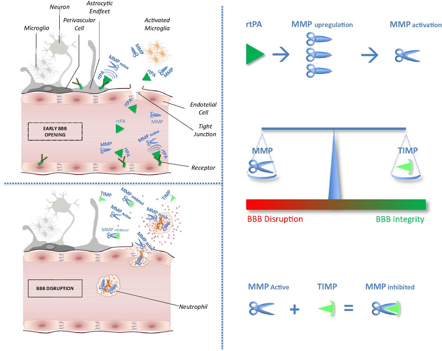
After cerebral ischemia, the general neuronal response to excitotoxic injury determines the release of pro-inflammatory cytokines that stimulate the local production of MMPs and TIMPs (4). In experimental models of brain ischemia, MMPs and MMP/TIMP unbalance play a detrimental role related to blood–brain barrier (BBB) disruption leading to hemorrhagic transformation and edema of an ischemic brain lesion (5). Circulating levels of MMP9 have been proved associated with poor outcomes in stroke patients treated with tissue plasminogen activator (rtPA) (6, 7). Furthermore, recent studies suggest that rtPA adverse effects may be mediated through MMPs upregulation and activation (2). No clinical study has hitherto considered selectively the effect of the balance between MMPs and their physiological inhibitor related to stroke outcomes after thrombolysis. Theoretical effects of rtPA on MMP/TIMP unbalance have been shown in Figure 1.
FIGURE 1
www.frontiersin.org Figure 1. Impact of tissue plasminogen activator on MMP/TIMP unbalance at the neurovascular unit level. After acute ischemic stroke, rtPA may cross blood–brain barrier (BBB), enter the brain parenchyma, and thereby damage neurovascular unit components by promoting metalloproteinase (MMPs) production and activation. Indeed, unbalance between MMPs and their natural inhibitors (tissue inhibitors of metalloproteinases, TIMPs) may exacerbate BBB disruption leading to hemorrhagic transformation and edema of an ischemic brain lesion.
The aim of this study was to evaluate the effect of MMPs/TIMPs ratio on outcomes of ischemic stroke in the same cohort of the biological markers associated with acute ischemic stroke (MAGIC) study. Because MMP inhibition is considered a possible therapeutic target for stroke patients (8), a clearer understanding of MMP/TIMP interplay, compared with the effect of MMPs only, would have important implications for acute stroke therapies.

More at link.