Use the labels in the right column to find what you want. Or you can go thru them one by one, there are only 34,148 posts. Searching is done in the search box in upper left corner. I blog on anything to do with stroke. DO NOT DO ANYTHING SUGGESTED HERE AS I AM NOT MEDICALLY TRAINED, YOUR DOCTOR IS, LISTEN TO THEM. BUT I BET THEY DON'T KNOW HOW TO GET YOU 100% RECOVERED. I DON'T EITHER BUT HAVE PLENTY OF QUESTIONS FOR YOUR DOCTOR TO ANSWER.
Changing stroke rehab and research worldwide now.Time is Brain!trillions and trillions of neuronsthatDIEeach day because there areNOeffective hyperacute therapies besides tPA(only 12% effective). I have 523 posts on hyperacute therapy, enough for researchers to spend decades proving them out. These are my personal ideas and blog on stroke rehabilitation and stroke research. Do not attempt any of these without checking with your medical provider. Unless you join me in agitating, when you need these therapies they won't be there.
What this blog is for:
My blog is not to help survivors recover, it is to have the 10 million yearly stroke survivors light fires underneath their doctors, stroke hospitals and stroke researchers to get stroke solved. 100% recovery. The stroke medical world is completely failing at that goal, they don't even have it as a goal. Shortly after getting out of the hospital and getting NO information on the process or protocols of stroke rehabilitation and recovery I started searching on the internet and found that no other survivor received useful information. This is an attempt to cover all stroke rehabilitation information that should be readily available to survivors so they can talk with informed knowledge to their medical staff. It lays out what needs to be done to get stroke survivors closer to 100% recovery. It's quite disgusting that this information is not available from every stroke association and doctors group.
Showing posts with label initiate research. Show all posts
Showing posts with label initiate research. Show all posts
Will your competent? doctor and hospital get followup research initiated that will create protocols that prevent neurodegeneration?
Do you
prefer your doctor, hospital and board of director's incompetence NOT
KNOWING? OR NOT DOING? Your choice; let them be incompetent or demand
action!
From gut-primed T cells to
microglial signaling and persistent gene-regulatory states, researchers
map an intricate immune network that connects the brain with the rest of
the body.
Immune
cells communicate across a continuous, bidirectional circuit between
the CNS and periphery. Immune cells from across the body engage CNS
tissues and, in turn, are shaped by signals returning from the brain.
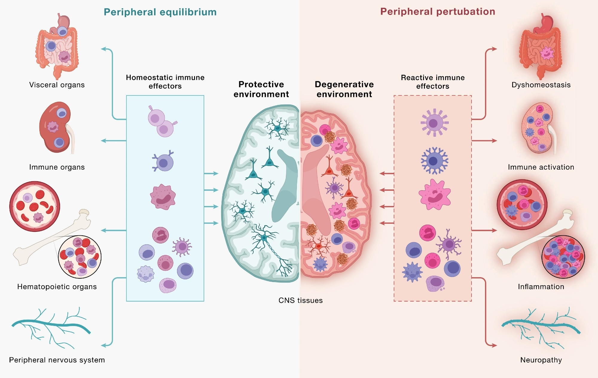
(Left) Under peripheral equilibrium, immune effectors mobilized from
visceral organs, secondary lymphoid organs, hematopoietic tissues, and
the peripheral nervous system (PNS) - including CD4+ and CD8+
T cells, B cells, monocytes (top 3 cell types), NK cells, dendritic
cells, mast cells, neutrophils, and innate lymphoid cells (bottom
cluster of cells) - engage the CNS in a homeostatic dialogue that
supports neural cell health, plasticity, and repair, sustaining a
protective environment. (Right) Age-associated dysfunction across the
same peripheral compartments reshapes this circuit: altered immune
effectors enter the CNS and contribute to shifting the local environment
toward a degenerative state in which microglia adopt disease-associated
programs, astrocytes become reactive, and neurons accumulate pathology.
This model underscores how the same circuitry can yield protective or
pathogenic outcomes and that neurodegeneration is downstream of
dysfunctional brain-immune crosstalk. Study: Neurodegeneration as a dysregulation of neuroimmune crosstalk
A recent perspective published in the journal Cell
synthesizes scientific evidence suggesting that neurodegeneration
involves intricate crosstalk between neurons and immune cells, linking
the brain to peripheral immunity through bidirectional exchange.
Strategies that restore immune homeostasis or recalibrate neuroimmune
signaling may potentially slow neurodegeneration and promote recovery.
Historically, immune dysregulation has often been considered a
consequence of neurodegenerative disorders. Recent studies, however, are
beginning to change this scientific mindset, suggesting that disordered
communication between the brain and immune cells may also contribute to
disease onset and progression. The authors describe immune dysfunction
as a “concause” of neurodegeneration, meaning it may interact with
neuronal and glial vulnerabilities without necessarily being the initial
trigger. It is essential to advance understanding of the
pathophysiology of neurodegenerative diseases to inform therapeutic
development and the development of immune-based strategies.
In this perspective, researchers examined brain-immune interactions
and their potential role in neurodegeneration. They organized emerging
evidence into three frameworks: “outside-in” effects driven by
peripheral immunity, “inside-out” signaling coordinated by
brain-resident microglia, and “locked-in” gene regulatory programs that
can stabilize maladaptive neuroimmune states.
The brain-immune communication network
The brain continuously communicates with peripheral immune networks. Components of the CNS,
including the choroid plexus, meninges, and lymphatic and vascular
structures, interact with immune cells to relay signals related to
neural needs.
Helper and cytotoxic T cells can enter CNS border regions and, under
defined conditions, the brain parenchyma. Brain-immune communication
supports neural integrity but can promote pathology when dysregulated.
Microglia and BAMs provide surveillance, while lymphocytes confer antigen specificity and immunological memory.
Cytokines, complement, and MHC-I
are traditionally linked to immunity, but CNS cells also produce or
sense these molecules during neural activity. Innate lymphoid cells in
the dura can respond to injury, while the choroid plexus helps regulate
inflammatory signaling. In mice, increased neuronal activity may draw
antibody-secreting B-lineage cells into the hippocampus during synaptic
remodeling.
The gut also influences brain immunity. T cells educated in
gut-associated immune tissues can subsequently traffic to the borders of
the CNS and, under certain conditions, into the brain, while plasma
cells secreting IgA antibodies
protect blood vessels in the meninges. In addition, changes in the gut
microbiome could influence immune activity and microglial function.
Through the GBA, the gut and brain
are in constant dialogue with each other. The vagus nerve conveys
immunity-related information from the intestines to the brain.
Reward-related neural pathways can, in turn, influence peripheral immune
activity.
Brain-immune interactions in neurodegenerative disease
T cell activity has been implicated in PD, AD, ALS, and dementia with Lewy bodies (DLB).
In ALS4, an inherited form of ALS, cytotoxic T cells are detected early
in the blood and brain and expand as the disease progresses, consistent
with antigen-driven responses.
Repeating the reporting on this since repetition may be the only way to get thru the thick heads in your stroke hospitals! Print this out and slap your doctor with it, asking when s/he will initiate research in humans to prove this out!
Do you
prefer your doctor, hospital and board of director's incompetence NOT
KNOWING? OR NOT DOING? Your choice; let them be incompetent or demand
action!
The brain evidently can regenerate itself better than previously assumed after injuries or certain autoimmune diseases. Using a mouse model, researchers at the University of Zurich have demonstrated that special supporting and nourishing cells repopulate damaged areas of the brain by initially sending only newly formed cell nuclei there.
Glial cells are supporting and nourishing cells in the brain. Star-shaped glial cells called astrocytes are vital to the functioning of neurons. They supply the nerve cells with nutrients, help to regulate blood flow and keep brain tissue healthy. It had long been assumed that when astrocytes are lost – as happens, for instance, in brain injuries or autoimmune diseases such as rare neuromyelitis optica spectrum disorder, in which the body's own antibodies destroy these cells – the adult brain cannot fully replace them.
Regenerative astrocytes repair damaged tissue
A new study by co-lead authors Marina Herwerth and Matthias Wyss from the Institute of Pharmacology and Toxicology at the University of Zurich (UZH) has now overturned that assumption: their research team headed by Bruno Weber discovered a specialized group of "regenerative" astrocytes in the brains of living mice that step in on the perimeter of the damaged area of the brain to rebuild the cells.
The findings of our study reveal a previously unknown ability of the adult brain to repair itself. They point toward new ways of supporting recovery from ailments involving the loss of astrocytes."
Bruno Weber, University of Zurich
Only cell nuclei migrate
The researchers used two-photon microscopy to observe the brains of living mice in real time over a period of several weeks and mapped which genes switch on in which areas of the brain. This way they were able to identify the special astrocytes that take care of rebuilding injured tissue. But those cells don't just divide, they also perform a remarkable feat: "they send the newly formed nuclei of their daughter cells gliding across long distances to repopulate the damaged area of the brain and knit the astrocyte network back together," Weber explains.
Starting points for targeted regeneration
The discovery of how adult brain cell nuclei migrate through the long star-shaped extensions of astrocytes to injured tissue expands comprehension of how the brain organizes and regenerates itself after certain injuries. If those mechanisms can be selectively activated, that could help to more effectively repair damaged brain tissue, restore astrocyte networks and thus improve recovery after certain brain disorders. "We were able to identify numerous genes and signaling pathways that are temporarily activated during repair. They could serve as starting points in the future for influencing post-disease and -injury regeneration processes," Weber stresses.
Herwerth, M., et al. (2026). Focal astrocyte loss reveals nuclear translocation during lesion repopulation. Nature Neuroscience. DOI: 10.1038/s41593-026-02354-5. https://www.nature.com/articles/s41593-026-02354-5
Since you are massively sedentary during your hospital stay will your doctor at least prescribe cocoa? Oh NO, you DON'T have a functioning stroke doctor, do you? Doesn't read or implement research; just a flaccid walking/talking doctor impersonator! But first needs to initiate research in stroke subjects!
Sitting time is high in older adults and has been shown to temporarily impair endothelial function and blood pressure (BP). Flavanols, plant-derived compounds, acutely enhance endothelial function and reduce BP in older adults. The aim of this study was to investigate whether acute ingestion of cocoa flavanols can improve peripheral endothelial function and BP during prolonged sitting in healthy older adults. In a randomised, double-blinded, within-subject, cross-over, placebo-controlled human study, 20 apparently healthy, older adults (age, 72.4 ± 5.0 years; 7 males, 13 females) consumed a high-flavanol (695 mg) and a low-flavanol (5.6 mg) cocoa beverage immediately before a 2-hour sitting bout. Flow-mediated dilation (FMD) of the superficial femoral (SFA; primary outcome) and brachial (BA) arteries, and BP, were assessed before and after sitting. Microvasculature haemodynamics were assessed in the gastrocnemius before, during, and after sitting. Sitting reduced both SFA FMD (Δ= −0.7%;p= 0.005) and BA FMD (Δ= −0.7%;p= 0.016) in the low-flavanol condition. The high-flavanol intervention prevented the decline in both SFA and BA FMD following sitting, with FMD measures remaining similar to pre-sitting (p> 0.3). Sitting increased both systolic (Δ= 6.1 mm Hg,p= 0.001) and diastolic BP (Δ= 2.6 mm Hg,p= 0.001), with no benefit from flavanol intake. Sitting increased muscle oxygenation resting levels (p< 0.001) and haemoglobin content (p< 0.001), and decreased muscle oxygen consumption during SFA occlusion (p< 0.001). Flavanols had no effect on the muscle microvasculature. These findings indicate that flavanol-rich foods may be efficacious nutritional strategies to counteract sitting-induced endothelial impairments during prolonged sitting in older adults, but do not alleviate sitting-induced increases in BP.
Two hours of sitting impaired upper and lower limb endothelial function (flow-mediated dilation; FMD) and increased blood pressure in healthy older adults. Consuming flavanols prior to sitting preserved endothelial function in both conduit arteries.
We’re sharing this article early to provide faster access to peer-reviewed, accepted research. It is citable and carries a permanent DOI. This version is subject to further edits and will be replaced automatically by the final Version of Record. All legal disclaimers apply.
Abstract
Background
Action observation (AO) and motor imagery (MI) represent promising, non-invasive strategies for promoting neuroplasticity in motor rehabilitation by engaging the shared neural substrates of actual movement. However, their translation to swallowing rehabilitation, particularly for neurogenic dysphagia, lacks a robust neurophysiological foundation. A critical barrier is the absence of direct, multimodal evidence comparing how swallowing-specific static AO (SAO), dynamic AO (DAO), and MI differentially engage the cortical swallowing network. Specifically, their immediate effects on corticobulbar excitability and hemodynamic activation within key sensorimotor regions remain unquantified and poorly contrasted, limiting the rationale for their targeted clinical application.
Objective
This study employed a dual-modal neuroimaging approach to precisely quantify and compare the immediate neurophysiological effects of SAO, DAO, and MI on the human swallowing sensorimotor system. We aim to evaluate their modulatory effects on bilateral suprahyoid motor cortical excitability and intracortical inhibitory/facilitatory circuitry using transcranial magnetic stimulation (TMS), and map their hemodynamic activation patterns within core sensorimotor cortices compared to motor execution (ME) using functional near-infrared spectroscopy (fNIRS).
Methods
Thirty-two healthy adults underwent integrated assessments using transcranial magnetic stimulation (TMS) and functional near-infrared spectroscopy (fNIRS). TMS measured motor-evoked potentials (MEPs), short-interval intracortical inhibition (SICI), and intracortical facilitation (ICF) in bilateral suprahyoid motor cortices during rest, SAO, DAO, and MI. fNIRS mapped hemodynamic changes in dorsal/ventral precentral (dPreCG/vPreCG) and postcentral gyri (dPoCG/vPoCG), superior/middle frontal gyri (SFG/MFG) during swallowing-specific SAO, DAO, MI and ME.
Results
MI reduced bilateral SICI and increased left ICF, concurrently activating bilateral dPoCG, left SFG/MFG, and right dPreCG/vPreCG/vPoCG—regions overlapping with ME-activated networks (bilateral vPreCG/vPoCG/MFG), with left MFG, right vPreCG/vPoCG as coactivating areas. In contrast, DAO reduced left SICI but elicited no hemodynamic activation, while SAO showed no significant effects.
Conclusion
MI enhances the excitability of the swallowing motor cortex and activates the key sensorimotor cortical areas governed ME of swallowing. MI shows greater superiority over AO and may become a promising effective rehabilitation strategy for neurogenic dysphagia.
Trial registration Chinese Clinical Trial Registry, ChiCTR2000036715. Registered on 24 August 2020, https//www.chictr.org.cn/bin/home.
1. Department of Neurology, Hainan General Hospital, Hainan Affiliated Hospital of Hainan Medical University, Haikou, China
2. Clinical Research Center, Affiliated Chinese Medicine, Hainan Medical University, Haikou, China
Abstract
Background:
Excessive neuroinflammation mediates blood–brain barrier (BBB) disruption and poor outcomes after acute ischemic stroke (AIS). Cyclophilin A (CypA), when released into the extracellular space (designated as eCypA), may participate in inflammatory reactions and vascular dysfunction. However, its role in regulating neuroinflammation and BBB injury in AIS, as well as the therapeutic potential of targeting eCypA remain unclear.
Methods:
ELISA was used to detect eCypA release in serum from 22 AIS patients (17 mild, 5 severe; 13 males, 9 females; mild: age 65.41 ± 10.20 years, NIHSS 2.88 ± 1.45; severe: age 64.00 ± 5.04 years, NIHSS 9.40 ± 3.29; blood sampled within 48 h of onset), in serum/cerebrospinal fluid (CSF) from transient middle cerebral artery occlusion (tMCAO) rats, and in supernatants from BV2 (microglia) or bEnd.3 (brain microvascular endothelial cells) exposed to oxygen–glucose deprivation/reoxygenation (OGD/R) or lipopolysaccharide (LPS). Nine-week-old male Sprague–Dawley rats (n = 5 per group) underwent 1.5 h of tMCAO via the intraluminal suture method followed by 24 h of reperfusion before sampling. These rats received intracerebroventricular injection of cyclophilin A-binding heptameric peptide (C46) before tMCAO establishment. Cerebral infarct volume was measured via TTC staining. BBB permeability was assessed by Evans blue extravasation. Western blot was employed to determine protein levels of tight junction (TJ) proteins, matrix metalloproteinases (MMPs) and proinflammatory mediators. Microglial activation was evaluated by immunofluorescence.
Results:
eCypA levels were significantly elevated in AIS patient serum (1.74 ± 0.23 ng/mL in mild, 2.39 ± 0.09 ng/mL in severe vs. 1.30 ± 0.19 ng/mL in healthy controls, p < 0.001), in tMCAO rat serum (2.57 ± 0.14 vs. 1.62 ± 0.07 ng/mL, p < 0.001) and CSF (2.14 ± 0.23 vs. 1.47 ± 0.19 ng/mL, p < 0.001), as well as in the supernatants of OGD/R-challenged BV2 (0.92 ± 0.01 vs. 0.55 ± 0.03 ng/mL, p < 0.001) and bEnd.3 cells (1.10 ± 0.05 vs. 0.52 ± 0.03 ng/mL, p < 0.001) and LPS-induced BV2 cells (1.12 ± 0.08 vs. 0.56 ± 0.13 ng/mL, p < 0.001) compared with their respective control groups. The eCypA inhibitory peptide C46 effectively improved neurological function, reduced cerebral infarct volume and edema in tMCAO rats. Moreover, C46 mitigated BBB permeability, preserved the expression levels of TJ proteins, and suppressed the activation of MMPs in tMCAO rats and OGD/R-treated bEnd.3 cells. Meanwhile, C46 administration inhibited microglial activation and downregulated the expression of proinflammatory mediators both in vivo (tMCAO rats) and in vitro (OGD/R- or LPS-induced BV2 microglia).
Conclusion:
eCypA, released by microglia and brain microvascular endothelial cells under ischemic–hypoxic and inflammatory conditions, serves as a critical pathogenic mediator that drives neuroinflammation and BBB disruption in AIS. Targeting eCypA with C46 peptide effectively abrogates these pathological cascades, thereby supporting eCypA as a novel therapeutic target for AIS.
With your risk of Parkinsons post stroke, IS YOUR DOCTOR SMART ENOUGH TO INSTITUTE THIS PROTOCOL FOR YOU? NO? Your doctor doesn't know about it and will do nothing! Fire that doctor and find someone intelligent!
Will your doctor and hospital help initiate research that will cure and prevent Parkinsons?
Researchers have been chasing the causes of Parkinson’s disease for more than a century, yet its hallmark feature — microscopic protein clumps called Lewy bodies — remains enigmatic: Patients with just a few Lewy bodies can be devastatingly ill, while others with neurons packed full of them sometimes show milder symptoms.
Another culprit has emerged. Nanoscale protein assemblies called alpha-synuclein oligomers had been hiding in plain sight, too small to detect with conventional microscopy. A group of researchers can now look more closely at oligomers — literally — because they developed a way to see them in postmortem brain tissue for the first time. They detail the discovery in a recent paper published in Nature Biomedical Engineering.
When the team captured their first clear images, the reaction was immediate.
Steven Lee, PhD
“We said, wow, isn’t this fantastic,” said Steven Lee, PhD, a microscopy specialist at the University of Cambridge in Cambridge, England, and co-lead author of the study. “We can see these small clumps; they have all different sizes, and they appear to be everywhere.”
It had been a long road. Despite extensive in vitro data suggesting oligomers existed, many researchers remained skeptical that they were actually present in the brain. Lee recalls Professor Sir John Hardy, FRS, an eventual co-author on the study and a geneticist who’d been working on Parkinson’s his whole career, standing up at an internal funder conference for the Aligning Science Across Parkinson’s initiative. “He said, ‘We have been looking for [these] oligomers for 30 years, and we haven’t found them.’”
By that point, Lee’s team had already been working on imaging. He chimed in. “I said, ‘I think the reason you haven’t is not biological; it’s technological.’”
Seeing Stars in Daylight
Lewy bodies don’t appear out of nowhere — they are an end product of a progressive assembly line.
The process starts when single misfolded units of the alpha-synuclein protein begin sticking together, forming tiny intermediate clumps, acting like bridges between solitary proteins and the massive fibrils that eventually become Lewy bodies.
In countless experiments across cell cultures and animal models, researchers demonstrated that these small, soluble oligomers kill neurons with ruthless efficiency, while their larger cousins — the fully formed Lewy bodies — leave cells relatively unharmed.
This disease-associated subpopulation of alpha-synuclein oligomers is much more common than the Lewy bodies traditionally used to diagnose Parkinson’s, making them the faint stars of a hidden galaxy of Parkinson’s pathology, while the Lewy bodies were merely the handful of planets visible to the naked eye.
The challenge in visualizing them, Lee explained, wasn’t building a more powerful microscope. It was more fundamental than that.
“If you look up at the blue sky, you know that starlight is there, right?” he said. “You can’t see stars in the daytime because the Rayleigh scatter — the blue sky — raises the background to a point where they’re invisible. The same thing is true in brain tissue.”
Human brains are, to put it technically, extremely messy at the molecular level. Tissue autofluorescence — the natural glow from cellular components like lipids and other biological matter under laser light — creates a high background intensity, or “noise floor,” that obscures faint signals. The oligomers, being nanoscale, can only bind to a few of the fluorescent antibodies that scientists use to tag and visualize them under a microscope, making their signal incredibly dim. It’s like trying to spot a candle flame during a firework show.
The solution, which the team calls Advanced Sensing of Aggregates-Parkinson’s Disease, involved a two-part strategy analogous to stargazing: Make the background “night sky” in the brain darker and use a more powerful microscope to capture the lighter, toxic aggregates in the foreground.
The research team used Sudan Black B, a fat-soluble dye, to quench much of this obfuscating autofluorescence. An optimized 10-minute incubation with the dye suppressed an incredible 93% of the tissue’s background glow, effectively switching off the lights and turning day into night.
That wasn’t enough. To achieve the sensitivity to detect the faint light from the oligomers, the team used a high numerical aperture objective lens, a piece of advanced optics typically used for imaging single molecules. A higher numerical aperture allows the microscope to gather light from a much wider angle, dramatically increasing the signal collected from the sample.
The combination dropped the noise floor low enough that the oligomers, faint as they were, finally became visible.
The Twist: Alpha-Synuclein Oligomers in Healthy Brains
What started as a 5-year project sometimes involved the team working around the clock to collect images in the lab of Sonia Gandhi, BMBCh, PhD, professor of neurology at the University College London, London, England, and co-lead author of the study. This eventually generated 12,028 high-resolution images and a dataset of 1.2 million individual protein aggregates, all of which are now online for researchers to search through. For their next paper, currently in review, the dataset was even bigger, equivalent to 6 months of continuous imaging, 24 hours a day, capturing 4.5 billion aggregates.
Science at this scale requires a certain obsessiveness.
“We always ask ourselves if what we see could be due to random chance,” explained Lucien Weiss, PhD, associate professor of engineering physics at Polytechnique Montreal, Montreal, Canada, and co-lead author of the study. “In this project, once we knew more about what to look for, we asked if the same signatures show up using other methods.”
Lucien Weiss, PhD
The team reran experiments with different methods and compared adjacent tissue slices, compared different reagents, and even sourced samples from a different brain bank. “Because we image many oligomers across many samples to get significant numbers," Weiss said, “we started to see really strong reproducibility.”
The findings broke down into three key discoveries, and the first one was a surprise. Yes, Parkinson’s brains were loaded with nanoscale alpha-synuclein assemblies — hundreds per cell. But so were healthy control brains.
“They’re there in healthy controls as well, which is really surprising and interesting,” Lee said. “We hadn’t quite expected that.” This wasn’t noise or artifact. The assemblies were there, reliably, in people who’d never had a tremor in their lives. But why do they turn toxic?
The second finding provided a clue. When the computational analysis parsed through the brightness distributions — a proxy for size — a distinct subpopulation emerged in the Parkinson’s samples. These assemblies were brighter, larger, and had chemical properties suggesting structural differences. They resisted enzymatic breakdown and showed “seed competence,” meaning they may act as a template to trigger a chain reaction, causing healthy proteins to misfold and join the unhealthy clump. And they clustered around neurons, astrocytes, and microglia in patterns never seen in healthy tissue.
The numbers were stark: These disease-specific assemblies made up about 10% of all oligomers in Parkinson’s brains but only 0.26% in healthy control brains.
Why Every Drug Has Failed
The implications ripple outward in uncomfortable directions. Said Lee, “So far, all attempts to develop disease-targeting drugs in Parkinson’s disease have failed. This tells us the standard model for drug discovery simply doesn’t work.”
Going forward, Lee’s team will use automated microscopy to screen drug libraries from a Japanese pharmaceutical company, testing whether compounds can disassemble, prevent, or accelerate the formation of the toxic alpha-synuclein oligomeric aggregates. He argued previous drug trials failed because they measured the average behavior of all proteins in the brain, most of which aren’t in this problematic oligomeric state.
There’s another problem, and it’s arguably worse. “There’s a massive misdiagnosis rate, about 20% both ways,” Lee revealed. Parkinson’s is diagnosed clinically, but the only definitive diagnosis comes from a postmortem brain biopsy. So roughly 1 in 5 patients enrolled in clinical trials doesn’t actually have Parkinson’s disease.
“You have to wait for them to pass away, check the brain, and go, ‘Oh, he didn’t have Parkinson’s, he had PSP [progressive supranuclear palsy],’” said Lee. “And all that data you’ve been collecting is difficult to make meaningful conclusions from.”
Meanwhile, they’re also working to detect these assemblies in living patients — in cerebrospinal fluid, blood, and even saliva. Early data suggest it’s possible. “We’ve shown there’s an increased aggregate load in cerebrospinal fluid of people with Parkinson’s,” Lee said. “We didn’t know whether they were present in the brain at that time. Now we know.”
This matters for two reasons. Firstly, a test that detects disease-specific oligomers could identify Parkinson’s before symptoms appear, when neurons are still salvageable. Secondly, clinical trials could track whether treatments reduce oligomer levels rather than waiting years to see if symptoms slow.
The Cure Question
On whether Parkinson’s could be cured in our lifetime, Weiss was cautious, pointing to the practical challenges of treating a disease of aging. “You don’t start treating someone until you have a reason to treat them. For people who have a genetic predisposition, you have a chance to intervene and do something about it and prolong the good years. But can it be cured forever? I’m not sure when you would know to start giving treatments to someone that may or may not develop the disease.”
Lee, however, was adamant. “I respectfully disagree completely, 100%,” he said. “Things that historically seemed incredibly difficult have been chipped at by incremental developments. It’s complex and difficult, but it’s not unsolvable.”
Funding will certainly play a part. This research itself was only possible because of funding by Aligning Science Across Parkinson’s, a philanthropic initiative supported by Sergey Brin, co-founder of Google, who has committed nearly half a billion dollars to Parkinson’s research. “We’re extremely lucky to be involved with a fantastic initiative,” Lee said.
Weiss added, “I get excited about solving challenges. We’re motivated by the idea that, if this works, we’re able to understand something that we couldn’t before. Ultimately, that knowledge grows and goes back into helping our communities. The public investment in science and understanding diseases is a long-term effort, but it’s really the best way we know of to solve these very complex problems. You have to put in this legwork now to solve the problem down the road.”
Get your competent? doctor and hospital to initiate research on EXACT DIETS NEEDED! Your doctor has known of this need for decades; why hasn't research been initiated already? Your doctor prefers incompetence since less work is involved? Serious question; ask your doctor that specific question and watch them squirm!
Diet quality has been proposed as a determinant of brain aging, which has attracted considerable attention given the current global demographic shift toward older age. Comprehensive global systematic reviews that have explored dietary patterns and brain aging highlight a recurrent theme. Any healthy dietary pattern that includes higher consumption of vegetables, fruits, legumes, nuts, fish and/or seafood, and unsaturated vegetable oils/fats and lower consumption of red and processed meats and sugar-sweetened beverages is associated with lower risk of age-related neurodegenerative disease. The biologic mechanism(s) underlying these cognitive protective effects are unknown. Furthermore, it is unlikely that consumption of a healthy dietary pattern alone will achieve clinically relevant success in reducing risk of cognitive decline and/or dementia given that there is no single risk factor that accounts for the variation in brain aging.
My PMR(physical medicine and rehabilitation) doctor knew nothing about stroke recovery. He just wrote three prescriptions for E.T.(Evaluate and Treat) to my PT, OT and ST. I could train a chimp to do that. If he knew anything about stroke recovery it didn't show.
As
the probability of getting strokes becomes higher, especially among
young adults, the need of the hour is a good rehabilitation centre. CE
speaks to experts at HCAH, a first-of-its-kind rehabilitation centre in the city.
Suffering
a stroke is an indescribably painful and difficult experience. But if
you are in the hands of experienced doctors who administer effective and
modern treatments, you will be back on your feet in no time. With this
in mind, HCAH, a first-of-its-kind rehabilitation centre in Hyderabad,
is ready to provide a dedicated Physical Medicine and Rehabilitation
(PMR) team for stroke recovery. They conducted a session to explain what
they do in the city, after which Dr Gaurav Thukral, co-founder and COO
of HCAH, and Dr Aastik Bhatt, a leading specialist in neuro
rehabilitation and physical medicine at HCAH, decoded it all for CE.
Dr
Gaurav said, “Hyderabad is known for its rehabilitation hospitals, with
25 to 30 such hospitals in the city. But they are not properly
equipped. What we are doing is strengthening our forces by getting
doctors who are trained to aid in recovery and rehabilitation. These
doctors, called physical medicine and rehab specialists, are not
therapists but are doctors who have done their MD.”
He added that
these doctors are trained to treat patients with strokes, spinal
injuries, traumatic brain injuries, and head injuries. The doctor guides
the team on which part of the body needs which type of therapy, such as
speech therapy for speech-related issues, physiotherapy for
strengthening the limbs, and cognitive therapy for rewiring the brain.
“We
use AI to predict and facilitate recovery.(AI is almost completely worthless until the underlying research for 100% recovery is there!) Unlike regular physiotherapy
sessions, these interactive robotics give patients confidence in their
recovery. They also stimulate the brain, leading to a speedier
recovery,” he explained, adding that there are three or four subsets of
patients who will use this. The first group is stroke patients, the
second is head injury patients, the third is spinal injury patients, and
the fourth includes those with painful conditions such as frozen
shoulder, lower back pain, and cervical pain.
Dr
Gaurav pointed out that they have machines such as Rapid, Exo-Skeleton,
and Balance Lab. “Usually, hospitals which have high-end doctors and
equipment also offer costlier treatments. However, it is the opposite at
HCAH because patients stay for fewer days at the centre, reducing
expenses. These doctors and machines are also available on an OPD basis,
allowing patients to avoid room rental costs,” he stated.
Elaborating
on how this rehab helps stroke patients, he said, “Nobody predicts a
stroke or a road accident, but when it happens, we find ourselves
trapped in our own bodies, confined to a bed, longing to walk again and
return to our jobs. The two main goals are regaining independence and
reducing medical expenses, both of which are possible here.”
Regarding
whether there would be complete recovery or any side effects, he noted,
“What we want is complete recovery,(Have you initiated the research to get to 100% recovery? Right now only 10% fully recover, all the rest of this paragraph is a lie!) but that is only possible if you
come to the recovery centre as soon as possible. After a stroke, there
is what we call the ‘golden period’ — the sooner you arrive, the sooner
your brain starts rewiring and sending signals to the muscles that need
to be activated. This process is combined with medication, equipment,
and medical supervision. However, if patients go home and return after
six months, the golden period is already over, making complete recovery
much more difficult.”
Dr Aastik Bhatt stressed the need to expand
rehabilitation services, explaining, “Timely rehabilitation is crucial
for stroke recovery, yet many centres remain inaccessible or
unaffordable. At HCAH, we have built a specialised PMR team, integrating
AI-powered rehabilitation tools and robotic-assisted therapy to deliver
precise, customised treatment. This ensures that every stroke patient
receives holistic, multidisciplinary care tailored to their recovery
needs.”