Changing stroke rehab and research worldwide now.Time is Brain! trillions and trillions of neurons that DIE each day because there are NO effective hyperacute therapies besides tPA(only 12% effective). I have 523 posts on hyperacute therapy, enough for researchers to spend decades proving them out. These are my personal ideas and blog on stroke rehabilitation and stroke research. Do not attempt any of these without checking with your medical provider. Unless you join me in agitating, when you need these therapies they won't be there.

What this blog is for:

My blog is not to help survivors recover, it is to have the 10 million yearly stroke survivors light fires underneath their doctors, stroke hospitals and stroke researchers to get stroke solved. 100% recovery. The stroke medical world is completely failing at that goal, they don't even have it as a goal. Shortly after getting out of the hospital and getting NO information on the process or protocols of stroke rehabilitation and recovery I started searching on the internet and found that no other survivor received useful information. This is an attempt to cover all stroke rehabilitation information that should be readily available to survivors so they can talk with informed knowledge to their medical staff. It lays out what needs to be done to get stroke survivors closer to 100% recovery. It's quite disgusting that this information is not available from every stroke association and doctors group.

Showing posts with label 3d brains. Show all posts

Thursday, August 6, 2026

3D Human Brain Tissue Model Replicates Alzheimer’s Pathology

 Now our stroke researchers CAN EXACTLY MODEL how a stroke causes dementia! Then they can create protocols that prevent that dementia from happening! But nothing will occur; THERE IS NO LEADERSHIP OR STRATEGY IN STROKE ANYPLACE! Don't have a stroke is their answer. 

3D Human Brain Tissue Model Replicates Alzheimer’s Pathology

Summary: Researchers introduced a highly reproducible three-dimensional human brain tissue model capable of replicating complex neurodegenerative processes in Alzheimer’s disease. Developed over nine years using human stem cells, the self-organizing tissue spheroids integrate functional neurons, astrocytes, and microglial immune cells into micro-architectures the size of half a pinhead.

The engineered tissue model faithfully expresses key Alzheimer’s-relevant genes and proteins, forms functional synaptic networks, and exhibits active microglial surveillance. The team demonstrated the platform’s clinical relevance by inducing characteristic amyloid beta aggregates and subsequently dissolving them using newly approved therapeutic antibodies.

Key Facts

  • Tri-Culture Cellular Interplay: Unlike traditional two-dimensional cultures, the 3D spheroid model incorporates neurons forming functional synapses, astrocytes providing metabolic support, and microglial immune cells performing active tissue surveillance.
  • Rapid Self-Organization: Driven by a proprietary differentiation cocktail and nutrient medium, differentiated stem cells assemble and self-organize into functional mini-tissue spheroids within one week.
  • Transcriptomic & Proteomic Fidelity: Comprehensive testing confirmed that all primary genes and proteins associated with Alzheimer’s pathology and cell-to-cell signaling are fully active within the engineered tissues.
  • Validation via Plaque Dissolution: The researchers successfully triggered human amyloid beta aggregate formation within the tissue spheroids and verified that current anti-amyloid Alzheimer’s therapeutics, mediated by active microglia, successfully cleared the pathology.
  • Robotic Automation & Industrial Scaling: The laboratory is currently adapting the platform for automated robotic manufacturing to produce thousands of identical, diseased tissue spheroids for large-scale industrial drug testing.

Source: LMU

How can Alzheimer’s research be made faster, better, and more effective? After decades of intensive research worldwide and despite recent therapeutic advances, scientists have not managed to fully arrest the progress of the disease.

“What we are still lacking is three-dimensional models that accurately replicate the complex interactions in human brain tissue with Alzheimer’s disease,” explains Dominik Paquet, Professor of Neurobiology at the Institute for Stroke and Dementia Research at LMU University Hospital.

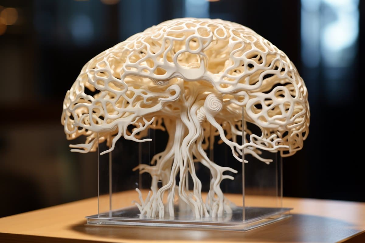
This shows a brain model.
Researchers bioengineered a 3D human brain tissue model containing neurons, astrocytes, and microglia that successfully replicates Alzheimer’s amyloid clearance and supports automated drug screening. Credit: Neuroscience News
This is precisely the area in which his team has now made major progress – with potentially far-reaching consequences for the development of new drugs against Alzheimer’s disease.

The group reported the exciting new results in the journal Nature Neuroscience.

For laypeople, it always seems a bit like magic when researchers take stem cells and grow tissues that resemble the human original. According to Dominik Paquet, however, it takes a mixture of scientific creativity, technical skills, and patience: “It took us nine years,” says the neuroscientist, “to develop our new, three-dimensional model of human brain tissue before it worked at all necessary levels.”

What does he mean by all necessary levels, we might ask? For Alzheimer’s research, the interaction of different cell types and their biochemical functions are the most important thing – much more so than the exact replication of the structure of the brain.

The right recipe for genuine interplay

The starting material for the new 3D tissue model is human stem cells, which can be converted into various brain cell types – in this case, neurons, astrocytes, and microglial cells. To obtain this set, the stem cells have to be treated with a cocktail of different substances “according to a very specific recipe we developed.”

In a special nutrient solution, Paquet continues, the differentiated cells connect and adhere to each other. “Within a week, they form little tissue balls about the size of half a pinhead. These spheroids self-organize and take on key functions of the brain.”

Almost everything just like in a real human brain

Their neurons form extensions and connect with functional synapses. The astrocytes supply their neighbors with nutrients. And the microglial cells – the immune cells of the brain – monitor their environment and ensure that no dead cells or foreign matter that does not belong there can accumulate. “We also tested whether all genes and proteins that are important for the study of Alzheimer’s disease are active in our tissue model,” says the neuroscientist. “And that was indeed the case.

Reproducible, modifiable – and disease-relevant

Another major advantage of the system is its reproducibility. That is to say, if you follow the instructions of the Munich researchers, you will reliably obtain tissue structures with the same composition and the same functions.

“However, we can also modify the generated brain tissue from the outside,” says Dominik Paquet. “For example, we can trigger symptoms of a disease like Alzheimer’s, test potential drugs, and so forth.” In fact, the researchers have successfully induced the formation of the Alzheimer’s-typical amyloid aggregates – and then dissolved them again with new drugs that are already available. The microglia that play a key role in the disease were demonstrably active in the process.

Next step: automation for efficient drug development

“Our system,” says Dominik Paquet, “could help accelerate the development of new drugs.”

With this goal in mind, his team is currently working on automating and scaling the manufacture of the tissue models using robots. This would mean manufacturing hundreds or even thousands of tissues with the same disease symptoms. This is particularly important for applications in industrial-scale medicine – for example, to be able to efficiently test many new substances in a human system for their effectiveness against Alzheimer’s.

Key Questions Answered:

Q: Why are three-dimensional tissue models better than traditional cell cultures for Alzheimer’s research?

A: Traditional 2D cell cultures grow flat on plastic dishes and lack the complex structural interactions, spatial organization, and multi-cell communication found in a living brain. The 3D spheroid model brings neurons, astrocytes, and microglia together in a spatial framework that allows microglial immune cells to actively clear dead cells and pathology just as they would in human brain tissue.

Q: What specific brain cell types are included in the new LMU Munich tissue model?

A: The model uses human stem cells differentiated into three primary cell populations: neurons (which build functional synaptic connections), astrocytes (which support cell metabolism and structure), and microglia (the resident immune cells responsible for monitoring brain health and clearing toxic protein aggregates).

Q: How does this development accelerate the discovery of new Alzheimer’s drugs?

A: Because the system is highly reproducible and being adapted for robotic automation, pharmaceutical companies will be able to manufacture thousands of standardized human brain tissue models exhibiting Alzheimer’s symptoms. This allows researchers to test thousands of potential drug candidates rapidly in a realistic human cellular environment before moving to animal or clinical trials.

Editorial Notes:

  • This article was edited by a Neuroscience News editor.
  • Journal paper reviewed in full.
  • Additional context added by our staff.

About this Alzheimer’s disease research news

Author: Dominic Anders
Source: LMU
Contact: Dominic Anders – LMU
Image: The image is credited to Neuroscience News

Monday, May 11, 2026

Lifelike 3D-Printed ‘Training Brains’ React Like Real Organs

 Our doctors could make a 3d model of your damaged brain, thus knowing EXACTLY WHERE YOUR DEAD GRAY AND WHITE MATTER IS. Thus simplifying recommending the EXACT PROTOCOLS THAT FIX SUCH DAMAGE!

Nothing will occur, there is NO leadership or strategy anywhere in stroke!

Lifelike 3D-Printed ‘Training Brains’ React Like Real Organs

Imagine what brain surgeons could do with a three-dimensional (3D)-printed model of the brain. Not a structural model printed in plastic and uniform throughout, but one that mirrors the heterogeneity of real human brain tissue — its mechanical, thermal, and electromagnetic properties.

photo of Christopher O Bryan PhD
Christopher O’ Bryan, PhD, MS

“If somebody has a brain tumor, you could take an MRI of their brain and make a 3D model from it that includes the tumor, and then practice surgery on that model,” said Christopher O’ Bryan, PhD, MS, assistant professor of mechanical and aerospace engineering at the University of Missouri (UM) in Columbia, Missouri. “You could learn how sound, electromagnetic waves, or physical insults interact with the brain to better understand concussions and traumatic brain injury.”

photo of 3D brains
     A scaled-down 3D model of the brain, about 15% of a real brain’s size.

O’Bryan and colleagues in UM’s College of Engineering recently published a study in the journal Materialia describing how the researchers designed a unique 3D printer ink made from a modified polymer — called a photo-crosslinkable poly(vinyl alcohol) methacrylate polymer — and then used it to print a scaled-down 3D model of the brain, about 15% of a real brain’s size. The scientists relied on a method called embedded 3D printing, crosslinking the polymer in a microgel bath, to achieve a model that mimicked the soft tissue and the structural complexities of a real human brain.

How They Did It

To provide a broader picture of why they conducted the study, O’Bryan described his lab’s goals. “The focus areas in our lab include the designing of soft materials, the testing of these materials, and then how to actually manufacture things out of these soft materials. And that’s especially challenging when you take into account that most soft materials are hydrogels and biopolymers which start out in a liquid phase ,” he said.

Tuesday, March 24, 2026

Lifelike 3D-Printed ‘Training Brains’ React Like Real Organs

 With ANY BRAINS AT ALL in our stroke medical 'professionals' we could do research that tells us exactly what interventions work to stop the neuronal cascade of death in the first week! Putting the brain on a chip with brain organoids and this and we could solve a lot of stroke recovery questions.  But nothing will occur; OUR 'PROFESSIONALS' ARE FUCKING INCOMPETENT! 

Lifelike 3D-Printed ‘Training Brains’ React Like Real Organs

Imagine what brain surgeons could do with a three-dimensional (3D)-printed model of the brain. Not a structural model printed in plastic and uniform throughout, but one that mirrors the heterogeneity of real human brain tissue — its mechanical, thermal, and electromagnetic properties.

photo of Christopher O Bryan PhD
Christopher O’ Bryan, PhD, MS

“If somebody has a brain tumor, you could take an MRI of their brain and make a 3D model from it that includes the tumor, and then practice surgery on that model,” said Christopher O’ Bryan, PhD, MS, assistant professor of mechanical and aerospace engineering at the University of Missouri (UM) in Columbia, Missouri. “You could learn how sound, electromagnetic waves, or physical insults interact with the brain to better understand concussions and traumatic brain injury.”

photo of 3D brains
     A scaled-down 3D model of the brain, about 15% of a real brain’s size.

O’Bryan and colleagues in UM’s College of Engineering recently published a study in the journal Materialia describing how the researchers designed a unique 3D printer ink made from a modified polymer — called a photo-crosslinkable poly(vinyl alcohol) methacrylate polymer — and then used it to print a scaled-down 3D model of the brain, about 15% of a real brain’s size. The scientists relied on a method called embedded 3D printing, crosslinking the polymer in a microgel bath, to achieve a model that mimicked the soft tissue and the structural complexities of a real human brain.

How They Did It

To provide a broader picture of why they conducted the study, O’Bryan described his lab’s goals. “The focus areas in our lab include the designing of soft materials, the testing of these materials, and then how to actually manufacture things out of these soft materials. And that’s especially challenging when you take into account that most soft materials are hydrogels and biopolymers which start out in a liquid phase ,” he said.

photo of 3D brains
Researchers used embedded 3D printing, crosslinking the polymer with a gel bath to mimic brain tissue.

The researchers’ embedded 3D printing approach uses “sacrificial support baths,” jelly-like baths, which act as a supportive matrix during the printing phase. “What this allows us to do is to print materials as liquids and keep them as liquids until we’ve completely printed our structure, and then we crosslink the printed structure using heat or UV [ultraviolet] light to make it solid. Then we take it out of the gel bath, and it’s become a soft, deformable structure — meaning it can change shape when a force is applied,” O’Bryan said.

Tuesday, February 11, 2025

Innovative 3D blood-brain barrier model enhances understanding of neuroinflammation

 Now our non-existent stroke leadership can direct our researchers to solve and prevent neuroinflammation post stroke. But since there is NO leadership in stroke, NOTHING WILL OCCUR! You're screwed along with all the 10 million yearly stroke survivors now and into perpetuity.

Innovative 3D blood-brain barrier model enhances understanding of neuroinflammation

A 3D model accurately mimicking the Blood-Brain Barrier (BBB) in a laboratory environment has been successfully developed by research teams led by Professor Jinah Jang from the Departments of Mechanical Engineering, Life Sciences, IT Convergence Engineering, and the Graduate School of Convergence at POSTECH, and Professor Sun Ha Paek from the Department of Neurosurgery at Seoul National University Hospital. This study was recently published in Biomaterials Research, an international academic journal on materials science.

Neurodegenerative diseases, including Alzheimer's, Parkinson's disease, and amyotrophic lateral sclerosis (ALS), result from the progressive decline of brain and nervous system functions, primarily due to aging. Chronic neuroinflammation, a key driver of these disorders, arises from the intricate interactions between cerebral blood vessels and neural cells, where the BBB plays a pivotal regulatory role. However, existing BBB models have been unable to replicate the complex three-dimensional 3D structure of cerebral blood vessels, posing significant challenges for research and drug development.

To address these limitations, the research team developed a cerebrovascular-specific bioink using "decellularized extracellular matrix" (CBVdECM), derived from porcine brain and blood vessels. Additionally, the team applied 3D bioprinting technology to construct a tubular vascular model that precisely replicates the anatomical structure and function of the human BBB.

A key feature of this model is the spontaneous formation of a dual-layered structure without external stimuli. When "HBMEC (human brain microvascular endothelial cells)" and "HBVP (human brain vascular pericytes)" were incorporated into the CBVdECM bioink and printed, the endothelial cells self-assembled into the inner vascular wall, while pericytes formed a surrounding layer. This resulted in the creation of a dual-layered structure that closely resembles the architecture of actual blood vessels.

Further, the research team successfully replicated the arrangement and organization process of "tight junction proteins," a component typically absent in conventional 2D models. Additionally, BBB permeability and inflammatory responses were observed following exposure to inflammation-inducing substances (TNF-α and IL-1β). This approach enabled the precise modeling of neuroinflammatory mechanisms, yielding critical insights into the role of BBB dysfunction and inflammation in the pathophysiology of neurodegenerative diseases.

Professor Sun Ha Paek of Seoul National University Hospital commented, "This study provides a crucial platform for investigating the pathological mechanisms of neuroinflammation and developing novel therapeutic strategies."

We aim to integrate additional cell types, such as glial cells, neurons, and immune cells, to refine methods for quantifying inflammatory responses and permeability, while also expanding to patient-specific disease models."

Professor Jinah Jang of POSTECH 

This research was supported by Ministry of Trade, Industry & Energy and the Korea Planning & Evaluation Institute of Industrial Technology's Industrial Technology Alchemist Project, as well as the National Research Foundation of Korea's University-Focused Research Institute Support Program.

Source:
Journal reference:

Han, H., et al. (2024). Cerebrovascular-Specific Extracellular Matrix Bioink Promotes Blood–Brain Barrier Properties. Biomaterials Research. doi.org/10.34133/bmr.0115.

Sunday, February 4, 2024

Revolutionary 3D-Printed Brain Tissue Mimics Human Function

Which built brain should our researchers be using?. I expect our researchers to be using the best one.

Nearly complete human brain grown in US lab: scientist August 2015

Multiregional brain on a chip  Jan 2017
Draper Laboratory developing “Brain-on-a-Chip”  October 2012

"Alzheimer's-in-a-Dish" Docs Win Top Smithsonian Ingenuity Award Nov. 2015 

A patient’s budding cortex — in a dish?  June 2015 


,Cell cultures in petri dishes open new doors to brain research  April 2017

Scientists create 3D-printed brain-like tissue from stem cells July 2017

3D Mini-Brains Accelerate Research for Repairing Brain Function December 2017

 The latest here:

Revolutionary 3D-Printed Brain Tissue Mimics Human Function

Summary: Researchers developed the world’s first 3D-printed brain tissue that grows and behaves similarly to natural brain tissue, marking a significant leap forward for neurological and neurodevelopmental disorder research.

This novel 3D-printing technique uses a horizontal layering approach and a softer bio-ink, allowing neurons to interconnect and form networks akin to human brain structures.

The ability to precisely control cell types and arrangements provides unparalleled opportunities to study brain functions and disorders in a controlled environment, offering new avenues for drug testing and understanding brain development and diseases like Alzheimer’s and Parkinson’s.

Key Facts:

  1. The 3D-printed brain tissue can form networks and communicate through neurotransmitters, similar to human brain interactions.
  2. This new printing method allows for precise control over cell types and arrangements, surpassing the capabilities of traditional brain organoids.
  3. The technique is accessible to many labs, not requiring special equipment or culture methods, and can significantly impact the study of various neurological conditions and treatments.

Source: University of Wisconsin

A team of University of Wisconsin–Madison scientists has developed the first 3D-printed brain tissue that can grow and function like typical brain tissue.

It’s an achievement with important implications for scientists studying the brain and working on treatments for a broad range of neurological and neurodevelopmental disorders, such as Alzheimer’s and Parkinson’s disease.

“This could be a hugely powerful model to help us understand how brain cells and parts of the brain communicate in humans,” says Su-Chun Zhang, professor of neuroscience and neurology at UW–Madison’s Waisman Center.

This shows a brain.
“Our tissue stays relatively thin and this makes it easy for the neurons to get enough oxygen and enough nutrients from the growth media,” Yan says. Credit: Neuroscience News

“It could change the way we look at stem cell biology, neuroscience, and the pathogenesis of many neurological and psychiatric disorders.”

Printing methods have limited the success of previous attempts to print brain tissue, according to Zhang and Yuanwei Yan, a scientist in Zhang’s lab. The group behind the new 3D-printing process described their method today in the journal Cell Stem Cell.

Instead of using the traditional 3D-printing approach, stacking layers vertically, the researchers went horizontally. They situated brain cells, neurons grown from induced pluripotent stem cells, in a softer “bio-ink” gel than previous attempts had employed.

“The tissue still has enough structure to hold together but it is soft enough to allow the neurons to grow into each other and start talking to each other,” Zhang says.

The cells are laid next to each other like pencils laid next to each other on a tabletop.

“Our tissue stays relatively thin and this makes it easy for the neurons to get enough oxygen and enough nutrients from the growth media,” Yan says.

The results speak for themselves — which is to say, the cells can speak to each other. The printed cells reach through the medium to form connections inside each printed layer as well as across layers, forming networks comparable to human brains.

The neurons communicate, send signals, interact with each other through neurotransmitters, and even form proper networks with support cells that were added to the printed tissue.

“We printed the cerebral cortex and the striatum and what we found was quite striking,” Zhang says. “Even when we printed different cells belonging to different parts of the brain, they were still able to talk to each other in a very special and specific way.”

The printing technique offers precision — control over the types and arrangement of cells — not found in brain organoids, miniature organs used to study brains. The organoids grow with less organization and control.

“Our lab is very special in that we are able to produce pretty much any type of neurons at any time. Then we can piece them together at almost any time and in whatever way we like,” Zhang says.

“Because we can print the tissue by design, we can have a defined system to look at how our human brain network operates. We can look very specifically at how the nerve cells talk to each other under certain conditions because we can print exactly what we want.”

That specificity provides flexibility. The printed brain tissue could be used to study signaling between cells in Down syndrome, interactions between healthy tissue and neighboring tissue affected by Alzheimer’s, testing new drug candidates, or even watching the brain grow.

“In the past, we have often looked at one thing at a time, which means we often miss some critical components. Our brain operates in networks. We want to print brain tissue this way because cells do not operate by themselves. They talk to each other. This is how our brain works and it has to be studied all together like this to truly understand it,” Zhang says.

“Our brain tissue could be used to study almost every major aspect of what many people at the Waisman Center are working on. It can be used to look at the molecular mechanisms underlying brain development, human development, developmental disabilities, neurodegenerative disorders, and more.”

The new printing technique should also be accessible to many labs. It does not require special bio-printing equipment or culturing methods to keep the tissue healthy, and can be studied in depth with microscopes, standard imaging techniques and electrodes already common in the field.

The researchers would like to explore the potential of specialization, though, further improving their bio-ink and refining their equipment to allow for specific orientations of cells within their printed tissue..

“Right now, our printer is a benchtop commercialized one,” Yan says. “We can make some specialized improvements to help us print specific types of brain tissue on-demand.”

Funding: This study was supported in part by NIH-NINDS (NS096282, NS076352, NS086604), NICHD (HD106197, HD090256), the National Medical Research Council of Singapore (MOH-000212, MOH-000207), Ministry of Education of Singapore (MOE2018-T2-2-103), Aligning Science Across Parkinson’s (ASAP-000301), the Bleser Family Foundation, and the Busta Foundation.

About this neurotech research news

Author: Emily Leclerc
Source: University of Wisconsin
Contact: Emily Leclerc – University of Wisconsin
Image: The image is credited to Neuroscience News

Original Research: Open access.
3D bioprinting of human neural tissues with functional connectivity” by Su-Chun Zhang et al. Cell Stem Cell


Abstract

3D bioprinting of human neural tissues with functional connectivity

Highlights

  • Functional human neural tissues assembled by 3D bioprinting
  • Neural circuits formed between defined neural subtypes
  • Functional connections established between cortical-striatal tissues
  • Printed tissues for modeling neural network impairment

Summary

Probing how human neural networks operate is hindered by the lack of reliable human neural tissues amenable to the dynamic functional assessment of neural circuits. We developed a 3D bioprinting platform to assemble tissues with defined human neural cell types in a desired dimension using a commercial bioprinter.

The printed neuronal progenitors differentiate into neurons and form functional neural circuits within and between tissue layers with specificity within weeks, evidenced by the cortical-to-striatal projection, spontaneous synaptic currents, and synaptic response to neuronal excitation.

Printed astrocyte progenitors develop into mature astrocytes with elaborated processes and form functional neuron-astrocyte networks, indicated by calcium flux and glutamate uptake in response to neuronal excitation under physiological and pathological conditions.

These designed human neural tissues will likely be useful for understanding the wiring of human neural networks, modeling pathological processes, and serving as platforms for drug testing.

Tuesday, May 12, 2020

A 3D human brain–like tissue model of herpes-induced Alzheimer’s disease

With any brains at all in stroke we could reuse this to simulate strokes and see what interventions stop the neuronal cascade of death. But that assumes there are two functioning neurons to rub together in stroke leadership. I have seen nothing that suggests those two neurons exist. 

A 3D human brain–like tissue model of herpes-induced Alzheimer’s disease