Changing stroke rehab and research worldwide now.Time is Brain! trillions and trillions of neurons that DIE each day because there are NO effective hyperacute therapies besides tPA(only 12% effective). I have 523 posts on hyperacute therapy, enough for researchers to spend decades proving them out. These are my personal ideas and blog on stroke rehabilitation and stroke research. Do not attempt any of these without checking with your medical provider. Unless you join me in agitating, when you need these therapies they won't be there.

What this blog is for:

My blog is not to help survivors recover, it is to have the 10 million yearly stroke survivors light fires underneath their doctors, stroke hospitals and stroke researchers to get stroke solved. 100% recovery. The stroke medical world is completely failing at that goal, they don't even have it as a goal. Shortly after getting out of the hospital and getting NO information on the process or protocols of stroke rehabilitation and recovery I started searching on the internet and found that no other survivor received useful information. This is an attempt to cover all stroke rehabilitation information that should be readily available to survivors so they can talk with informed knowledge to their medical staff. It lays out what needs to be done to get stroke survivors closer to 100% recovery. It's quite disgusting that this information is not available from every stroke association and doctors group.

Showing posts with label glutamate poisoning. Show all posts

Thursday, September 4, 2025

Excessive glutamate stimulation and neurodegeneration

 Your incompetent? doctor has done nothing on glutamate poisoning!
  • glutamate poisoning (7 posts to July 2020)
  • And I'm sure they DID NOTHING with this either!

    generic drug candesartan (brand name: ATACAND®) Blood Pressure Drug Helps Alzheimer's June 2018

     This line from there is instructive:

    The scientists found that candesartan prevented glutamate-induced neuronal death. 

    Absolute proof the entire stroke medical world is blitheringly stupid!

    Excessive glutamate stimulation and neurodegeneration


    Lena Kabulashvili, Mariam Pipia

    Abstract


    Glutamate is the main excitatory neurotransmitter in the central nervous system and plays a key role in synaptic transmission, learning, and memory processes. However, excessive glutamate release and overstimulation of excitatory receptors (NMDA, AMPA) lead to an overload of calcium ions in neurons. This, in turn, promotes the formation of free radicals, mitochondrial damage, and the activation of cellular death pathways.

    Tuesday, April 8, 2025

    Scientists Capture How Glutamate Opens Neuron Signaling Channels

     Your incompetent doctor and hospital have done nothing to stop the glutamate poisoning part of the neuronal cascade of death I bet! And this research won't trigger ways to stop that problem, will it?

  • glutamate poisoning (6 posts to July 2020)
  • And I'm sure they DID NOTHING with this either!

    generic drug candesartan (brand name: ATACAND®) Blood Pressure Drug Helps Alzheimer's June 2018

     This line from there is instructive:

    The scientists found that candesartan prevented glutamate-induced neuronal death. 

    The latest here:

    Scientists Capture How Glutamate Opens Neuron Signaling Channels

    In an effort to understand how brain cells exchange chemical messages, scientists say they have successfully used a highly specialized microscope to capture more precise details of how one of the most common signaling molecules, glutamate, opens a channel and allows a flood of charged particles to enter. The finding, which resulted from a study led by Johns Hopkins Medicine researchers, could advance the development of new drugs that block or open such signaling channels to treat conditions as varied as epilepsy and some intellectual disorders. 

    A report on the experiments, funded by the National Institutes of Health and in collaboration with scientists at UTHealth Houston, was published March 26 in the journal Nature

    Neurons are the cellular foundation of the brain, and the ability to experience our environment and learn depends on [chemical] communications between neurons."

    Edward Twomey, Ph.D., assistant professor of biophysics and biophysical chemistry at the Johns Hopkins University School of Medicine

    Scientists have long known that a major molecule responsible for neuron-to-neuron communications is the neurotransmitter glutamate, a molecule abundant in the spaces between neurons. Its landing place on neurons is a channel called an AMPA receptor, which interacts with glutamate, and then acts like a pore that takes in charged particles. The ebb and flow of charged particles creates electrical signals that form communications between neurons. 

    To figure out details of the miniscule movements of AMPA receptors (at the level of single atoms), researchers used a very high-powered microscope to image these channels during specific steps in the communications processes. For the study, the scientists used a cryo-electron microscope (cryo-EM) in a facility at the Johns Hopkins University School of Medicine. 

    Typically, scientists find it easier to study cell samples that are chilled, a state that provides a stable environment. But at normal body temperature, Twomey's team found that the AMPA receptors and glutamate activity increased, providing more opportunities to capture this process in cryoEM images. 

    To that end, the scientists purified AMPA receptors, taken from lab-grown human embryonic cells that are used widely in neuroscience research to produce such proteins. Then, they heated the receptors to body temperature (37 degrees Celsius or 98.6 degrees Fahrenheit) before exposing them to glutamate. Immediately after this, the receptors were flash frozen and analyzed with cryoEM to get a snapshot of the AMPA receptors bound to the major signaling molecule, glutamate. 

    Twomey's previous research has shown that drugs such as perampanel, used to treat epilepsy, act as a door stopper around the AMPA receptor to limit the channel from opening and reducing the abundance of activity known to happen in brain cells of people with epilepsy. 

    Twomey says the findings could be used to develop new drugs that bind to AMPA receptors in different ways that either open or close the signaling channels of brain cells. 

    "With each new finding, we are figuring out each of the building blocks that enable our brains to function," says Twomey. 

    Additional scientists who contributed to the work are Anish Kumar Mondal from Johns Hopkins and Elisa Carrillo and Vasanthi Jayaraman from UTHealth Houston.

    Funding for the research was provided by the National Institutes of Health (R35GM154904, R35GM122528), the Searle Scholars Program and the Diana Helis Henry Medical Research Foundation.

    Source:
    Journal reference:

    Mondal, A. K., et al. (2025). Glutamate gating of AMPA-subtype iGluRs at physiological temperatures. Nature. doi.org/10.1038/s41586-025-08770-0.

    Wednesday, March 19, 2025

    Candesartan Mitigates Paclitaxel-Induced Peripheral Neuropathy in Human Neuron Cells: A Comprehensive in Vitro Evaluation


    But shouldn't your doctor already be using Candesartan on you? Or are they that incompetent that they don't follow and implement research?

    Bacause of this piece of the neuronal cascade of death!

    glutamate poisoning


    generic drug candesartan (brand name: ATACAND®) Blood Pressure Drug Helps Alzheimer's June 2018

     This line from there is instructive:

    The scientists found that candesartan prevented glutamate-induced neuronal death.

    Candesartan Mitigates Paclitaxel-Induced Peripheral Neuropathy in Human Neuron Cells: A Comprehensive in Vitro Evaluation

    •          Karar H. Alfarttoosi Department of Pharmacology, College of Pharmacy, Ahl Al-Bayt University, Karbala, Iraq.
    • Ihsan S. Rabeea Department of Clinical Pharmacy and Therapeutics, College of Pharmacy, Kufa University, Najaf, Iraq

    DOI:

    https://doi.org/10.31351/vol33iss(4SI)pp67-76

    Abstract

    Paclitaxel (Taxol) belongs to the initial taxane category and is employed for treating diverse types of cancers, including ovarian, lung, and breast cancers. Paclitaxel is associated with the highest prevalence of peripheral neuropathy. The detection of PIPN may necessitate a reduction in dosage or discontinuation of treatment, which can have consequences for cancer care. Activation of AT2R has shown neuroprotective effects in different rodent models. this study aims to assess the potential impact of candesartan in mitigating the deleterious effect on human neuron cells. The methods involved in Immunocytochemistry to characterize the neuron cells depend on the expression of the B tubulin III protein. WST-8 test was used on human neuron cell culture to assess the individual and combined cytotoxicity and inhibition ratio (IR%) of Taxol and candesartan. The Chou-Talalay index (CI) equation was utilized to determine if the interaction was antagonistic, additive, or synergistic. As well as investigating the morphological change. The results, findings demonstrated a notable increase in the inhibitory ratio (IR%) when comparing Taxol to the control group at all serial concentrations, with statistical significance (P<0.05). on the other hand, candesartan did not show any significant difference compared to the control group (P>0.05). Interestingly, the combination treatment group showed a significant reduction in the inhibitory ratio (IR%) when compared to the Taxol group. the estimated Chou-Talalay indices (CI) values suggest a significant antagonistic interaction between Taxol and candesartan at different dosages in sequential concentrations (CI>1.1).The morphology imaging of neuron cells after treatment reveals enhanced neuronal cell morphology when using combination Taxol and candesartan. In conclusion, candesartan exhibits the potential to mitigate the cytotoxicity effect of Taxol chemotherapy.         

    How to Cite

    Karar H. Alfarttoosi, Ihsan S. Rabeea. Candesartan Mitigates Paclitaxel-Induced Peripheral Neuropathy in Human Neuron Cells: A Comprehensive in Vitro Evaluation. Iraqi Journal of Pharmaceutical Sciences [Internet]. 2025 Feb. 15 [cited 2025 Feb. 17];33((4SI):67-76. Available from: https://bijps.uobaghdad.edu.iq/index.php/bijps/article/view/3529

    Saturday, September 14, 2024

    In Vitro and in vivo characterization of nasal pH-Responsive in-situ hydrogel of Candesartan-loaded invasomes as a potential stroke treatment


    But shouldn't your doctor already be using Candesartan on you? Or are they that incompetent that they don't follow and implement research?

    Bacause of this piece of the neuronal cascade of death!

    glutamate poisoning


    generic drug candesartan (brand name: ATACAND®) Blood Pressure Drug Helps Alzheimer's June 2018

     This line from there is instructive:

    The scientists found that candesartan prevented glutamate-induced neuronal death.

    In Vitro and in vivo characterization of nasal pH-Responsive in-situ hydrogel of Candesartan-loaded invasomes as a potential stroke treatment

      Drug Delivery and Translational Research Aims and scope Submit manuscript

    Abstract

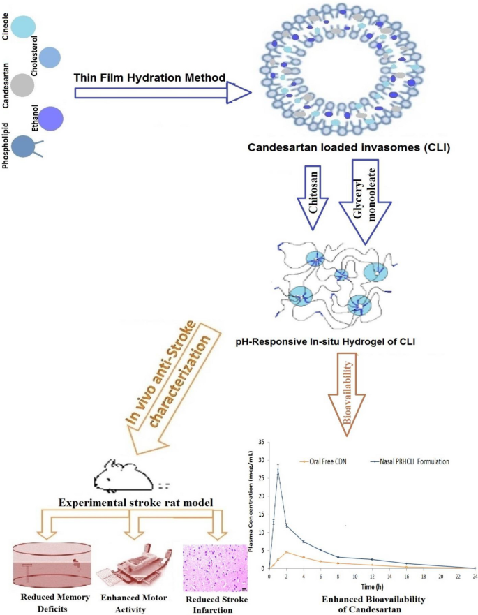
    Candesartan (CDN) is a useful anti-stroke medication because it lowers blood pressure, inflammation, oxidative stress, angiogenesis and apoptosis. However, CDN has limited efficacy due to its low solubility and poor bioavailability. This study set out to develop nasal pH-responsive in situ hydrogel of CDN-loaded invasomes a (PRHCLI) for enhancing CDN’s release, penetration, bioavailability, and effectiveness as a possible treatment for stroke. Based on the results of the pre-formulation investigation, the optimum CLI formulation for intravasomal delivery of CDN was determined to be 3% of phospholipid, 0.16% of cholesterol, 3% of ethanol, and 1% of cineole. The optimum formulation significantly enhanced CDN permeation and release by 2.06-fold and 59.06%, respectively. The CLI formulation was added to a mixture of chitosan (0.67%w/v) and glyceryl monooleate (0.27%v/v) to develop PRHCLI. The PRHCLI formulation enhanced the release and permeation of CDN relative to free CDN by 2.15 and 2.76 folds, respectively. An experimental rat stroke model was utilized for in vivo studies to evaluate the bioavailability, effectiveness, and toxicity of the PRHCLI formulation. The nasal PRHCLI drops increased the CDN’s bioavailability by 3.20-fold compared to oral free CDN. Increased grip strength and decreased flexion, spontaneous motor activity, and Morris Water Maze scores in comparison to oral free CDN showed that nasal PRHCLI drops have better anti-stroke activity. The toxicity evaluation revealed the safety of nasal PRHCLI. Hence, nasal PRHCLI drops may represent a promising avenue as a stroke therapy.

    Graphical abstract

    This is a preview of subscription content, log in via an institution to check access.

    Wednesday, August 24, 2022

    Mentally exhausted? Study blames buildup of key chemical in brain

     WHOM  in stroke will look at this and question what research is needed to see if this is causing mental exhaustion post stroke? It will never occur, there is NO stroke leadership and NO stroke strategy. All you stroke survivors can just pound sand.

    But since  glutamate poisoning is already suggested as one of the 5 causes of the neuronal cascade of death in the first days. just maybe this is a following result.  And I'm obviously stroke-addled to even think that I might know more that all these Ph.D. researchers.

    Mentally exhausted? Study blames buildup of key chemical in brain

    Toxicity of excess glutamate may contribute to cognitive fatigue, but some experts are skeptical

    Weary student in a classroom
    SolisImages/iStock

    You know the feeling. You’ve been cramming for a test or presentation all day, when suddenly you can’t remember simple things, like what you ate for breakfast, or where exactly Belize is. Now, a study hints at why we get so unraveled after hours of hard mental labor: a toxic buildup of glutamate, the brain’s most abundant chemical signal.

    The study isn’t the first to try to explain cognitive fatigue—and it is bound to stir up controversy, says Jonathan Cohen, a neuroscientist at Princeton University who wasn’t involved with the work. Many scientists once thought doing difficult mental tasks used up more energy than easy tasks, exhausting the brain like exercise can do to muscles. Some even suggested drinking a sugary milkshake would make you mentally sharper than an artificially sweetened one, he says. But Cohen and many others in the field are skeptical of such simplistic explanations. “It's all been debunked,” he says.

    In the new study, researchers looked at whether levels of glutamate are related to behavior that so often manifests when we’re mentally exhausted. Seeking easy, immediate gratification, for example, or acting impulsively. Glutamate typically excites neurons, playing key roles in learning and memory, but too much of it can wreak havoc on brain function, causing problems ranging from cell death to seizures.

    The scientists used a noninvasive technique called magnetic resonance spectroscopy, which can detect glutamate through a combination of radio waves and powerful magnets. They chose to focus on a brain region called the lateral prefrontal cortex, which helps us stay focused and make plans. When a person becomes mentally exhausted, this region becomes less active.

    The researchers divided 39 paid study participants into two groups, assigning one to a series of difficult cognitive tasks that were designed to induce mental exhaustion. In one, participants had to decide whether letters and numbers flashing on a computer screen in quick succession were green or red, uppercase or lowercase, and other variations. In another, volunteers had to remember whether a number matched one they’d seen three characters earlier. The experiment lasted for about 6 hours, with two 10-minute breaks and a simple lunch of a sandwich and piece of fruit. In the second group, people did much easier versions of the same tasks.

    As the day dragged on, the researchers repeatedly measured cognitive fatigue by asking participants to make choices that required self-control—deciding to forgo cash that was immediately available so they could earn a larger amount later, for example. The group that had been assigned to more difficult tasks made about 10% more impulsive choices than the group with easier tasks, the researchers observed. At the same time, their glutamate levels rose by about 8% in the lateral prefrontal cortexa pattern that did not show up in the other group, the scientists report today in Current Biology.

    Advertisement

    “We’re still far from the point where we can say that working hard mentally causes a toxic buildup of glutamate in the brain,” says the study’s first author, Antonius Weihler, a computational psychiatrist at the GHU Paris Psychiatry and Neurosciences. But if it does, it underscores the well-known restorative powers of sleep, which “cleanses” the brain by flushing out metabolic waste. It might be possible to use glutamate levels in the prefrontal cortex to detect severe fatigue and monitor recovery from conditions such as depression or cancer, the team suggests.

    Abnormal glutamate signaling occurs in many brain disorders. There are already drugs that target the neuronal receptors for glutamate, including esketamine, a form of the anesthetic ketamine which is used to treat depression, and memantine, which is used to treat the symptoms of Alzheimer’s disease. Researchers are also exploring glutamate-based therapies for a number of other disorders, such as schizophrenia and epilepsy.

    One important limitation of the study is that the scanners used aren’t powerful enough to distinguish between glutamate and another closely related molecule, glutamine, notes Alexander Lin, a clinical spectroscopist at Brigham and Women’s Hospital. But the findings “provide the basis for examining how glutamate could potentially be modulated by medications or devices such as neurostimulation,” he says.

    Sebastian Musslick, a neuroscientist at Brown University, doubts metabolic waste will turn out to be a key contributor to cognitive fatigue. He suspects instead that the uptick in glutamate as the brain tires serves a purpose. The organs in our bodies are in constant communication with our brains, letting us know when we need to eat, sleep, drink water, and go to the bathroom. Maybe the prefrontal cortex’s glutamate is sending a similar status update to the brain’s internal monitoring system, Musslick suggests.

    For Cohen, the most compelling reason to be skeptical of the idea that waste products play an important role in cognitive fatigue is that it can’t explain the human ability to often push through cognitive fatigue, or effortlessly perform demanding computational tasks such as face recognition that require megawatts of energy for computers to perform. To juggle this many demanding tasks, the brain has to have a more sophisticated computational system for allocating effort than the simple buildup or depletion of metabolic byproducts, he says. “It just can’t be that easy.”

    Friday, November 26, 2021

    Recovery After Stroke: New Insight to Promote Brain Plasticity

    Quit using the word neuroprotection, it gives no sense of urgency, whereas the neuronal cascade of death sounds important. Do you tell your patients; 'We were unable to get neuroprotection working for you'? Or do you truthfully tell them; ' We were unable to stop the neuronal cascade of death, leaving billions of neurons to die'? Which one will cause your stroke hospital to solve the problem? Because I would start telling patients to bill the hospital $1000 for every dead neuron. THAT would get action going.

    Recovery After Stroke: New Insight to Promote Brain Plasticity

    • Neurological Sciences and Cerebrovascular Research Laboratory, Department of Neurology and Stroke Center, La Paz University Hospital, Neuroscience Area of Hospital La Paz Institute for Health Research (IdiPAZ), Universidad Autónoma de Madrid, Madrid, Spain

    Introduction

    The recovery of the patient is the goal of the neuroscientists after a stroke. To achieve this recovery, there is a crucial need for increasing the understanding of the pathophysiological mechanisms that spontaneously engage early after stroke, which involve excitotoxicity, free radical damage, increased glutamate concentrations, and inflammation, leading to cell death.(The neuronal cascade of death, known since a Rockefeller University report in 2009) To assume that only neurons are vulnerable to these pathophysiological responses is simplistic. Stroke affects all components of the neurovascular unit, which consists of endothelial cells, pericytes, neurons, glial cells, white matter fiber tracts, myelin, and extracellular matrix proteins. It is, therefore, important to focus on brain protection as a whole rather than neuroprotection in isolation (1).

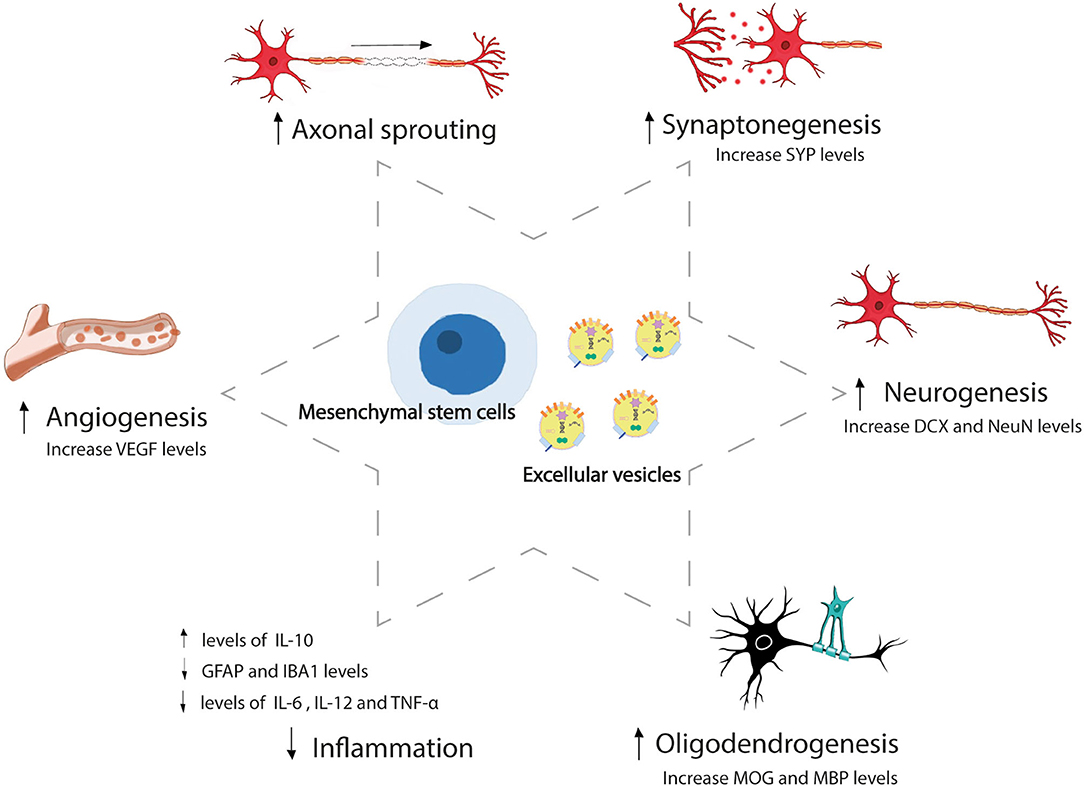
    In the last 50 years, we have gained significant insights into the molecular mechanisms of recovery after stroke, in addition to the damage mechanisms. Due to its plasticity, the brain has the ability to reorganize its function and structure, which involves processes of self-protection and self-repair. Brain plasticity is a very complex process that involves adaptive structural and functional changes in the brain, including neurogenesis, synaptogenesis, angiogenesis, oligodendrogenesis, and astrogliosis modulation, and promotes collateral circulation, processes that begin immediately after stroke (24). Such is the self-repair ability brain that the stroke-induced neurogenesis is not only limited to the subventricular zone and hippocampal dentate gyrus. It has also been revealed that several additional areas of the brain promote mammalian adult neurogenesis, which include the hypothalamus, striatum, substantia nigra, cortex, and amygdala (5). The neural stem cells of the neurogenic areas generate new neural cells that migrate to the lesion site and become mature neurons, orchestrating neurological repair through nerve repair, neuron polarization, axonal sprouting and pruning, neurite outgrowth, and myelin repair, promoting post-stroke recovery (4). However, in the unfavorable microenvironment that occurs in the lesion, most new cells do not survive (4). Given that this self-repair capacity is limited, there has been a growing interest in the potential for brain plasticity-inducing interventions to enhance post-stroke recovery through rehabilitation, trophic factors, cell therapy, and extracellular vesicles. These therapeutic approaches currently hold great promise by targeting the mechanisms involved in brain plasticity (2, 68) (Figure 1). The beneficial effects of rehabilitation therapies in stroke recovery are well-known (9), although there are still certain aspects to be clarified, such as the optimal time to start rehabilitation, as well as its intensity and duration. Other approaches that involve brain stimulation, such as transcranial magnetic and electrical stimulation, can enhance recovery and post-stroke plasticity (10), and innovations with exoskeletons and brain-machine interfaces (11) have opened up new research lines. However, we would like to focus on other novel and promising strategies, such as the administration of trophic factors, stem cells, and extracellular vesicles.

    FIGURE 1
    www.frontiersin.org

    Figure 1. Mesenchymal stem cell and extracellular vesicles-induced brain repair process after stroke. Mesenchymal stem cells and extracellular vesicles administration-induced brain plasticity that involves adaptive structural and functional changes, including angiogenesis, axonal sprouting, synaptogenesis, neurogenesis, oligodendrogenesis, and inflammation modulation after stroke. SYP, synaptophysin; DCX, doublecortin; NeuN, neuronal nuclear protein; MOG, myelin-oligodendrocyte glycoprotein; MBP, myelin basic protein; IL, interleukin; GFAP, glial fibrillary acidic protein; IBA, ionized calcium-binding adapter molecule; TNF, tumor necrosis factor; VEGF, vascular endothelial growth factor.

    Trophic Factors as Therapeutic Strategy to Promote Brain Plasticity

    In recent years, trophic factors have generated a great deal of interest in the clinical context, given that their administration is not restricted to a narrow therapeutic window. Several trophic factors, including erythropoietin, brain-derived neurotrophic factor, granulocyte-colony-stimulating factor, vascular endothelial growth factor, fibroblast growth factor, epidermal growth factor, and heparin-binding epidermal growth factor, have anti-inflammatory and anti-excitotoxic protective properties and have demonstrated efficacy in promoting neurogenesis and angiogenesis, stimulating progenitor cell proliferation, preventing blood-brain barrier (BBB) disruption and ultimately promoting functional recovery in experimental stroke models (1215). However, a number of these approaches were lost in translation from a bench to a bedside (14, 15), while others have not yet been tested in clinical trials.

    Cell Therapy: The Factory of Trophic Factors and Key Molecules to Improve Recovery

    But why settle for administering a single factor when we can administer the whole arsenal? This is where stem cell therapy plays an important role. Stem cells can secrete various trophic factors and key molecules, promote brain plasticity, and reduce overall inflammation. In particular, mesenchymal stem cells (MSCs) from bone marrow or adipose tissue have demonstrated efficacy in experimental animal stroke models (1623). These positive findings are translated to clinical trials, where MSCs and other cells (such as bone marrow mononuclear cells) have demonstrated safety in patients with stroke (2430) and even efficacy in promoting improvement in white matter injuries at 1 year (31). Perhaps, one of the most stimulating findings in stem cell translational research is the discovery of an abundant quantity of MSCs within adipose tissue. Adipose tissue-derived MSCs (AD-MSCs) are of special interest, not only due to their abundance but also their relative ease of obtention through procedures such as liposuction and abdominoplasty, which obviate the ethical concerns with embryonic MSCs. Due to immunoprivileged characteristics of this cell type, allogeneic administration is possible, in our opinion (32). Treatment can, therefore, be administered at an early stage, which is crucial for a disease where delays result in irrevocable loss of brain function. The administration of AD-MSCs in the acute phase could inhibit the aforementioned pathophysiological mechanisms that are activated early after stroke, thereby participating not only in the repair processes of the neurovascular unit but also in its protection. In terms of clinical feasibility, the intravenous route for delivering AD-MSCs is attractive, given its low invasiveness, low risk, and greater comfort for the patient. Using this route, AD-MSCs are unable to reach the brain due to their lack of ability in crossing the BBB; however, they exert their beneficial therapeutic actions by delivering their secretome from the peripheral organs where they are confined (19). MSCs exert their action by the release of key molecules or extracellular vesicles (EVs) by paracrine effects (33), rather than through differentiation to replace damaged neurons (16, 18).

    Extracellular Vesicles: The Novel Strategy to Enhance Brain Recovery

    Extracellular vesicles are released from all cell types, harbor important molecules such as proteins, DNA, lipids, mRNAs, and microRNAs, and participate in cell-to-cell communication. EVs can act as an active principle promoting several mechanisms of recovery after stroke, including brain plasticity. It has been shown that the intravenous administration of MSC-derived EVs promotes functional recovery and brain plasticity in an ischemic stroke rat model (3437). Moreover, our group found that an intravenous administration of EVs-improved outcomes by promoting the processes involved in white matter repair in subcortical stroke in rats (35). Other authors have also shown that EVs induce higher axonal density and neurite remodeling (34), new formation of endothelial cells (36), sprouting of new capillaries, and higher endothelial integrity (37). MSC-derived EVs are, therefore, a promising approach for repairing the components of the neurovascular unit to promote overall post-stroke recovery (38, 39).

    MSC-derived EVs are able to go one step farther than MSCs as a therapeutic strategy, given that MSC-EVs can cross the BBB, resolve cell-related problems, such as immune compatibility, tumor formation, and vascular occlusion, and can be stored in hospital settings without the need for toxic cryopreservative agents, offering an approach for acute ischemic stroke. Due to their small size, MSC-EVs can be saved from phagocytosis by macrophages. In addition, selective manipulation of their cargo by bioengineering can lead to individualized medicine (40, 41). There is, currently, only one ongoing clinical trial aimed at assaying the efficacy of the allogeneic administration of MSC-derived EVs enriched by miR-124 for improving the recovery of patients with acute ischemic stroke, registered in clinicaltrials.gov (Identifier: NCT03384433). A previous study using an animal model of stroke demonstrated that the administration of EVs loaded with miR-124 promoted cortical neural progenitors and neurogenesis (42). These functions of the microRNA content of EVs make them important not only as treatment but also as biomarkers. We showed that circulating EVs from patients with stroke contain miRNA and proteins related to risk factors and etiology, post-ischemic immune response, endogenous protection, and angiogenesis (43). We also observed differences in the levels of the microRNA content of EVs according to the topography of the stroke (subcortical and cortical-subcortical ischemic stroke) and related to improved recovery after stroke (44). Given the progress of research on EVs, further information on brain-derived EVs under stroke conditions is necessary (45). The content of EVs can be used as biomarkers that improve our understanding of the mechanisms by which EVs act in stroke to help develop new therapeutic strategies and find new molecular targets for this neurological disease.

    Discussion

    The experimental studies in animal model and clinical trials have indicated that intravenously administered MSC therapy seems to be a promising therapeutic strategy after stroke (1623, 2628, 30). However, there are still some unresolved issues that have to be investigated such as the most appropriate administration timing, the doses required for successful recovery (46), and the dose regimen (single-dose or repeat-doses) that reach the higher threshold of brain repair after stroke. Likewise, the limitations of cell therapy will be determined by the results of clinical trials.

    Moreover, MSC-derived EVs have some important advantages that can be exploited when translating a therapeutic strategy for stroke. EVs provide a great feature as a drug delivery system, given their ability to cross the BBB (40). Thanks to the development of delivery system technologies, MSC-derived EVs can be engineered and designed to carry specific therapeutic molecules (47) according to brain tissue repair needs, avoiding molecules that could induce adverse effects, moving toward personalized medicine.

    Beyond the proven beneficial outcomes with the MSC-derived EV treatment in experimental animal models of stroke (3439), many aspects are yet to be resolved about the production of EVs for their use in clinical practice, such us large-scale production, conditions in different physiologically environments, and standardized experimental protocol for extracting EVs to provide batch uniformity according to GMP regulations (40, 48, 49).

    In conclusion, we are facing a disease that has a high incidence and prevalence and results in major disability. However, we are also finding new and promising therapeutic options, such as trophic factors, cell therapy, and EVs that can positively contribute to stroke recovery by improving brain plasticity. For EVs, their ability to cross the BBB and their editable cargo provide live information on molecules that participate in damage and post-stroke repair, which could lead to personalized and precision medicine.


    Thursday, December 3, 2020

    A Neurotoxic Ménage-à-trois: Glutamate, Calcium, and Zinc in the Excitotoxic Cascade

     Since excitotoxicity has been known for decades our fucking failures of our stroke associations  have excelled in DOING NOTHING to solve it. Yes it is hard to solve but leaders solve problems. WHERE THE FUCK ARE OUR STROKE LEADERS? Cowering somewhere they don't have to talk to survivors? You can email me, I have my address in the right column under 'email me'. See how easy it is to talk to a survivor? Or are you afraid of me?

    A Neurotoxic Ménage-à-trois: Glutamate, Calcium, and Zinc in the Excitotoxic Cascade

     
    • 1Sue and Bill Gross Stem Cell Research Center, University of California, Irvine, Irvine, CA, United States
    • 2Center for Advanced Sciences and Technology (CAST), University “G. d’Annunzio” of Chieti−Pescara, Chieti, Italy
    • 3Department of Neuroscience, Imaging, and Clinical Sciences (DNISC), Laboratory of Molecular Neurology, University “G. d’Annunzio” of Chieti−Pescara, Chieti, Italy
    • 4Department of Science and Technology, University of Sannio, Benevento, Italy
    • 5Institute for Memory Impairments and Neurological Disorders, University of California, Irvine, Irvine, CA, United States

    Fifty years ago, the seminal work by John Olney provided the first evidence of the neurotoxic properties of the excitatory neurotransmitter glutamate. A process hereafter termed excitotoxicity. Since then, glutamate-driven neuronal death has been linked to several acute and chronic neurological conditions, like stroke, traumatic brain injury, Alzheimer’s, Parkinson’s, and Huntington’s diseases, and Amyotrophic Lateral Sclerosis. Mechanisms linked to the overactivation of glutamatergic receptors involve an aberrant cation influx, which produces the failure of the ionic neuronal milieu. In this context, zinc, the second most abundant metal ion in the brain, is a key but still somehow underappreciated player of the excitotoxic cascade. Zinc is an essential element for neuronal functioning, but when dysregulated acts as a potent neurotoxin. In this review, we discuss the ionic changes and downstream effects involved in the glutamate-driven neuronal loss, with a focus on the role exerted by zinc. Finally, we summarize our work on the fascinating distinct properties of NADPH-diaphorase neurons. This neuronal subpopulation is spared from excitotoxic insults and represents a powerful tool to understand mechanisms of resilience against excitotoxic processes.

    Introduction

    Excitotoxicity is a form of neuronal death triggered by excessive and/or sustained exposure to the amino acid glutamate, the primary excitatory neurotransmitter in the brain. Evidence accumulated in the past four decades indicates that excitotoxicity is a critical contributor to the neuronal demise occurring upon acute and chronic neurological conditions, like stroke, Alzheimer’s disease (AD), Huntington’s disease (HD), Amyotrophic Lateral Sclerosis (ALS), and Parkinson’s disease (PD) (Mehta et al., 2013).

    Although, 50 years have passed since the first description of glutamate’s neurotoxic activity (Olney, 1969), therapeutic strategies set at counteracting these processes have been only partially exploited. In that regard, the targeting of upstream mechanisms of glutamate-driven neurotoxicity has produced, in the late 80s, an early wave of enthusiasm and fueled a level of optimism that has not been corroborated in the following years. These approaches have been found promising in preclinical models (Lee et al., 1999) but failed in clinical trials (Lee et al., 1999; Ikonomidou and Turski, 2002; Chamorro et al., 2016; Choi, 2020). Only riluzole and memantine, two drugs that target glutamate-driven neuronal death, have been approved for the treatment of ALS and AD, respectively.

    Although, most of the preclinical findings failed “the bench to bed” translation, this experimental evidence has significantly helped dissect the molecular underpinnings of excitotoxicity. These studies have also helped provide support for the excitotoxic cascade hypothesis (Zivin and Choi, 1991; Choi, 2020). The construct posits that excitotoxic neuronal death is primarily mediated by the glutamate-driven activation of N-methyl-D-aspartate receptors (NMDARs) and the subsequent toxic intraneuronal accumulation of calcium (Ca2+). The NMDAR-driven Ca2+ overload is, in fact, a mandatory step in the process as most of the downstream mechanisms of the cascade, like the generation of reactive oxygen species (ROS; of mitochondrial and non-mitochondrial origin), or reactive nitrogen species (RNS), the concurrent mitochondrial dysfunction, metabolic impairment, as well as the activation of necrotic/apoptotic pathways, are all Ca2+-dependent processes (Lee et al., 1999; Lai et al., 2014; Bano and Ankarcrona, 2018; Choi, 2020; Swanson and Wang, 2020). However, Ca2+ is not alone, and other cations find a way to participate in the death banquet. Zinc (Zn2+) is, for instance, a VIP guest.

    In the review, we provide a brief overview of the role of Zn2+ in the brain and discuss its neurotoxic properties and how they intertwine with the excitotoxic cascade. Finally, we focus on the distinct features of the NADPH-diaphorase neurons, a subpopulation spared from excitotoxic insults offering an intriguing model to further our understanding of neuroprotective mechanisms.

    Zinc Homeostasis and Its Role in Brain Functions

    After iron, Zn2+ is the most abundant metal ion in the brain. The cation can be found in either structural or labile forms (Sensi et al., 2009). Structural Zn2+ is tightly bound to proteins/peptides and acts as a critical component for proper protein folding or as the catalytic/co-catalytic element required for several enzymes (McCall et al., 2000).

    Labile, free Zn2+ is either stored in the lumen of intracellular organelles, like synaptic “zinkergic” vesicles, mitochondria, lysosomes, the endoplasmic reticulum (ER), and the Golgi apparatus, or bound to metallothioneins (MTs), a class of metal-binding redox-sensitive proteins (Maret, 1994). Under physiological conditions, cytosolic Zn2+ concentrations are kept in a picomolar to a low nanomolar range (Outten and O’Halloran, 2001) through the carefully orchestrated activity of Zn2+ transporters (ZnTs), Zrt-, Irt-related proteins (ZIPs), Zn2+-stores and binding proteins (Sekler et al., 2007; Sensi et al., 2009).

    Zn2+ within synaptic vesicles is released, along with glutamate, during excitatory neurotransmission (Sensi et al., 2009). Once released in the synaptic cleft, the cation shapes the post-synaptic glutamate responses by modulating the activity of glutamatergic receptors, like NMDARs and the α-amino-3-hydroxy-5-methyl-4-isoxazolepropionic acid receptors (AMPARs) (Paoletti et al., 1997; Kalappa et al., 2015). Zn2+ exerts an inhibitory effect on NMDARs by acting on a high (nanomolar) and a low (micromolar) affinity site that is located on the GluN2A and GluN2B subunits, respectively (Rachline et al., 2005). As for AMPARs, the cation has been proposed to exert an inhibitory activity by acting on the histidine residues of the receptor ligand-binding domain (Kalappa et al., 2015). However, direct structural evidence for this interaction is still missing. Of note, recent findings indicate that ZnT1-dependent cation extrusion in the proximity of synaptic NMDARs is required for the Zn2+-dependent inhibition of the receptor (Mellone et al., 2015; Krall et al., 2020). A novel post-synaptic mechanism that may put under a new light the cation’s modulating activities as the metal has been so far thought to only act via its presynaptic release. Extracellular Zn2+ is also implicated in the modulation of neurotrophic signaling as the cation is critical for the activity of the matrix metalloproteinases (MMPs), a class of enzymes involved in matrix remodeling and the maturation of the brain-derived neurotrophic factor (BDNF) from its precursor form proBDNF (Hwang et al., 2005), a process activated by Zn2+ supplementation and impaired by metal chelation (Corona et al., 2010; Frazzini et al., 2018). The proBDNF/BDNF balance is critical for neuronal functioning as the two peptides exert opposite effects. BDNF affects long-term memory processes and neuronal survival. On the contrary, proBDNF inhibits GABAergic neurotransmission (Riffault et al., 2014), facilitates long-term depression (Woo et al., 2005), and activates neuronal death-related pathways (Teng et al., 2005; Mizui et al., 2016). Zn2+ has also been shown to activate the BDNF receptor TrkB directly. This process, called transactivation, is triggered by synaptically-released Zn2+ and/or ROS-driven intracellular Zn2+ elevations (Huang et al., 2008; Huang and McNamara, 2012). However, the mechanisms described “in vitro” settings do not entirely translate in “in vivo” conditions as, surprisingly, TrkB activation has been found to be unaffected in transgenic mice devoid of vesicular Zn2+ (Helgager et al., 2014).

    Unlike what is known about vesicular Zn2+, the intracellular labile pools’ physiological significance has been only partially unraveled. Along with its role as a metal reservoir, compelling evidence indicates that releasable Zn2+ can affect mitochondria and lysosomal functioning and, in close analogy with Ca2+, act as a signaling molecule (Yamasaki et al., 2007).

    Most importantly, like Ca2+, when dysregulated, Zn2+ can turn into a potent neurotoxin (Sensi et al., 2009).

    Zinc as Neurotoxin

    The contribution of Zn2+ in neurodegenerative processes has been extensively investigated. In conditions characterized by the overactivation of excitatory signaling, synaptically released Zn2+ can flux into post-synaptic neurons through Zn2+ permeable channels (Sensi et al., 2009). Zn2+ entry occurs primarily through voltage-gated Ca2+ channels (VGCCs) and Ca2+ permeable AMPARs (CP-AMPARs) (Sensi et al., 1997, 1999b; McDonald et al., 1998; Colvin et al., 2000; Sheline et al., 2002). AMPAR permeability to Ca2+ and Zn2+ ions is restricted to certain neuronal populations or occurs upon disease associated challenges. The distinct expression pattern and the high permeability to Ca2+ and Zn2+ contribute to the unique role of CP-AMPAR in selective neurodegeneration (see Weiss, 2011 for an extensive review on the topic). NMDARs are poorly permeable to Zn2+ ions (Sensi et al., 1997).

    Additional routes of entry are the Na+-Zn2+ exchanger and transient receptor potential channels (TRP); however, their contribution to the cation’s toxic accumulation is still mostly unexplored (Bouron and Oberwinkler, 2014). The exact amount of Zn2+ released from presynaptic terminals and the degree of its contribution to cation accumulation in the post-synaptic neurons are also not completely clear and have been matters of lively debates (Kay and Toth, 2008; Vergnano et al., 2014).

    Zn2+ released from intracellular pools also participates in the cation’s cytosolic build-up (McCord and Aizenman, 2014). In this regard, MTs are a significant source of intracellular Zn2+ (Maret, 1994; Aizenman et al., 2000). MTs mobilize a large amount of Zn2+ (ranging 10–100 nM) in response to Ca2+-driven generation of ROS/RNS as well as in conditions of mild acidosis, a combination found in excitotoxic settings and several neurodegenerative conditions. The critical role played by Zn2+ released from MTs in the production of neuronal death is supported by the ability of oxidative agents [i.e., 2,2′-dithiodipyridine (DTDP) or N-ethylmaleimide (NEM)] to trigger widespread apoptotic neuronal death (Aizenman et al., 2000; Gibon et al., 2010). This process is mostly reduced by Zn2+chelators and independent of Ca2+ load. Intraneuronal Zn2+ rises are not the final step of the toxic cascade but are critical to trigger mitochondrial and lysosomal dysfunction, as well as the activation of neurotoxic pathways in the cytosol (Sensi et al., 2009; Ji et al., 2019; Koh et al., 2019).

    Mitochondria are a primary target of intracellular Zn2+ as the cation accumulates in the organelles thanks to their steep electrochemical gradient (Δp). Once sequestered, Zn2+, along with Ca2+, contributes to Δp loss and promotes ROS generation (Sensi et al., 1999a; Ji and Weiss, 2018). Zn2+ mobilization is an essential prerequisite to trigger irreversible mitochondrial dysfunction as the cation, by acting in close synergy with Ca2+ damaging effects, promote the full demise of the organelles and, eventually, cell death (Jiang et al., 2001; Granzotto and Sensi, 2015). Within mitochondria, Zn2+ acts by inhibiting complexes of the electron transport chain (ETC) and α-ketoglutarate dehydrogenase (αKGDH) of the Krebs cycle, thereby promoting aberrant ROS production and metabolic failure (Sensi et al., 2009; Ji et al., 2019). Zn2+ interactions with αKGDH and the matrix-facing complexes of the ETC support the presence of the cation in the mitochondrial matrix. Moreover, recent findings indicate that mitochondrial Zn2+ uptake through the activation of the mitochondrial Ca2+ uniporter (MCU) participates in producing the neuronal death found in preclinical models of brain ischemia (Ji et al., 2019, 2020). Zn2+ also triggers the permeabilization of the mitochondrial membrane through the activation of the mitochondrial permeability transition pore (MPTP; a key promoter of cell death; Bernardi et al., 2015), thereby generating the release/production of pro-apoptotic factors [like cytochrome c, apoptosis-inducing factor (AIF), and ΔN-Bcl-XL] (Jiang et al., 2001; Bossy-Wetzel et al., 2004; Bonanni et al., 2006; Ji et al., 2019).

    In addition, Zn2+ elevations target lysosomes (Koh et al., 2019). Lysosomal Zn2+ rises, coupled with the accumulation of lipid peroxidation byproducts (4-hydroxinonenal), are instrumental for organelle membrane permeabilization (LMP). LMP results in cation release in the cytosol, along with the activation of lysosomal degrading enzymes. These events are critical for neuronal and astrocyte death when exposed to oxidative challenges (Lee and Koh, 2010; Koh et al., 2019).

    Zn2+ also affects many cytosolic pathways to promote demise in the CNS cells, including activation of apoptotic/necrotic pathways, modulation of plasma membrane channels, depletion of metabolic substrates, and the induction of cytosolic oxidative enzymes. In neurons and astrocytes, the metal contributes to NADPH oxidase activation, resulting in aberrant O2 generation (Noh and Koh, 2000; Brennan et al., 2009; Swanson and Wang, 2020). Similarly, Zn2+ activates the neuronal isoform of the nitric oxide synthase (nNOS), thereby promoting increased production of nitric oxide (NO) (Kim and Koh, 2002). These two pathways converge in a process in which O2 + NO generate ONOO (peroxynitrite), a potently neurotoxic RNS (Bossy-Wetzel et al., 2004). Of note, the Zn2+-driven ROS/RNS production promotes further metal release from intracellular redox-sensitive stores (like MTs), thereby exacerbating a vicious feed-forward loop of cation dyshomeostasis (Corona et al., 2011; Slepchenko et al., 2017). At the cytosolic level, Zn2+ promotes NAD+ depletion, thereby resulting in glyceraldehyde-3-phosphate dehydrogenase (GAPDH, a critical enzyme in the glycolytic pathway) inhibition, ATP breakdown, and eventually energetic neuronal failures (Sheline et al., 2000). This NAD+ depletion may critically impinge on mechanisms that are relevant to aging (Lautrup et al., 2019). Zn2+ also targets and promotes activation of PARP (Kim and Koh, 2002), cyclin-dependent kinase 5 (Cdk5; Tuo et al., 2018), and AMPK (Kim et al., 2020), three molecules involved in cell death pathways.

    Finally, Zn2+ mobilization, by contributing to the activation of the CamKII/p38/syntaxin/calcineurin axis, promotes outward potassium (K+) currents, a critical step in the production of neuronal apoptosis (Yu et al., 1997; McCord and Aizenman, 2013; Shah and Aizenman, 2014; Aizenman et al., 2020).

    The Resilience of nNOS (+) Neurons: a Model to Investigate Excitotoxic Mechanisms

    Intriguingly, some neuronal subpopulations are mostly insensitive to excitotoxicity. The phenomenon is present in oculomotor neurons, Onuf’s nucleus neurons, and NADPH-diaphorase neurons (Koh et al., 1986; Brockington et al., 2013). NADPH-diaphorase neurons are a subset of medium-sized aspiny interneurons that are largely spared following excitotoxic hits (Koh et al., 1986; Koh and Choi, 1988; Uemura et al., 1990; Weiss et al., 1994; Granzotto and Sensi, 2015). The subpopulation is characterized by the overexpression of nNOS [also known as NOS1; hereafter termed nNOS (+) neurons (Dawson et al., 1991; Hope et al., 1991)]. nNOS (+) neurons are present with a relatively more significant percentage in the striatum but are also expressed in good numbers in the hippocampus and the cerebral cortex. The subpopulation encompasses various cellular subtypes characterized by distinct morphological, transcriptomic, and functional features (Tricoire and Vitalis, 2012). Early studies have shown that these neurons survive instead of the widespread neuronal loss documented by brain autopsy of AD, HD, and PD patients, three conditions characterized by a robust glutamatergic overdrive (Ferrante et al., 1985; Graveland et al., 1985; Mufson and Brandabur, 1994).

    Our group has recently exploited this neuronal subpopulation’s unique features to evaluate the mechanisms that promote resilience to excitotoxicity. Employing an array of single-cell imaging and biochemical approaches, we have demonstrated that nNOS (+) neurons fail to generate ROS in response to excitotoxic stimuli (Canzoniero et al., 2013; Granzotto and Sensi, 2015), a critical step that contributes to their resilience and enhanced survival upon glutamate-driven neurodegeneration.

    The investigation of these processes has indicated an intriguing scenario in which the ROS-dependent release of intracellular Zn2+ acts as a critical intermediate step of the excitotoxic process (Granzotto and Sensi, 2015). Thus, experimental data support the notion that Zn2+ participates, with glutamate and Ca2+, in a neurotoxic ménage-à-trois.

    Overactivation of NMDARs is the first mandatory step in the excitotoxic cascade; compelling evidence indicates that the receptor triggers the activation of early signaling pathways involving PSD95 and nNOS recruitment as well as aberrant Ca2+-driven induction of nNOS (Szydlowska and Tymianski, 2010; Fricker et al., 2018; Wu and Tymianski, 2018). Disruption of the NMDAR/PSD95/nNOS axis prevents excitotoxic damage in in vitro and in vivo preclinical models of cerebral ischemia (Aarts et al., 2002). Functional, transcriptomic, and biochemical analysis, however, indicate that nNOS (+) neurons express fully operational NMDARs that do not differ from the ones present in the general population of nNOS (−) neurons (Price et al., 1993; Landwehrmeyer et al., 1995; Canzoniero et al., 2013; Granzotto and Sensi, 2015; Granzotto and Sensi, 2015, observations). Interestingly, additional studies have also indicated that nNOS (+) neurons are positive to cobalt staining, a maneuver employed to identify CP-AMPARs, thereby suggesting that these cells possess a significant number of these glutamate receptor subtypes (Weiss et al., 1994). This set of findings supports the notion that NMDAR-driven Ca2+ overload and nNOS activation are necessary but not sufficient steps for the initiation and development of the excitotoxic cascade. Additional downstream processes are required, and Zn2+ participates in these mechanisms with a leading role.

    Mitochondria, the Final Common Pathway

    Early studies indicated that mitochondria are critical hubs for the development of the excitotoxic cascade (Ankarcrona et al., 1995). The organelles participate in the clearance of NMDAR-driven cytosolic Ca2+ raises and are instrumental for the activation of apoptotic and necrotic processes (Ankarcrona et al., 1995; Schinder et al., 1996). Mitochondrial Ca2+ overload results in organelle dysfunction, aberrant ROS generation, and, ultimately, neuronal loss (Dugan et al., 1995; Stout et al., 1998; Duchen, 2012; Rizzuto et al., 2012).

    Mitochondria of nNOS (+) cells are insensitive to excitotoxicity and have emerged as a critical switch to turn off the injurious process (Canzoniero et al., 2013; Granzotto and Sensi, 2015; Figure 1). Although, mitochondria of these neurons take up large amounts of Ca2+, the organelles respond with minimal Δp losses and negligible generation of ROS (Canzoniero et al., 2013; Granzotto and Sensi, 2015). Early studies have shown that, to counteract the detrimental effects linked to peroxynitrite generation, nNOS (+) neurons express high levels of SOD2, the ROS quenching enzyme that is strategically localized inside of mitochondria (Gonzalez-Zulueta et al., 1998). Therefore, it is conceivable that this constitutive overexpression of SOD2 makes the subpopulation better equipped to cope with the oxidative surge produced by the excitotoxic challenges.

    FIGURE 1
    www.frontiersin.org

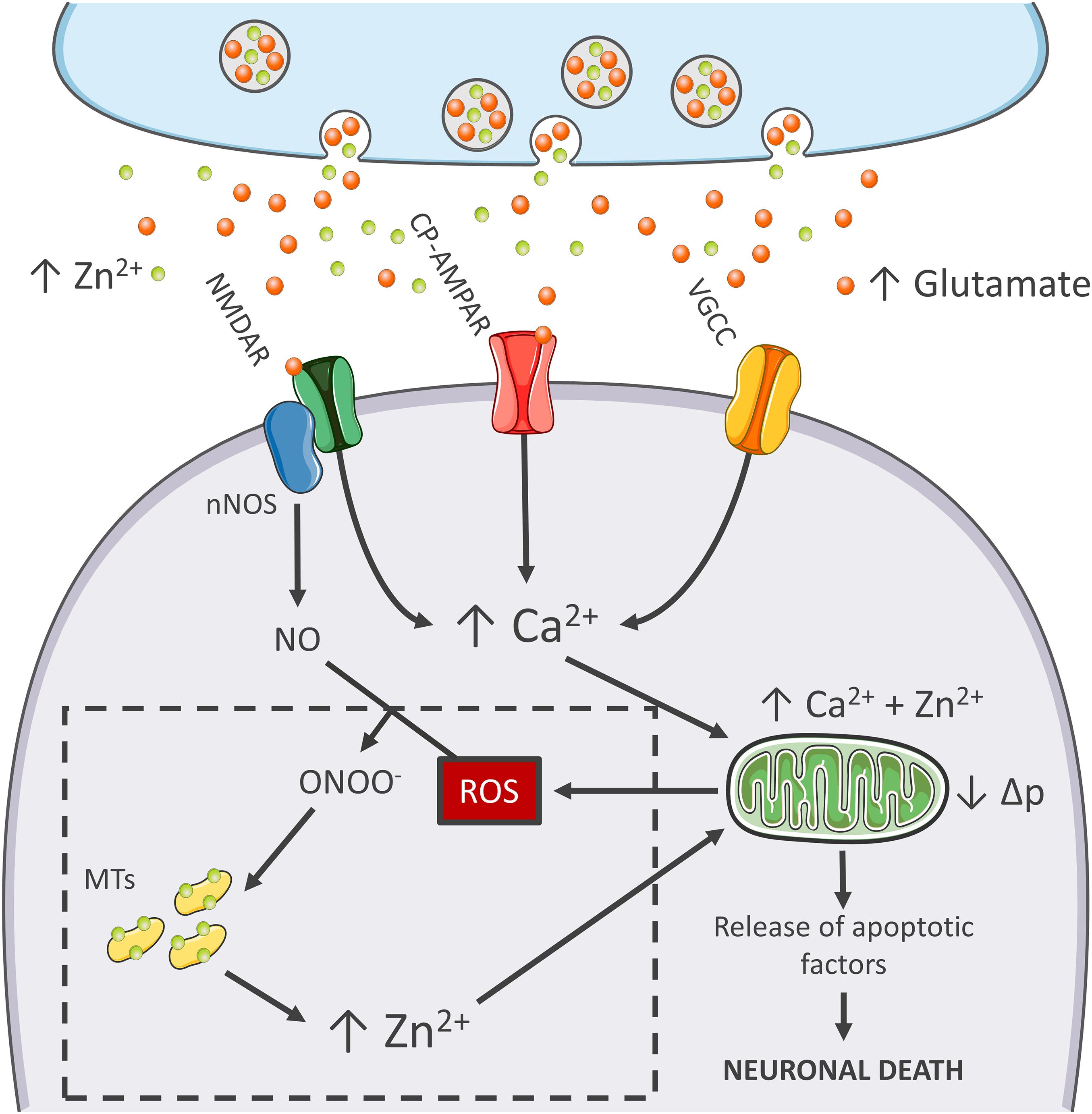
    Figure 1. Zn2+ in the excitotoxic cascade. Aberrant release of glutamate from presynaptic terminals triggers NMDAR activation, which, in turn, promotes Ca2+ entry and generation of RNS and ROS of mitochondrial and extramitochondrial origin. The surge of ROS and RNS is required for intraneuronal Zn2+ mobilization from metallothioneins (MTs; Zn2+ buffering redox-sensitive proteins prone to release Zn2+ following oxidative stimuli). Intraneuronal Zn2+ rises target mitochondria and, along with Ca2+, contribute to the organelle impairment. Dysfunctional mitochondria fail to cope with Ca2+ clearance and further exacerbate Ca2+ dysregulation and ROS production. The lack of ROS generation in nNOS (+) neurons is a critical point of divergence in the excitotoxic cascade. By missing the injurious interaction between ROS and RNS, the subpopulation fails to mobilize neurotoxic Zn2+, prevents mitochondrial failure, and eventually neuronal death (dashed line box). In the general population of nNOS (−) neurons, pharmacological Zn2+ chelation prevents the full development of the excitotoxic cascade and mimics nNOS (+) cells’ behavior. These findings lend support to the idea that intraneuronal Zn2+ release is a critical regulator of excitotoxicity.

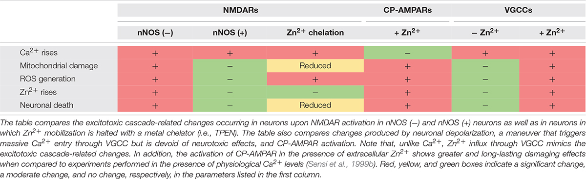
    The idea that mitochondrial dysfunction and oxidative stress are prerequisites for NMDA-driven neuronal loss is in line with the “source-specific” hypothesis of excitotoxicity. The construct posits that the neurotoxic cascade depends on the route of Ca2+ entry, mainly NMDARs, rather than the magnitude of cation load (Wu and Tymianski, 2018). In agreement with this view, abundant Ca2+ entry through VGCCs, a maneuver devoid of neurotoxic effects, fails to trigger ROS and Δp changes (Table 1). This phenomenon shows great analogies with the effects of Ca2+ rises observed in nNOS (+) neurons following NMDAR activation (Granzotto and Sensi, 2015). Although, NMDAR and VGCC activation produces large Ca2+ rises, differences can be found when dissecting the temporal progression of the two stimuli. Unlike VGCC-driven Ca2+ entry, NMDAR overactivation promotes a prolonged and sustained build-up of Ca2+, a phenomenon likely due to impaired cation handling. Conceivably, the NMDAR-driven generation of RNS and ROS can severely affect the mitochondrial Ca2+ buffering as well as the defective extrusion of the cation.

    TABLE 1
    www.frontiersin.org

    Table 1. The functional hallmarks of excitotoxicity.

    RNS/ROS can per se contribute to mitochondrial damage (Murphy, 2009). However, an alternative angle is offered by the mitotoxic properties of Zn2+. The cation represents a critical point of convergence between Ca2+, ROS, and mitochondrial failure. By missing ROS generation, nNOS (+) neurons fail to mobilize intracellular Zn2+ upon NMDAR overactivation (Granzotto and Sensi, 2015). Chelation experiments support the hypothesis that NMDAR-triggered Zn2+ rises are required for the full development of the excitotoxic cascade. In that respect, chelation prevents Zn2+ rises without affecting the upstream mechanisms of the cascade (i.e., Ca2+ entry or the Ca2+-driven generation of RNS/ROS). In nNOS (−) neurons, blockade of excitotoxic-driven Zn2+ elevations results in reduced mitochondrial dysfunction and improved intracellular Ca2+ cycling; two functional changes that closely match the ones observed in the nNOS (+) subpopulation (Granzotto and Sensi, 2015) and provide neuroprotection in excitotoxic settings (Wang and Thayer, 2002). On the contrary, nNOS (+) neurons are extremely vulnerable to pharmacological maneuvers that promote Zn2+ elevations, thereby suggesting that the damaging effects of the cation can override the protective machinery of nNOS (+) neurons (Granzotto and Sensi, 2015, and unpublished observations).

    These results are in line with several studies showing that Zn2+ chelation is highly neuroprotective as the maneuver prevents mitochondrial failure, irreversible dysregulation of Ca2+ homeostasis, and eventually neuronal demise (Jiang et al., 2001; Bossy-Wetzel et al., 2004; Medvedeva et al., 2009; Vander Jagt et al., 2009; Clausen et al., 2013; Medvedeva and Weiss, 2014; Ji and Weiss, 2018; Ji et al., 2020; Table 1).

    The peculiar milieu offered by nNOS (+) neurons replicates these mechanisms in a naïve, patho-physiologically relevant setting and allows inference on the central role played by Zn2+ in the excitotoxic process (Figure 1). Zn2+ is, therefore, not an accomplice or an amplifier of Ca2+-driven toxicity but rather the downstream executioner. Zn2+ actively promotes mitochondrial dysfunction, Ca2+ dysregulation and, eventually, neuronal death.

    More at link.