Use the labels in the right column to find what you want. Or you can go thru them one by one, there are only 34,408 posts. Searching is done in the search box in upper left corner. I blog on anything to do with stroke. DO NOT DO ANYTHING SUGGESTED HERE AS I AM NOT MEDICALLY TRAINED, YOUR DOCTOR IS, LISTEN TO THEM. BUT I BET THEY DON'T KNOW HOW TO GET YOU 100% RECOVERED. I DON'T EITHER BUT HAVE PLENTY OF QUESTIONS FOR YOUR DOCTOR TO ANSWER.
Changing stroke rehab and research worldwide now.Time is Brain!trillions and trillions of neuronsthatDIEeach day because there areNOeffective hyperacute therapies besides tPA(only 12% effective). I have 523 posts on hyperacute therapy, enough for researchers to spend decades proving them out. These are my personal ideas and blog on stroke rehabilitation and stroke research. Do not attempt any of these without checking with your medical provider. Unless you join me in agitating, when you need these therapies they won't be there. Did your doctor somehow miss the recovery protocol that Pedro Bach-y-Rita used to recover fully back in 1958 with only a partial brain?
What this blog is for:
My blog is not to help survivors recover, it is to have the 10 million yearly stroke survivors light fires underneath their doctors, stroke hospitals and stroke researchers to get stroke solved. 100% recovery. The stroke medical world is completely failing at that goal, they don't even have it as a goal. Shortly after getting out of the hospital and getting NO information on the process or protocols of stroke rehabilitation and recovery I started searching on the internet and found that no other survivor received useful information. This is an attempt to cover all stroke rehabilitation information that should be readily available to survivors so they can talk with informed knowledge to their medical staff. It lays out what needs to be done to get stroke survivors closer to 100% recovery. It's quite disgusting that this information is not available from every stroke association and doctors group.
Your competent? doctor can tell you all about chemokines and their role in your recovery, right? Or are you going to say nothing and let incompetence fester? And not initiating human testing is an even worse offense!
Chemokines are traditionally known for their roles in immune cell recruitment during inflammation, but emerging evidence suggests that they may also directly regulate cellular states within the central nervous system. Specifically, it remains unclear whether CXCL16 affects microglial functional states in ischemic stroke. Here, we demonstrated that recombinant CXCL16 (rCXCL16) modulated the expression of inflammation- and repair-associated markers in primary microglia and in the ischemic brain. Functionally, microglia pretreated with rCXCL16 increased HT-22 cell viability and reduced apoptosis in an indirect co-culture system. Consistently, in vivo administration of rCXCL16 reduced infarct size, restored neurobehavior performance, and suppressed apoptosis in experimental stroke in mice. These findings identify rCXCL16 as a modulator of microglial responses and suggest that its neuroprotective effects are associated with reduced inflammatory marker expression and attenuation of apoptotic injury after ischemic stroke.
Astrocytes are central regulators of neural homeostasis, synaptic function, and neuroinflammatory responses in the central nervous system (CNS). Upon pathological stimuli, astrocytes undergo reactive transformations, producing pro-inflammatory cytokines, reactive oxygen species (ROS), and chemokines, which exacerbate neuronal injury. Flavonoids, a diverse class of polyphenolic compounds found in fruits, vegetables, and medicinal plants, have emerged as potent modulators of astrocyte activity, promoting neuroprotection and cognitive enhancement. These compounds, including quercetin, hesperetin, rutin, casticin, and anthocyanins, attenuate astrocyte-mediated neuroinflammation by suppressing NF-κB, MAPK, TLR, and NLRP3 inflammasome signaling while activating antioxidant pathways such as Nrf2 and PI3K/Akt. Flavonoid-mediated modulation also enhances the synthesis and release of neurotrophic factors, including BDNF, GDNF, NGF, and TGF-β1, which support synaptic plasticity, dendritic spine formation, and network connectivity. By preserving astrocytic homeostasis, reducing gliosis, and regulating astrocyte–microglia crosstalk, flavonoids mitigate cytokine-mediated neuronal damage(Well them tell us EXACTLY HOW TO DO THAT! Oh, your research wasn't good enough, was it?), restore synaptic integrity, and improve learning and memory in models of neurodegeneration, ischemia, and neuroinflammation. Preclinical evidence suggests that flavonoids can cross the blood–brain barrier, exhibit low toxicity, and synergize with other neuroprotective interventions. Understanding the molecular mechanisms of flavonoid–astrocyte interactions provides insight into precision therapeutic strategies aimed at alleviating neuroinflammation and enhancing CNS resilience, offering promising avenues for the prevention and treatment of cognitive and neurodegenerative disorders.
ummary: A new study reveals astrocyte
dysfunction may be a driver of cognitive decline and memory loss in
those with dementia. This may be caused by a protein build-up in the
astrocytes that appears to trigger abnormal antiviral activity that
results in memory loss.
Source: Weill Cornell Medicine
People
with dementia have protein build-up in astrocytes that may trigger
abnormal antiviral activity and memory loss, according to a preclinical
study by a team of Weill Cornell Medicine investigators.
Dysfunction
in cells called neurons, which transmit messages throughout the brain,
has long been the prime suspect in dementia-related cognitive deficits.
But a new study, published in Science Advances on April 19,
suggests that abnormal immune activity in non-neuronal brain cells
called astrocytes is sufficient to cause cognitive deficits in dementia.
The
discovery could lead to new treatments that reduce excess immune
activity in astrocytes and their detrimental effects on other brain
cells and cognition.
“Astrocyte dysfunction alone can drive
memory loss, even when neurons and other cells are otherwise healthy,”
said co-senior author Dr. Anna Orr, the Nan and Stephen Swid Assistant
Professor of Frontotemporal Dementia Research in the Feil Family Brain
and Mind Research Institute and a member of the Helen and Robert Appel
Alzheimer’s Disease Research Institute at Weill Cornell Medicine.
“We
found, in mice, that astrocytes can cause cognitive decline through
their antiviral activities, which can make neurons hyperactive.”
While
neurons have been intensively studied in dementia and other diseases,
much less research has focused on astrocytes, which many scientists
viewed as playing only supporting roles to neurons in brain health.
“We
are very interested in the roles of astrocytes in cognitive and
behavioral disorders,” she said. “These cells are prevalent in the brain
and perform various key functions, but their involvement in
neurocognitive disorders like dementia are poorly understood.”
When
the investigators, including first author Dr. Avital Licht-Murava, a
former postdoctoral associate in the Orr lab, examined tissue samples
from deceased individuals who were diagnosed with either Alzheimer’s
disease or frontotemporal dementia, they found an accumulation of a
protein called TDP-43 in astrocytes within the hippocampus, a brain
region crucial for memory.
To understand the effects of this
protein build-up, the team conducted a series of experiments in mouse
models and brain cells grown in the laboratory. Other senior
investigators that contributed to the study include Dr. Robert
Schwartz at Weill Cornell Medicine and Dr. Robert Froemke at New York
University.
In mice, the build-up of TDP-43 in astrocytes was
sufficient to cause progressive memory loss but not other behavioral
changes. “Astrocytes in the hippocampus seem to be more vulnerable to
this pathology.” she said.
To understand the causes of memory loss
at the molecular level, co-senior author Dr. Adam Orr, an assistant
professor of research in neuroscience in the Feil Family Brain and Mind
Research Institute and a member of the Appel Alzheimer’s Disease
Research Institute at Weill Cornell Medicine, analyzed gene expression
and found high levels of antiviral gene activities, even though no virus
was present in the brain.
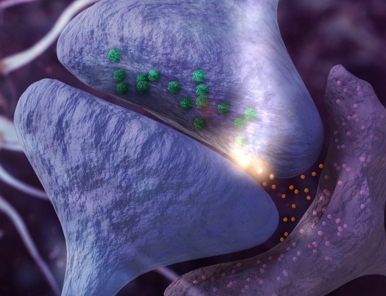
Synapses
are composed of presynaptic and postsynaptic terminals (blue) and are
contacted by astrocytes (purple), a crucial non-neuronal cell type.
Dementia-associated protein buildup in astrocytes causes abnormal
release of immune factors (orange) that disturb presynaptic function and
cause neuronal hyperactivity and cognitive decline in mice. Credit:
Original 3D by BROKENGRID
Astrocytes produced
excessive amounts of immune messengers called chemokines, which can
activate CXCR3 chemokine receptors typically found on infiltrating
immune cells. To their surprise, the team discovered that CXCR3 receptor
levels were elevated in hippocampal neurons, and that excessive CXCR3
receptor activity made neurons “hyperactive,” Dr. Anna Orr said.
“Blocking
CXCR3 reduced neuronal firing in individual neurons and eliminating
CXCR3 in mice by genetic engineering alleviated cognitive deficits
caused by astrocytic TDP-43 build-up,” Dr. Adam Orr said. These
experiments demonstrate that impaired astrocytes can have a detrimental
role in dementia, he said.
Both investigators were excited by the potential clinical implications of their findings.
“For effective therapeutics, we need to consider astrocytes along with neurons,” Dr. Anna Orr said.
Drugs
that target the identified immune pathways might help improve cognitive
function in people with dementia. She noted that scientists are already
testing CXCR3 blockers to treat arthritis and other inflammatory
conditions in clinical trials. These drugs could be tested and
potentially repurposed for dementia.
This study may also provide
insights into how antiviral immune responses can cause cognitive
dysfunction. Previous research has linked viral infections to
Alzheimer’s disease and to long-term neurocognitive effects such as
memory loss and brain fog. Abnormal immune activity in astrocytes might
contribute to these cognitive effects as well as increase individuals’
susceptibility to viral infections, which could further worsen brain
health and promote some cases of dementia.
The team is currently
studying how TDP-43 alters antiviral activities in astrocytes and
whether these changes increase brain susceptibility to viral
pathogens.
“Astrocytes can promote resilience or vulnerability
to brain disease,” Dr. Anna Orr said. “Understanding how they enable
cognitive function or cause cognitive decline will be critical to
understanding brain health and developing effective therapies.”
About this neuroscience and memory research news
Author: Barbara Prempeh Source: Weill Cornell University Contact: Barbara Prempeh – Weill Cornell University Image: The image is an Original 3D by BROKENGRID
Astrocytic TDP-43 dysregulation impairs memory by modulating antiviral pathways and interferon-inducible chemokines
Transactivating
response region DNA binding protein 43 (TDP-43) pathology is prevalent
in dementia, but the cell type–specific effects of TDP-43 pathology are
not clear, and therapeutic strategies to alleviate TDP-43–linked
cognitive decline are lacking.
We found that patients with
Alzheimer’s disease or frontotemporal dementia have aberrant TDP-43
accumulation in hippocampal astrocytes.
In mouse models, induction
of widespread or hippocampus-targeted accumulation in astrocytic TDP-43
caused progressive memory loss and localized changes in antiviral gene
expression. These changes were cell-autonomous and correlated with
impaired astrocytic defense against infectious viruses.
Among the
changes, astrocytes had elevated levels of interferon-inducible
chemokines, and neurons had elevated levels of the corresponding
chemokine receptor CXCR3 in presynaptic terminals. CXCR3 stimulation
altered presynaptic function and promoted neuronal hyperexcitability,
akin to the effects of astrocytic TDP-43 dysregulation, and blockade of
CXCR3 reduced this activity. Ablation of CXCR3 also prevented
TDP-43–linked memory loss.
Thus, astrocytic TDP-43 dysfunction
contributes to cognitive impairment through aberrant chemokine-mediated
astrocytic-neuronal interactions.
What EXACTLY is your stroke doctor, stroke hospital and stroke association doing to get this completely tested in human clinical trials? And why are they doing nothing?
Laziness? Incompetence? Or just don't care? No leadership? No strategy? Not my job? The board of directors didn't tell them that totally solving stroke was their job, not just lazily relying on the status quo?
› Author AffiliationsFunding This
work was supported by Deutsche Forschungsgemeinschaft (DFG) grant
SFB1123-A03 to J.B. and A.K., SFB1123-B03 to Y.A. and by DFG within the
framework of Munich Cluster for Systems Neurology (EXC 1010 SyNergy) to
J.B.
Chemokines orchestrate leukocyte recruitment in atherosclerosis and
their blockade is a promising anti-atherosclerotic strategy, but few
chemokine-based approaches have advanced into clinical trials, in part
owing to the complexity and redundancy of the chemokine network.
Macrophage migration inhibitory factor (MIF) is a pivotal mediator of
atherosclerotic lesion formation. It has been characterized as an
inflammatory cytokine and atypical chemokine that promotes atherogenic
leukocyte recruitment and lesional inflammation through interactions
with the chemokine receptors CXCR2 and CXCR4, but also exhibits
phase-specific CD74-mediated cardioprotective activity. The unique
structural properties of MIF and its homologue MIF-2/D-DT offer
intriguing therapeutic opportunities including small molecule-,
antibody- and peptide-based approaches that may hold promise as
inhibitors of atherosclerosis, while sparing tissue-protective classical
chemokine pathways. In this review, we summarize the pros and cons of
anti-MIF protein strategies and discuss their molecular characteristics
and receptor specificities with a focus on cardiovascular disease.
MIF -
chemokine receptor -
atypical chemokine -
small molecule drug compound -
peptide -
antibody
Introduction
Atherosclerosis is a chronic inflammatory disease of our arteries
that is characterized by the development of lipid-rich inflamed plaques
in the vessel wall. Lesion progression and plaque rupture may result in
detrimental cardiovascular events such as acute myocardial infarction
and ischaemic stroke,[1]
[2] the leading causes of death worldwide.[3]
Influenced by genetic and environmental risk factors such as
hyperlipidaemia, atherosclerosis is initiated by endothelial
dysfunction, followed by an accumulation of oxidized low-density
lipoproteins (oxLDLs) and an inflammatory cell infiltrate dominated by
monocytes and T cells into the atherogenic vessel wall. Infiltrating
monocytes differentiate into macrophages and lipid-laden foam cells.
Lesion progression also involves vascular smooth muscle cell (VSMC)
proliferation, necrotic core formation and wall remodelling that may
eventually lead to plaque destabilization, rupture and thrombosis.[4]
These processes are mediated by inflammatory cytokines and chemokines
at all stages. Some 50 classical chemokines interact with 18
G-protein-coupled receptor (GPCR)-type chemokine receptors. This network
is characterized by a high degree of redundancy and promiscuity and
chemokines are divided into CC-, CXC-, CX3C- and C-type sub-classes and correspondingly termed receptors.[5]
[6]
Due to their causal role in atherogenesis, anti-cytokine/-chemokine
approaches are pursued as therapeutic strategies to attenuate
atherosclerosis.[7]
Several chemokine-blocking antibodies and chemokine receptor-inhibiting
small molecule drug (SMD) compounds are in advanced pre-clinical
testing and (early) clinical trial phases.[7]
[8]
[9]
[10]
[11]
Importantly, the promising results obtained with an interleukin-1β
(IL-1β)-blocking antibody in the CANTOS trial have validated the
inflammatory hypothesis in atherosclerosis and demonstrated the power of
cytokine-based anti-inflammatory drugs in patients with established
atherosclerotic disease.[12]
Macrophage migration inhibitory factor (MIF) is an inflammatory
cytokine with chemokine-like characteristics and unique structural
properties and is classified as a prototypical member of the emerging
family of atypical chemokines (ACKs).[13]
[14]
[15]
[16] ACKs lack the typical chemokine-fold and conserved N-terminal cysteines of classical chemokines,[6] but exhibit chemotactic activity and bind to classical chemokine receptors.[16] MIF is up-regulated in human atherosclerotic lesions[17] and its levels correlate with coronary artery disease (CAD).[18]
[19] Mif gene deletion (Mif-KO)
and antibody-based neutralization of MIF in experimental
atherosclerosis suggest it is a major driver of atheroprogression during
several stages of the disease.[14]
[18]
Here, we discuss molecular strategies to inhibit MIF and its
structural homologue D-dopachrome tautomerase (D-DT), also termed MIF-2,
in atherosclerosis and other inflammatory diseases. We cover
antibody-based strategies, small molecules directed at the unique MIF
catalytic pocket around N-terminal proline-2 or at allosteric sites and
emerging peptide-based approaches. The pros and cons of these
strategies, potential side effects and envisaged receptor pathway
specificities are compared.
They ask a great question. Who is being given the task to answer it?Shit, no one will, it will just drop by the wayside like all the other promising stroke questions. http://journal.frontiersin.org/Journal/10.3389/fnins.2014.00029/full?
Ann M. Turnley*, Harleen S. Basrai and Kimberly J. Christie
Department of Anatomy and Neuroscience, The University of Melbourne, Parkville, VIC, Australia
After two decades of research the existence of adult neural precursor cells and the phenomenon of adult neurogenesis is well established. However, there has been little or no effective harnessing of these endogenous cells to promote functional neuronal replacement following neural injury or disease. Neural precursor cells can respond to neural damage by proliferating, migrating to the site of injury, and differentiating into neuronal or glial lineages. However, after a month or so, very few or no newborn neurons can be detected, suggesting that even though neuroblasts are generated, they generally fail to survive as mature neurons and contribute to the local circuitry. Is this lack of survival and integration one of the major bottlenecks that inhibits effective neuronal replacement and subsequent repair of the nervous system following injury or disease? In this perspective article the possibility that this bottleneck can be targeted to enhance the integration and subsequent survival of newborn neurons will be explored and will suggest some possible mechanisms that may need to be modulated for this to occur. Introduction
Two decades of research has demonstrated that a surprisingly wide variety of factors can influence adult neural precursor cell biology (Christie and Turnley, 2012). This includes extrinsic factors, such as growth factors, cytokines, chemokines, neurotrophins, steroids and extracellular matrix molecules as well as cell intrinsic factors such as transcription factors and signal transduction pathway regulators (Christie and Turnley, 2012; Christie et al., 2013a). In general, endogenous adult neural precursor cells can be quite easily induced to proliferate and migrate, and depending on the context, differentiate into neuronal or glial cell types. However, fewer factors have been identified that induce newborn neurons to integrate into the local circuitry and survive more than a few weeks after their birth. Indeed at least 50% of newborn neurons fail to survive longer than a month or two after their generation (Petreanu and Alvarez-Buylla, 2002; Dayer et al., 2003). This makes sense under normal physiological conditions, where newborn neurons replenish local neurons lost due to normal turnover, to homeostatically maintain neuron numbers (Valley et al., 2009). Addition of newborn neurons to existing circuitry has specific functional outcomes. In the olfactory bulb, addition of new neurons is required for short-term olfactory memory, perceptual learning, and for innate olfactory responses (Breton-Provencher et al., 2009; Moreno et al., 2009; Sakamoto et al., 2011). In the hippocampus, adult neurogenesis plays roles in anxiety and affective behaviors, cognition and spatial memory (Ming and Song, 2011), and is proposed to be vital for forgetting of hippocampal-dependent short-term memories (Frankland et al., 2013). However, in instances of larger neuronal loss, such as following injury or disease, this failure of newborn neurons to increase their integration and survival in conjunction with increases in proliferation and redirected migration means that the full potential of adult neural progenitor cells (NPCs) to repair the damage may not be realized. This perspective article will explore some of the mechanisms and factors that may be targeted to enhance newborn neuron survival, summarized in Table 1.
Neural
stem/progenitor cells (NSC) respond to injury after brain injuries
secreting IL-1, IL-6, TNF-α, IL-4 and IL-10, as well as chemokines
members of the CC and CXC ligand families. CXCL12 is one of the
chemokines secreted at an injury site and is known to attract
NSC-derived neuroblasts, cells that express CXCL12 receptor, CXCR4.
Activation of CXCR4 by CXCL12 depends on two domains located at the
N-terminal of the chemokine. In the present work we aimed to investigate
if the N-terminal end of CXCL12, where CXCR4 binding and activation
domains are located, was sufficient to induce NSC-derived neuroblast
chemotaxis. Our data show that a synthetic peptide analogous to the
first 21 amino acids of the N-terminal end of CXCL12, named PepC-C
(KPVSLSYRCPCRFFESHIARA), is able to promote chemotaxis of neuroblasts in vivo, and stimulate chemotaxis and proliferation of CXCR4 + cells in vitro, without affecting NSC fate. We also show that PepC-C upregulates CXCL12 expression in vivo and in vitro.
We suggest the N-terminal end of CXCL12 is responsible for a positive
feedback loop to maintain a gradient of CXCL12 that attracts neuroblasts
from the subventricular zone into an injury site.
I can just see asking your doctor that you want microneedles stuck into your brain to induce neurogenesis. I would have it done. http://www.hindawi.com/journals/sci/2013/205878/
Abstract
We tested the hypothesis that transient microinjury to the brain elicits cellular and humoral responses that stimulate hippocampal neurogenesis. Brief stereotaxic insertion and removal of a microneedle into the right hippocampus resulted in (a) significantly increased expression of granulocyte-colony stimulating factor (G-CSF), the chemokine MIP-1a, and the proinflammatory cytokine IL12p40; (b) pronounced activation of microglia and astrocytes; and (c) increase in hippocampal neurogenesis. This study describes immediate and early humoral and cellular mechanisms of the brain’s response to microinjury that will be useful for the investigation of potential neuroprotective and deleterious effects of deep brain stimulation in various neuropsychiatric disorders.
Chemokines – chemotactic cytokines – are small secreted proteins that attract and activate immune and non-immune cells in vitro and in vivo.
It has been suggested that chemokines and their receptors play a role
in the central nervous system (CNS), in addition to their well
established role in the immune system. We focus here on three
chemokines—CXCL12 (C-X-C motif ligand 12), CCL2 (C-C motif ligand 2),
and CX3CL1 (C-X-3C motif ligand 1) – and their principal receptors –
CXCR4 (C-X-C motif receptor 4), CCR2 (C-C motif receptor 2) and CX3CR1
(C-X-3C motif receptor 1), respectively. We first introduce the
classification of chemokines and their G-protein coupled receptors and
the main signaling pathways triggered by receptor activation. We then
discuss the cellular distribution of CXCL12/CXCR4, CCL2/CCR2 and
CX3CL1/CX3CR1 in adult brain and the neurotransmission and
neuromodulation effects controlled by these chemokines in the adult CNS.
Changes in the expression of CXCL12, CCL2 and CX3CL1 and their
respective receptors are also increasingly being implicated in the
pathogenesis of CNS disorders, such as Alzheimer's disease, Parkinson's
disease, HIV-associated encephalopathy, stroke and multiple sclerosis,
and are therefore plausible targets for future pharmacological
intervention. The final section thus discusses the role of these
chemokines in these pathophysiological states. In conclusion, the role
of these chemokines in cellular communication may make it possible: (i)
to identify new pathways of neuron–neuron, glia–glia or neuron–glia
communications relevant to both normal brain function and
neuroinflammatory and neurodegenerative diseases; (ii) to develop new
therapeutic approaches for currently untreatable brain diseases.
This could explain why we are getting atherosclerosis. http://www.nanoscalereslett.com/content/pdf/1556-276X-7-394.pdf Background
Recent fabrication of nanostructured materials with different surface properties has generated a great deal of interest for developing implant materials, i.e., cardiovascular, dental, orthopedic, percutaneous, subcutaneous, and auditory [1-5]. The interface between nanostructured materials and biological tissues is likely to vary dependent upon the surface properties of the nanomaterial. Understanding the degree of toxicity induced by the unique cellular interaction of nanostructured materials is a major concern before utilization in biomedical applications [6-8]. Therefore, fabricating biocompatible materials which are designed to perform specific functions within living organisms has become a key component for generating nanodevices for biomedical applications, including implants. Macrophages play a critical role during innate and acquired immune responses through the phagocytosis of foreign material. During an immune response, macrophages are typically the first cell type to respond and will secrete proteins (cytokines and chemokines) in order to recruit more immune cells to the site of injury. Atherosclerosis is a pathological process that takes place in the major arteries and is the underlying cause of heart attacks, stroke, and peripheral artery disease. The earliest detectable lesions, called fatty streaks, contain macrophage foam cells that are derived from recruited monocytes. The formation of these foam cells correlates to inflammatory responses [9-11]. In particular, immune cells such as monocytes and macrophages play a key role in mediating host tissue response to implants in the foreign body reaction. One study demonstrated that the macrophage receptor with collagenous structure (MARCO) displayed limited expression in healthy cells but increased in expression around the synovial fluid following hip replacements [12]. This study indicated that the presence of a foreign body can generate an immune response, and the continued presence of the foreign body can potentially lead to macrophage buildup and production of foam cells. Recent reports have shown that microscaled landscapes are able to direct shape and migration of cultured cells. When cultured on ridges and grooves of nanoscale dimensions, cells migrate more extensively to the ridges than into the grooves. Cell shape is aligned and extended in the direction of the groove [13]. Osteoblasts grown on a fibrous matrix composed of multiwalled carbon nanofibers (100 nm in diameter) exhibit increased proliferation compared to those on flat glass surfaces [14-16]. Nanodots larger than 100 nm in diameter induced an apoptosis-like morphology for NIH-3T3 fibroblast cells [17]. Breast epithelial cells proliferate and form multicellular spheroids on interwoven polyamide fibers fabricated using electrospinning polymer solution onto a glass slide [18]. A 3-D nanofibrillar surface covalently modified with tenascin-C-derived peptides enhances neuronal growth in vitro [19]. The cardiomyoblast H9c2 shows induced cell adhesion and cytoskeleton organization on nanodot arrays smaller than 50 nm [20].
Recently, arrays of nanodots with defined diameter and depth have been fabricated using aluminum nanopores as a template during oxidation of tantalum thin films [21]. The pore size of aluminum oxide is controllable and uniformly distributed; the depth of dots depends on the
voltage applied; thus, it can serve as a convenient mold to fabricate tantalum into a nanodot array of specific diameter and depth. The structure containing nanodots of uniform size could serve as a comparable nanolandscape to probe cellular response at the molecular level.
Although many implant surface topographies are commercially available, there is generally a lack of detailed comparative histological studies at the nano-interface that document how these surfaces interact with living cells, in particular immune cells. In the present study, different sizes of nanodot arrays ranging from 10 to 200 nm were used to evaluate the growth
and inflammatory response of macrophages and foam cells.
Rest at the link, a total of 18 pages, baffle your doctor with questions from here.