Showing posts with label 60 Hertz flickering light. Show all posts
Showing posts with label 60 Hertz flickering light. Show all posts
Monday, September 21, 2026
EXPRESS: Visual gamma stimulation causes prolonged enhancement of low-frequency blood flow oscillations across superficial cortical regions in mice
Alzheimer’s disease may be combated by improving blood vessel health in the brain


Saturday, January 17, 2026
Gamma frequency entrainment attenuates amyloid load and modifies microglia
Ask your competent? doctor how to EXACTLY do this even before human testing occurs! No ability to extrapolate; PURE INCOMPETENCE!
Or maybe you want to do the 60 Hertz one, just ask your competent? doctor to clarify.
Already bought a 40H flickering light for dementia prevention, might need to get a 60H one. Don't do that unless you get a doctor's prescription.
Gamma frequency entrainment attenuates amyloid load and modifies microglia
Affiliations
- PMID: 27929004
- PMCID: PMC5656389
- DOI: 10.1038/nature20587
Erratum in
- Author Correction: Gamma frequency entrainment attenuates amyloid load and modifies microglia.Nature. 2018 Oct;562(7725):E1. doi: 10.1038/s41586-018-0351-4.PMID: 30046102
- Author Correction: Gamma frequency entrainment attenuates amyloid load and modifies microglia.Nature. 2024 Dec;636(8042):E2. doi: 10.1038/s41586-024-08343-7.PMID: 39592835 No abstract available.
Abstract
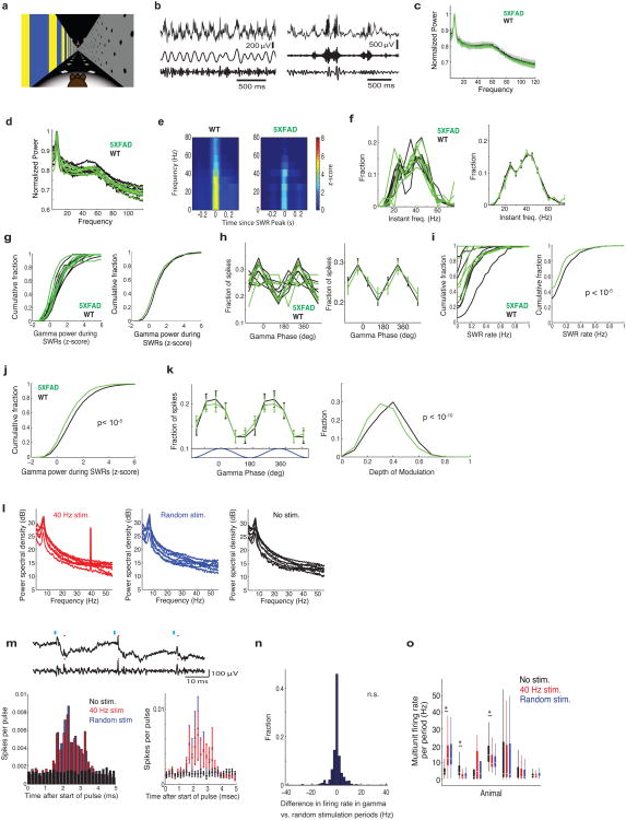
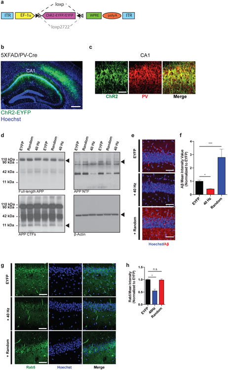
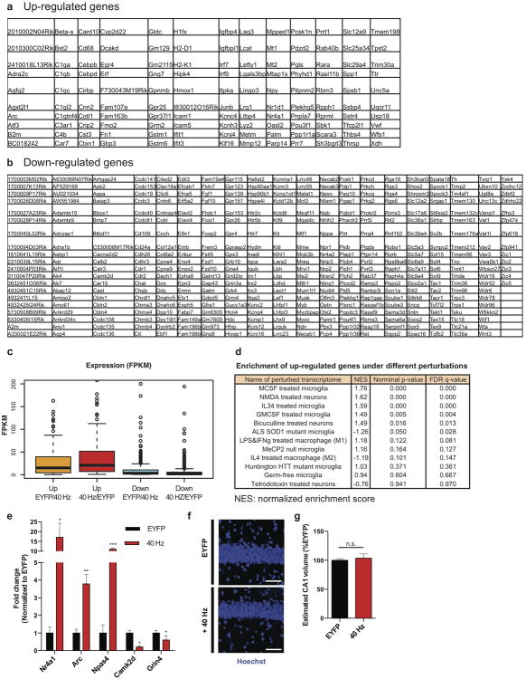
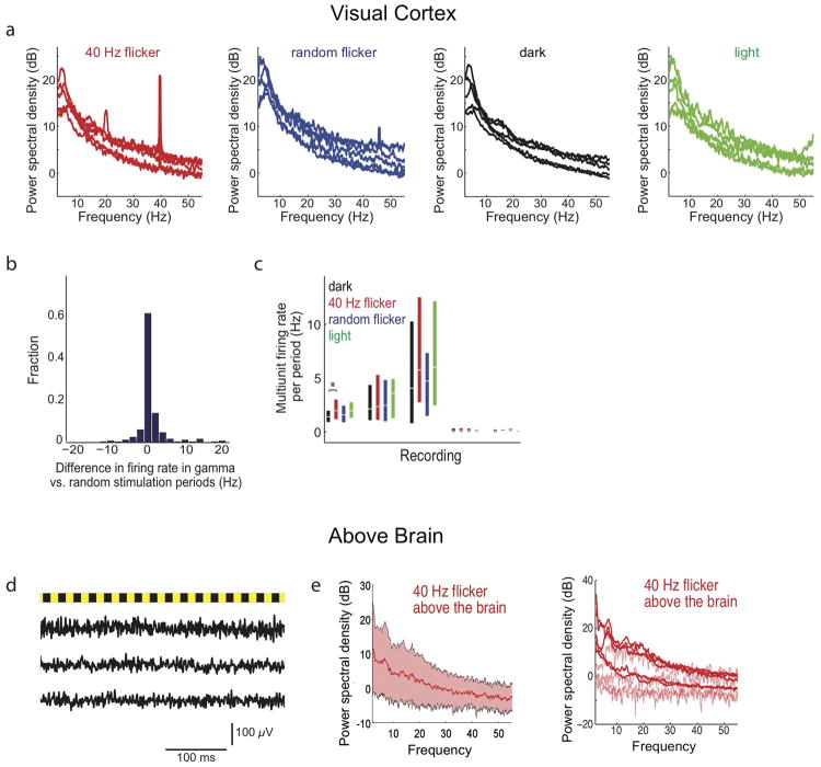
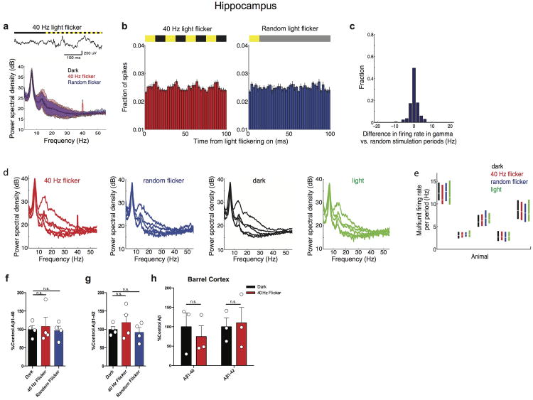
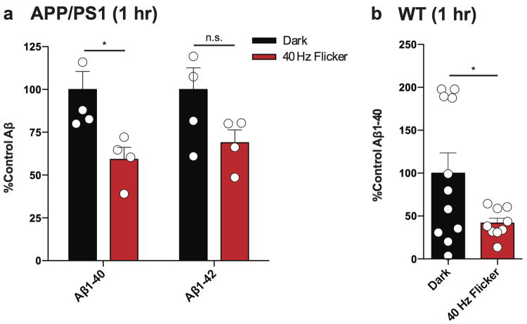
Changes in gamma oscillations (20-50 Hz) have been observed in several neurological disorders. However, the relationship between gamma oscillations and cellular pathologies is unclear. Here we show reduced, behaviourally driven gamma oscillations before the onset of plaque formation or cognitive decline in a mouse model of Alzheimer's disease. Optogenetically driving fast-spiking parvalbumin-positive (FS-PV)-interneurons at gamma (40 Hz), but not other frequencies, reduces levels of amyloid-β (Aβ)1-40 and Aβ 1-42 isoforms. Gene expression profiling revealed induction of genes associated with morphological transformation of microglia, and histological analysis confirmed increased microglia co-localization with Aβ. Subsequently, we designed a non-invasive 40 Hz light-flickering regime that reduced Aβ1-40 and Aβ1-42 levels in the visual cortex of pre-depositing mice and mitigated plaque load in aged, depositing mice. Our findings uncover a previously unappreciated function of gamma rhythms in recruiting both neuronal and glial responses to attenuate Alzheimer's-disease-associated pathology.
Figures
Comment in
Saturday, December 6, 2025
A Breakthrough in Stroke Rehabilitation: Non-Invasive Biophoton Therapy Promotes Neurofunctional Recovery in Chronic Stroke Patients
What does your doctor think? Better than these?
Biophoton
therapy is a form of complementary and alternative light therapy that
uses specific wavelengths of light to stimulate the body's natural
self-healing processes
. It is based on the theory that living
cells emit very low levels of light, or "biophotons," which play a role
in cellular communication and regulation.Yes, biophotons are real; they are extremely faint light particles (ultra-weak photon emissions or UPE) emitted by all living cells as byproducts of metabolic processes, detectable with sensitive equipment but invisible to the naked eye, and research suggests they play roles in cellular communication, signaling, and potentially even health indicators.
A Breakthrough in Stroke Rehabilitation: Non-Invasive Biophoton Therapy Promotes Neurofunctional Recovery in Chronic Stroke Patients
James Z Liu1,2*, Helen Y Gu1,2, Mariola Smotrys1, Seth Robinson2, Huixia Yu2, Devin R Liu2, and Sherry Liu1
1First Institute of All Medicines, 139 Pittsburgh Road, Butler, PA 16001, USA
2Tesla Bio Healing, LLC. 111 McCoy Street, Milford, DE 19963, USA
ABSTRACT
Background:
Effective treatments for chronic stroke remain a critical unmet medical need. Conventional rehabilitation approaches often yield limited
functional recovery, especially in patients with long-standing deficits. Emerging evidence suggests that biophoton-based therapies may offer a novel, non
invasive solution to support neuroregeneration.
Objective:
To evaluate the therapeutic efficacy and safety of Automatic Biophoton Generators (ABGs) in patients with chronic stroke using validated clinical,
neurological, and neurophysiological outcome measures.
Methods:
A randomized, triple-blind, placebo-controlled clinical trial was conducted with 46 chronic stroke patients who received either active ABG
treatment (n = 26) or placebo devices (n = 20) over two weeks. Participants in the placebo group were later crossed over to receive active treatment for
an additional four weeks. Each treatment group received therapy with 4 ABGs placed around a hotel bed. Stroke recovery was assessed using the Stroke
Impact Scale (SIS), SF-36 quality-of-life survey, clinician-led neurological examinations, and quantitative Electroencephalography (qEEG) with Event
Related Potentials (ERP). Additional evaluations included 3D Non-Linear Scanning (3D-NLS) for energetic brain imaging and Bio-Well analysis for organ
and meridian energy coherence.
Results:
Biophoton therapy resulted in statistically significant improvements in SIS scores, stroke recovery rate, neurological exam scores, and quality-of
life measures (p < 0.01). qEEG revealed increased posterior alpha frequencies, reduced theta/beta ratios, and improved ERP latencies, indicating enhanced
attention, memory processing, and cognitive speed. 3D-NLS imaging showed rapid cortical reactivation and restored hemispheric balance after only 6
days of therapy. Bio-Well analysis demonstrated systemic energy restoration across the brain, cardiovascular system, and endocrine organs. No adverse
events were reported.
Conclusion:
This study provides strong clinical and neurophysiological evidence that ABG-based biophoton therapy significantly improves functional
outcomes in patients with chronic stroke. The therapy was well tolerated and offers a promising, non-invasive approach to address long-standing neurological
impairments. These findings support the broader potential of biophoton quantum therapy in treating neurodegenerative and chronic inflammatory disorders.
Tuesday, October 28, 2025
Daily Light and Sound Therapy May Slow Alzheimer’s Decline
Didn't your competent? doctor set you up with this years ago?
Oh, YOU DON'T HAVE A COMPETENT DOCTOR, DO YOU?
40Hz sensory stimulation (3 posts to March 2021)
60 Hertz flickering light
(4 posts to July 2021) Or does your doctor believe the 60 Hz is better?
Daily Light and Sound Therapy May Slow Alzheimer’s Decline
Summary: A long-term study found that daily 40Hz light and sound stimulation may help slow cognitive decline in people with late-onset Alzheimer’s disease. After two years of treatment, participants maintained stronger cognitive performance than typical Alzheimer’s patients and showed reduced levels of tau protein, a key disease biomarker.
The noninvasive therapy, known as GENUS, works by synchronizing brain activity to a 40Hz gamma rhythm through light and sound. While early-onset patients saw fewer benefits, researchers say the results highlight the potential for a safe, at-home intervention to delay Alzheimer’s progression.
Key Facts
- Sustained Benefits: Late-onset Alzheimer’s participants maintained higher cognitive scores and improved sleep patterns over two years.
- Biomarker Reduction: Two participants showed significant decreases in plasma tau levels, linked to Alzheimer’s pathology.
- Safe and Noninvasive: The GENUS 40Hz light-and-sound therapy was well tolerated and feasible for daily home use.
Source: Picower Institute at MIT
A new research paper documents the outcomes of five volunteers who continued to receive 40Hz light and sound stimulation for around two years after participating in an MIT early-stage clinical study of the potential Alzheimer’s disease therapy.
The results show that for the three participants with late-onset Alzheimer’s disease, several measures of cognition remained significantly higher than comparable Alzheimer’s patients in national databases.
Moreover, in the two late-onset volunteers who donated plasma samples, levels of Alzheimer’s biomarker tau proteins were significantly decreased.
The three volunteers who experienced these benefits were all female. The two other participants, each of whom were males with early-onset forms of the disease, did not exhibit significant benefits after two years.
The dataset, while small, represents the longest-term test so far of the safe, non-invasive treatment method (called GENUS, for gamma entrainment using sensory stimuli), which is also being evaluated in a nationwide clinical trial run by MIT-spinoff company Cognito Therapeutics.
“This pilot study assessed the long-term effects of daily 40Hz multimodal GENUS in patients with mild AD,” the authors wrote in Alzheimer’s & Dementia: The Journal of the Alzheimer’s Association.
“We found that daily 40Hz audiovisual stimulation over 2 years is safe, feasible, and may slow cognitive decline and biomarker progression, especially in late-onset AD patients.”
Diane Chan, a former research scientist in The Picower Institute for Learning and Memory and neurologist at Massachusetts General Hospital, is the study’s lead and co-corresponding author. Picower Professor Li-Huei Tsai, director of The Picower Institute and the Aging Brain Initiative at MIT, is the study’s senior and co-corresponding author.
An “open label” extension
In 2020, MIT enrolled 15 volunteers with mild Alzheimer’s disease in an early-stage trial to evaluate whether an hour a day of 40Hz light and sound stimulation, delivered via an LED panel and speaker in their homes, could deliver clinically meaningful benefits.
Several studies in mice had shown that the sensory stimulation increases the power and synchrony of 40Hz gamma frequency brain waves, preserves neurons and their network connections, reduces Alzheimer’s proteins such as amyloid and tau, and sustains learning and memory. Several independent groups have also made similar findings over the years.
MIT’s trial, though cut short by the Covid-19 pandemic, found significant benefits after three months. The new study examines outcomes among five volunteers who continued to use their stimulation devices on an “open label” basis for two years.
These volunteers came back to MIT for a series of tests 30 months after their initial enrollment. Because four participants started the original trial as controls (meaning they initially did not receive 40Hz stimulation), their open label usage was six to 9 months shorter than the 30-month period.
The testing at 0, 3 and 30 months of enrollment included measurements of their brain wave response to the stimulation, MRI scans of brain volume, measures of sleep quality and a series of five standard cognitive and behavioral tests.
Two participants gave blood samples. For comparison to untreated controls, the researchers combed through three national databases of Alzheimer’s patients, matching thousands of them on criteria such as age, gender, initial cognitive scores, and retests at similar timepoints across a 30-month span.
Outcomes and outlook
The three female late-onset Alzheimer’s volunteers showed improvement or slower decline on most of the cognitive tests, including significantly positive differences compared to controls on three of them.
These volunteers also showed increased brain-wave responsiveness to the stimulation at 30 months and showed improvement in measures of circadian rhythms. In the two late-onset volunteers who gave blood samples, there were significant declines in phosphorylated tau (47 percent for one and 19.4 percent for the other) on a test recently approved by the FDA as the first plasma biomarker for diagnosing Alzheimer’s.
“One of the most compelling findings from this study was the significant reduction of plasma pTau217, a biomarker strongly correlated with AD pathology, in the two late-onset patients in whom follow-up blood samples were available,” the authors wrote in the journal.
“These results suggest that GENUS could have direct biological impacts on Alzheimer’s pathology, warranting further mechanistic exploration in larger randomized trials.”
Though the initial trial results showed preservation of brain volume at 3 months among those who received 40Hz stimulation, that was not significant at the 30-month timepoint.
And the two male early-onset volunteers did not show significant improvements on cognitive test scores. Notably, the early onset patients showed significantly reduced brain-wave responsiveness to the stimulation.
Though the sample is small, the authors hypothesize that the difference between the two sets of patients is likely attributable to the difference in disease onset rather than the difference in gender.
“GENUS may be less effective in early onset Alzheimer’s disease patients, potentially owing to broad pathological differences from late-onset Alzheimer’s disease that could contribute to differential responses,” the authors wrote. “Future research should explore predictors of treatment response, such as genetic and pathological markers.”
ease onset rather than the difference in gender.
“GENUS may be less effective in early onset Alzheimer’s disease patients, potentially owing to broad pathological differences from late-onset Alzheimer’s disease that could contribute to differential responses,” the authors wrote. “Future research should explore predictors of treatment response, such as genetic and pathological markers.”
40Hz sensory stimulation (3 posts to March 2021)
Or does your doctor believe the 60 Hz is better?
Daily Light and Sound Therapy May Slow Alzheimer’s Decline
Summary: A long-term study found that daily 40Hz light and sound stimulation may help slow cognitive decline in people with late-onset Alzheimer’s disease. After two years of treatment, participants maintained stronger cognitive performance than typical Alzheimer’s patients and showed reduced levels of tau protein, a key disease biomarker.
The noninvasive therapy, known as GENUS, works by synchronizing brain activity to a 40Hz gamma rhythm through light and sound. While early-onset patients saw fewer benefits, researchers say the results highlight the potential for a safe, at-home intervention to delay Alzheimer’s progression.
Key Facts
- Sustained Benefits: Late-onset Alzheimer’s participants maintained higher cognitive scores and improved sleep patterns over two years.
- Biomarker Reduction: Two participants showed significant decreases in plasma tau levels, linked to Alzheimer’s pathology.
- Safe and Noninvasive: The GENUS 40Hz light-and-sound therapy was well tolerated and feasible for daily home use.
Source: Picower Institute at MIT
A new research paper documents the outcomes of five volunteers who continued to receive 40Hz light and sound stimulation for around two years after participating in an MIT early-stage clinical study of the potential Alzheimer’s disease therapy.
The results show that for the three participants with late-onset Alzheimer’s disease, several measures of cognition remained significantly higher than comparable Alzheimer’s patients in national databases.
Moreover, in the two late-onset volunteers who donated plasma samples, levels of Alzheimer’s biomarker tau proteins were significantly decreased.
The three volunteers who experienced these benefits were all female. The two other participants, each of whom were males with early-onset forms of the disease, did not exhibit significant benefits after two years.
The dataset, while small, represents the longest-term test so far of the safe, non-invasive treatment method (called GENUS, for gamma entrainment using sensory stimuli), which is also being evaluated in a nationwide clinical trial run by MIT-spinoff company Cognito Therapeutics.
“This pilot study assessed the long-term effects of daily 40Hz multimodal GENUS in patients with mild AD,” the authors wrote in Alzheimer’s & Dementia: The Journal of the Alzheimer’s Association.
“We found that daily 40Hz audiovisual stimulation over 2 years is safe, feasible, and may slow cognitive decline and biomarker progression, especially in late-onset AD patients.”
Diane Chan, a former research scientist in The Picower Institute for Learning and Memory and neurologist at Massachusetts General Hospital, is the study’s lead and co-corresponding author. Picower Professor Li-Huei Tsai, director of The Picower Institute and the Aging Brain Initiative at MIT, is the study’s senior and co-corresponding author.
An “open label” extension
In 2020, MIT enrolled 15 volunteers with mild Alzheimer’s disease in an early-stage trial to evaluate whether an hour a day of 40Hz light and sound stimulation, delivered via an LED panel and speaker in their homes, could deliver clinically meaningful benefits.
Several studies in mice had shown that the sensory stimulation increases the power and synchrony of 40Hz gamma frequency brain waves, preserves neurons and their network connections, reduces Alzheimer’s proteins such as amyloid and tau, and sustains learning and memory. Several independent groups have also made similar findings over the years.
MIT’s trial, though cut short by the Covid-19 pandemic, found significant benefits after three months. The new study examines outcomes among five volunteers who continued to use their stimulation devices on an “open label” basis for two years.
These volunteers came back to MIT for a series of tests 30 months after their initial enrollment. Because four participants started the original trial as controls (meaning they initially did not receive 40Hz stimulation), their open label usage was six to 9 months shorter than the 30-month period.
The testing at 0, 3 and 30 months of enrollment included measurements of their brain wave response to the stimulation, MRI scans of brain volume, measures of sleep quality and a series of five standard cognitive and behavioral tests.
Two participants gave blood samples. For comparison to untreated controls, the researchers combed through three national databases of Alzheimer’s patients, matching thousands of them on criteria such as age, gender, initial cognitive scores, and retests at similar timepoints across a 30-month span.
Outcomes and outlook
The three female late-onset Alzheimer’s volunteers showed improvement or slower decline on most of the cognitive tests, including significantly positive differences compared to controls on three of them.
These volunteers also showed increased brain-wave responsiveness to the stimulation at 30 months and showed improvement in measures of circadian rhythms. In the two late-onset volunteers who gave blood samples, there were significant declines in phosphorylated tau (47 percent for one and 19.4 percent for the other) on a test recently approved by the FDA as the first plasma biomarker for diagnosing Alzheimer’s.
“One of the most compelling findings from this study was the significant reduction of plasma pTau217, a biomarker strongly correlated with AD pathology, in the two late-onset patients in whom follow-up blood samples were available,” the authors wrote in the journal.
“These results suggest that GENUS could have direct biological impacts on Alzheimer’s pathology, warranting further mechanistic exploration in larger randomized trials.”
Though the initial trial results showed preservation of brain volume at 3 months among those who received 40Hz stimulation, that was not significant at the 30-month timepoint.
And the two male early-onset volunteers did not show significant improvements on cognitive test scores. Notably, the early onset patients showed significantly reduced brain-wave responsiveness to the stimulation.
Though the sample is small, the authors hypothesize that the difference between the two sets of patients is likely attributable to the difference in disease onset rather than the difference in gender.
“GENUS may be less effective in early onset Alzheimer’s disease patients, potentially owing to broad pathological differences from late-onset Alzheimer’s disease that could contribute to differential responses,” the authors wrote. “Future research should explore predictors of treatment response, such as genetic and pathological markers.”
ease onset rather than the difference in gender.
“GENUS may be less effective in early onset Alzheimer’s disease patients, potentially owing to broad pathological differences from late-onset Alzheimer’s disease that could contribute to differential responses,” the authors wrote. “Future research should explore predictors of treatment response, such as genetic and pathological markers.”
Thursday, August 8, 2024
Light and Sound Therapy Maintains Myelin in Alzheimer’s
Doesn't your competent? doctor already have you using this? Oh, you DON'T HAVE A FUNCTIONING STROKE DOCTOR, DO YOU?
40 Hertz flickering light (2 posts to July 2021)
40Hz sensory stimulation (3 posts to March 2021)
Has your doctor verified that you had myelin damage from your stroke? What protocols are being used to fix such damage?
Your competent? doctor and hospital need to ensure human testing gets done. I already bought a 40 Hertz light, not waiting decades before it is confirmed in humans.
This one mentions a 60 Hertz flickering light so ask your competent? doctor which one is better. No knowledge of either is a fireable offense!
Repeated Ketamine Anesthesia Restarts Plasticity in the Brain July 2021
Light and Sound Therapy Maintains Myelin in Alzheimer’s
Summary: A new study finds that 40Hz light and sound therapy helps maintain myelin, a crucial brain structure, in Alzheimer’s patients. This therapy, which protects neurons and supports brain function, could offer new treatment avenues for neurodegenerative diseases.
Researchers discovered that this stimulation enhances neural connections and reduces harmful inflammation. The therapy also shows potential for treating other conditions involving myelin loss, such as multiple sclerosis.
Key Facts:
- 40Hz sensory stimulation preserves myelin in Alzheimer’s patients.
- The therapy enhances neural connections and reduces inflammation.
- Potential applications include treatment for multiple sclerosis.
- Source: Picower Institute at MIT
Early-stage trials in Alzheimer’s disease patients and studies in mouse models of the disease have suggested positive impacts on pathology and symptoms from exposure to light and sound presented at the “gamma” band frequency of 40 Hz.
A new study zeroes in on how 40Hz sensory stimulation helps to sustain an essential process in which the signal-sending branches of neurons, called axons, are wrapped in a fatty insulation called myelin. Often called the brain’s “white matter,” myelin protects axons and insures better electrical signal transmission in brain circuits.

“Previous publications from our lab have mainly focused on neuronal protection,” said Li-Huei Tsai, Picower Professor in The Picower Institute for Learning and Memory and the Department of Brain and Cognitive Sciences at MIT and senior author of the new study in Nature Communications. Tsai also lead’s MIT’s Aging Brain Initiative. “But this study shows that it’s not just the gray matter, but also the white matter that’s protected by this method.”
This year Cognito Therapeutics, the spin-off company that licensed MIT’s sensory stimulation technology, published phase II human trial results in the Journal of Alzheimer’s Disease indicating that 40Hz light and sound stimulation significantly slowed the loss of myelin in volunteers with Alzheimer’s.
Also this year Tsai’s lab published a study showing that gamma sensory stimulation helped mice withstand neurological effects of chemotherapy medicines, including by preserving myelin. In the new study, members of Tsai’s lab led by former postdoc Daniela Rodrigues Amorim used a common mouse model of myelin loss—a diet with the chemical cuprizone— to explore how sensory stimulation preserves myelination.
Amorim and Tsai’s team found that 40Hz light and sound not only preserved myelination in the brains of cuprizone-exposed mice, it also appeared to protect oligodendrocytes (the cells that myelinate neural axons), sustain the electrical performance of neurons, and preserve a key marker of axon structural integrity.
When the team looked into the molecular underpinnings of these benefits, they found clear signs of specific mechanisms including preservation of neural circuit connections called synapses; a reduction in a cause of oligodendrocyte death called “ferroptosis;” reduced inflammation; and an increase in the ability of microglia brain cells to clean up myelin damage so that new myelin could be restored.
“Gamma stimulation promotes a healthy environment,” said Amorim who is now a Marie Curie Fellow at the University of Galway in Ireland. “There are several ways we are seeing different effects.”
The findings suggest that gamma sensory stimulation may help not only Alzheimer’s disease patients but also people battling other diseases involving myelin loss, such as multiple sclerosis, the authors wrote in the study.
Maintaining myelin
To conduct the study, Tsai and Amorim’s team fed some male mice a diet with cuprizone and gave other male mice a normal diet for six weeks. Halfway into that period, when cuprizone is known to begin causing its most acute effects on myelination, they exposed some mice from each group to gamma sensory stimulation for the remaining three weeks.
In this way they had four groups: completely unaffected mice, mice that received no cuprizone but did get gamma stimulation, mice that received cuprizone and constant (but not 40Hz) light and sound as a control, and mice that received cuprizone and also gamma stimulation.
After the six weeks elapsed, the scientists measured signs of myelination throughout the brains of the mice in each group. Mice that weren’t fed cuprizone maintained healthy levels, as expected. Mice that were fed cuprizone and didn’t receive 40Hz gamma sensory stimulation showed drastic levels of myelin loss.
Cuprizone-fed mice that received 40Hz stimulation retained significantly more myelin, rivaling the health of mice never fed cuprizone by some, but not all, measures.
The researchers also looked at numbers of oligodendrocytes to see if they survived better with sensory stimulation. Several measures revealed that in mice fed cuprizone, oligodendrocytes in the corpus callosum region of the brain (a key point for the transit of neural signals because it connects the brain’s hemispheres) were markedly reduced. But in mice fed cuprizone and also treated with gamma stimulation, the number of cells were much closer to healthy levels.
Electrophysiological tests among neural axons in the corpus callosum showed that gamma sensory stimulation was associated with improved electrical performance in cuprizone-fed mice who received gamma stimulation compared to cuprizone-fed mice left untreated by 40Hz stimulation.
And when researchers looked in the anterior cingulate cortex region of the brain, they saw that MAP2, a protein that signals the structural integrity of axons, was much better preserved in mice that received cuprizone and gamma stimulation compared to cuprizone-fed mice who did not.
Molecular mechanisms
A key goal of the study was to identify possible ways in which 40Hz sensory stimulation may protect myelin.
To find out, the researchers conducted a sweeping assessment of protein expression in each mouse group and identified which proteins were differentially expressed based on cuprizone diet and exposure to gamma frequency stimulation. The analysis revealed distinct sets of effects between the cuprizone mice exposed to control stimulation and cuprizone-plus-gamma mice.
A highlight of one set of effects was the increase in MAP2 in gamma-treated cuprizone-fed mice. A highlight of another set was that cuprizone mice who received control stimulation showed a substantial deficit in expression of proteins associated with synapses. The gamma-treated cuprizone-fed mice did not show any significant loss, mirroring results in a 2019 Alzheimer’s 40Hz study that showed synaptic preservation.
This result is important, the researchers wrote, because neural circuit activity, which depends on maintaining synapses, is associated with preserving myelin. They confirmed the protein expression results by looking directly at brain tissues.
Another set of protein expression results hinted at another important mechanism: ferroptosis. This phenomenon, in which errant metabolism of iron leads to a lethal buildup of reactive oxygen species in cells, is a known problem for oligodendrocytes in the cuprizone mouse model.
Among the signs was an increase in cuprizone-fed, control stimulation mice in expression of the protein HMGB1, which is a marker of ferroptosis-associated damage that triggers an inflammatory response. Gamma stimulation, however, reduced levels of HMGB1.
Looking more deeply at the cellular and molecular response to cuprizone demyelination and the effects of gamma stimulation, the team assessed gene expression using single-cell RNA sequencing technology.
They found that astrocytes and microglia became very inflammatory in cuprizone-control mice but gamma stimulation calmed that response. Fewer cells became inflammatory and direct observations of tissue showed that microglia became more proficient at clearing away myelin debris, a key step in effecting repairs.
The team also learned more about how oligodendrocytes in cuprizone-fed mice exposed to 40Hz sensory stimulation managed to survive better. Expression of protective proteins such as HSP70 increased and as did expression of GPX4, a master regulator of processes that constrain ferroptosis.
In addition to Amorim and Tsai, the paper’s other authors are Lorenzo Bozzelli, TaeHyun Kim, Liwang Liu, Oliver Gibson, Cheng-Yi Yang, Mitch Murdock, Fabiola Galiana-Meléndez, Brooke Schatz, Alexis Davison, Md Rezaul Islam, Dong Shin Park, Ravikiran M. Raju, Fatema Abdurrob, Alissa J. Nelson, Jian Min Ren, Vicky Yang and Matthew P. Stokes.
Funding: Fundacion Bancaria la Caixa, The JPB Foundation, The Picower Institute for Learning and Memory, the Carol and Gene Ludwig Family Foundation, Lester A. Gimpelson, Eduardo Eurnekian, The Dolby Family, Kathy and Miguel Octavio, the Marc Haas Foundation, Ben Lenail and Laurie Yoler, and the National Institutes of Health provided funding for the study.
About this neurology and neuroscience research news
Author: David Orenstein
Source: Picower Institute at MIT
Contact: David Orenstein – Picower Institute at MIT
Image: The image is credited to Neuroscience News
Original Research: Open access.
“Multisensory gamma stimulation mitigates the effects of demyelination induced by cuprizone in male mice” by Li-Huei Tsai et al. Nature Communications
Subscribe to:
Posts (Atom)