Changing stroke rehab and research worldwide now.Time is Brain! trillions and trillions of neurons that DIE each day because there are NO effective hyperacute therapies besides tPA(only 12% effective). I have 523 posts on hyperacute therapy, enough for researchers to spend decades proving them out. These are my personal ideas and blog on stroke rehabilitation and stroke research. Do not attempt any of these without checking with your medical provider. Unless you join me in agitating, when you need these therapies they won't be there.

What this blog is for:

My blog is not to help survivors recover, it is to have the 10 million yearly stroke survivors light fires underneath their doctors, stroke hospitals and stroke researchers to get stroke solved. 100% recovery. The stroke medical world is completely failing at that goal, they don't even have it as a goal. Shortly after getting out of the hospital and getting NO information on the process or protocols of stroke rehabilitation and recovery I started searching on the internet and found that no other survivor received useful information. This is an attempt to cover all stroke rehabilitation information that should be readily available to survivors so they can talk with informed knowledge to their medical staff. It lays out what needs to be done to get stroke survivors closer to 100% recovery. It's quite disgusting that this information is not available from every stroke association and doctors group.

Showing posts with label any brains at all. Show all posts

Friday, May 22, 2026

Evaluating upper limb movement smoothness metrics derived from markerless 3D motion tracking in children with neurological disorders

 If this doesn't get in your hospital in the next month to objectively determine your recovery protocol needs; THEN EVERYONE IS MASSIVELY INCOMPETENT!

You need to get them all fired and the hospital reconstituted! Obviously no one in your hospital has any brains at all if this doesn't immediately trigger; 'Let's get this for stroke rehab.'

Evaluating upper limb movement smoothness metrics derived from markerless 3D motion tracking in children with neurological disorders

    We are providing an unedited version of this manuscript to give early access to its findings. Before final publication, the manuscript will undergo further editing. Please note there may be errors present which affect the content, and all legal disclaimers apply.

    Abstract

    Background

    Clinical assessment of movement smoothness during reaching in children is often based on subjective, ordinal-scaled therapist ratings. Markerless 3D motion tracking using an RGB-D camera offers a promising avenue for more objective and fine-grained smoothness evaluation. This study investigated the use of a new markerless RGB-D-based tracking method, SMPLify-KiDS, for the quantitative assessment of upper limb movement smoothness in a heterogeneous population of children with neurological impairments.

    Methods

    Thirty-eight children with neurological diagnoses performed three reaching tasks from the Melbourne Assessment 2 (MA2) MA2 with their more affected arm while being recorded with an RGB-D camera. Upper limb kinematics were extracted using SMPLify-KiDS, and two widely used smoothness metrics—the log dimensionless jerk (LDJ) and the modified spectral arc length (SPARC)—were computed. The same movement sequences were independently scored by two therapists according to MA2 guidelines. Correlations between quantitative smoothness metrics and therapist scores were evaluated using Spearman’s rank correlation coefficient, and smoothness metric differences between ordinal therapist score levels were analyzed using non-parametric statistical tests for individual items and averaged values.

    Results

    Quantitative smoothness metrics showed moderate to strong correlations with therapist scores per item (for LDJ:0.666 ≤ p ≤ 0.836; for SPARC:0.556 ≤ p ≤ 0.674  ), and very strong to excellent correlations for averaged values (for LDJ: p= 0.843, for SPARC:p= 0.755). Both metrics revealed statistically significant differences between ordinal therapist score levels, and LDJ strongly correlated with task duration.

    Conclusions

    Smoothness metrics derived from markerless RGB-D motion tracking are able to discriminate between clinically assigned smoothness levels in children with neurological impairments. LDJ showed closer alignment with therapist scores than SPARC, potentially due to its relationship with movement duration—a factor that also appears relevant to clinical perception of smoothness. These findings support the integration of markerless motion capture into clinical assessments to provide objective, quantitative, and fine-grained information on movement quality, complementing established therapist-based evaluations.

    Sunday, May 17, 2026

    Novel Antibody Repairs Acute Spinal Cord Lesions

     With ANY BRAINS AT ALL  in stroke leadership, this would trigger immediate research to see if it would work for stroke also!  That will never occur, there aren't two functioning neurons anywhere in stroke.

    Let's see how long your doctor has DONE NOTHING with nogo-A
    • nogo-A (14 posts to May 2012) 

    That's an impressive amount of incompetence displayed everywhere in your hospital!

    Novel Antibody Repairs Acute Spinal Cord Lesions

    Summary: A multinational clinical trial demonstrated that a novel antibody, NG101, successfully preserves existing nerve tissue and accelerates the regression of spinal cord lesions following acute injury.

    The antibody operates by neutralizing Nogo-A, a naturally occurring protein that structurally blocks the regeneration of damaged nerve fibers. By combining advanced magnetic resonance imaging (MRI) with clinical data, researchers have objectively visualized the therapeutic mechanics early in treatment, establishing an essential blueprint for restoring functional brain-to-muscle signal pathways.

    Key Facts

    • Removing the Healing Barrier: NG101 targets and neutralizes Nogo-A, an unhelpful protein found in the sheaths of nerve fibers within the brain and spinal cord that actively blocks damaged fibers from healing after an acute trauma.
    • Accelerated Lesion Regression: Advanced imaging methods confirmed for the first time that the antibody therapy speeds up the healing of spinal cord lesions, allowing nerve fibers to regenerate in the tissue surrounding the impact site.
    • Tissue Loss Interception: The therapy considerably slows down the loss of existing nerve tissue and offsets structural degradation by stimulating the regrowth of entirely new nerve fibers.
    • Functional Reconnection: The newly formed and surviving nerve fibers successfully navigate across or around the injury site, re-establishing vital connections with the spinal cord centers that control the peripheral nerves of the hands, arms, and legs.

    Source: University of Zurich

    Spinal cord injuries – often caused by sports or traffic accidents – can result in tetraplegia or paraplegia and severely limit independence.

    In late 2024, an international research group led by the University of Zurich (UZH) and Balgrist University Hospital completed a multinational clinical trial in which patients with acute spinal cord injuries were successfully treated with the novel antibody NG101.

    This shows an axon.
    By neutralizing the inhibitory Nogo-A protein within the nerve fiber sheaths, the antibody NG101 allows surviving and newly generated axons to effectively navigate around spinal lesions. Credit: Neuroscience News
    The results showed that NG101 accelerates the regression of spinal cord lesions and preserves existing nerve tissue.

    Antibody neutralizes unhelpful protein

    Discovered at UZH roughly 30 years ago, NG101 targets the protein Nogo-A, which is found in the sheaths of nerve fibers in the spinal cord and brain. This protein blocks the healing of damaged nerve fibers in the spinal cord following acute injury.

    By neutralizing Nogo-A, NG101 removes this barrier to growth and healing, thereby boosting nerve fiber regeneration and supporting the functional regeneration of spinal cord tissue.

    Visible results in the spinal cord

    The research team’s latest study has revealed another critical piece of the puzzle. “In our new study, we were able to use advanced imaging methods to show for the first time how this antibody therapy works directly in the spinal cord,” says Patrick Freund, UZH professor and head of the Spinal Cord Injury Center at Balgrist University Hospital.

    The magnetic resonance imaging data revealed two important effects. First, spinal cord injuries healed more quickly in the presence of NG101, which suggests that nerve fibers were able to regenerate in the tissue surrounding the injury. Second, the loss of nerve tissue slowed down considerably and was offset by the regrowth of new nerve fibers. Previous animal experiments conducted by the researchers had already established how critical this stage is.

    This is due to newly formed nerve fibers needing to find a way to navigate across or around the injury site in order to restore the pathways linking the brain and the spinal cord.

    New connections to peripheral nerves

    The group’s latest findings suggest that it is precisely this process that is supported by NG101.

    “This allows surviving and newly regenerated nerve fibers to re-establish connections with the spinal cord centers that control the hand, arm and leg nerves,” says Freund, who led the study.

    “These connections are essential for relaying signals from the brain to the muscles.” For some patients, this means a greater chance of recovering arm and hand function.

    NG101 not only improves the function of the spinal cord but has also been shown to alter its structure, which supports the regeneration of nerve tissue. This marks an important step toward new, effective treatments for spinal cord injuries.

    “We are now able to visualize the effect of the therapy early on and in an objective way,” says Freund. “This opens up the possibility of using future treatments more strategically and conducting a more reliable evaluation of their outcomes.”

    Key Questions Answered:

    Q: If someone is paralyzed from a car accident, can this drug make them walk again?

    A: While it is too early to guarantee full mobility for every patient, the clinical trial proved that NG101 helps surviving and newly grown nerve fibers reconnect with the spinal cord centers controlling the arms, hands, and legs. For patients with acute injuries, this significantly increases the mathematical probability of recovering crucial hand and arm functions.

    Q: Why doesn’t the spinal cord just heal itself naturally like a broken bone?

    A: The central nervous system contains a literal molecular brake pad. A specific protein called Nogo-A resides in the protective sheaths of your nerve fibers. Following a sudden trauma, this protein actively stops damaged nerve fibers from growing or repairing themselves. NG101 acts as a shield that shuts down Nogo-A so natural regeneration can take over.

    Q: How do doctors know the drug is actually repairing the spine and not just masking symptoms?

    A: Researchers used high-resolution MRI data to track the treatment on a cellular and structural level. The imaging objectively showed two clear visual markers: the physical holes (lesions) in the spinal cord shrank much faster, and the rapid degradation of delicate nerve tissue was replaced by the visible architecture of freshly sprouting nerve fibers.

    Editorial Notes:

    • This article was edited by a Neuroscience News editor.
    • Journal paper reviewed in full.
    • Additional context added by our staff.

    About this spinal cord injury and neurology research news

    Author: Kurt Bodenmueller
    Source: University of Zurich
    Contact: Kurt Bodenmueller – University of Zurich
    Image: The image is credited to Neuroscience News

    Characteristics of plantar pressure during walking in stroke patients with hemiplegia of different brunnstrom stages: a study based on five-region smart insoles

     How EXACTLY does this get survivors recovered? You, along with your mentor and senior researchers incompetently don't know stroke research is to get survivors recovered? I won't feel sorry for you when you are the 1 in 4 per WHO that has a stroke! 

    Characteristics of plantar pressure during walking in stroke patients with hemiplegia of different brunnstrom stages: a study based on five-region smart insoles

      We are providing an unedited version of this manuscript to give early access to its findings. Before final publication, the manuscript will undergo further editing. Please note there may be errors present which affect the content, and all legal disclaimers apply.

      Abstract

      Plantar pressure measurement is frequently employed to assess the gait characteristics of stroke patients. This study developed a gait assessment system for stroke patients utilizing a self-developed five-region smart insole, with the goal of objectively evaluating their gait characteristics. A total of 75 stroke patients (14 at Brunnstrom stage III, 18 at stage IV, 27 at stage V, and 16 at stage VI) and 24 healthy subjects (designated “Health”) were recruited. A smart insole was used to collect the subjects’ gait parameters and plot plantar pressure curves. The results showed the following: (1) There were statistically significant differences among the five groups in the durations of the gait cycle, the double support phase, the bilateral single support phases, and the swing phase, as well as in the ratio of peak pressure to body weight for the bilateral toe bone regions, medial metatarsal regions, lateral metatarsal regions, and heel regions. No statistically significant differences were found in the bilateral arch peak pressure-to-body weight ratio among the five groups. (2) Each of the five subject groups exhibited a unique plantar pressure curve profile. The smart insole used in this study can provide objective gait assessment for stroke patients and effectively differentiate the gait characteristics of patients at different Brunnstrom stages.(Which would mean if you had ANY BRAINS AT ALL, that you could easily develop an EXACT REHAB PROTOCOL to fix those gait problems found!)

      Sunday, March 29, 2026

      Stroke rehab goes high-tech: UP study reveals robotic therapy may restore lost grip

       Come back when you finish the job correctly! Will restore using these EXACT PROTOCOLS! 'May' is not a valid result!

      What will you do about those like me that have dead brain there and no longer have any signals coming thru? Do you actually have any brains at all that might be useful in solving stroke to 100% recovery?

      Stroke rehab goes high-tech: UP study reveals robotic therapy may restore lost grip

      MARCH 29, 2026 — The University of the Philippines has just dropped a study that could change how we look at stroke recovery in the country. Researchers Micah Angelo Bacani and Manuel Ramos Jr. from UP Diliman’s Electrical and Electronics Engineering Institute tested a robotic hand orthosis powered by surface electromyography (sEMG) signals — and achieved an impressive 86% response accuracy rate. In plain language: the device listens to muscle signals from the arm and translates them into movement, giving stroke survivors a shot at regaining control of their grip.

      Why does this matter? Stroke remains one of the leading causes of disability in the Philippines. Many survivors struggle with long-term loss of hand function, making everyday tasks — from holding a spoon to signing a document — frustratingly difficult. 

      Traditional rehab often relies on repetitive, passive exercises. But this study argues that recovery is stronger when patients actively participate, engaging their neurological pathways rather than just following preset motions. 

      That’s where robotics step in: machines that respond to the patient’s own muscle signals, not just programmed routines.

      The researchers explained, “Using surface electromyography (sEMG) signals from the arm, the recovering stroke patient can control the robotic assistive device for rehabilitation. This is the myoelectric hand orthosis.” 

      Additionally, the study found a direct, proportional relationship between the patient’s intended force and the device’s response. In other words, the harder the patient tries, the stronger the orthosis reacts.

      Now, that’s not just science — it’s empowerment. Imagine the psychological boost of seeing your effort instantly translated into movement, instead of waiting months for uncertain progress.

      Of course, this raises bigger questions for us. Will our healthcare system embrace robotic rehab, or will it remain locked in academic journals? Can public hospitals afford such technology, or will it be another innovation reserved for private clinics and the wealthy? And most importantly, will patients trust machines to help them heal?This study is a reminder that science is racing ahead, but policy and accessibility often lag behind. If robotics can truly help stroke survivors reclaim their independence, then the challenge is no longer technological — it’s social, economic, and political.

      When machines can help us heal, will the Philippines let technology be a lifeline for the many, or a luxury for the few?

      Tuesday, March 24, 2026

      Lifelike 3D-Printed ‘Training Brains’ React Like Real Organs

       With ANY BRAINS AT ALL in our stroke medical 'professionals' we could do research that tells us exactly what interventions work to stop the neuronal cascade of death in the first week! Putting the brain on a chip with brain organoids and this and we could solve a lot of stroke recovery questions.  But nothing will occur; OUR 'PROFESSIONALS' ARE FUCKING INCOMPETENT! 

      Lifelike 3D-Printed ‘Training Brains’ React Like Real Organs

      Imagine what brain surgeons could do with a three-dimensional (3D)-printed model of the brain. Not a structural model printed in plastic and uniform throughout, but one that mirrors the heterogeneity of real human brain tissue — its mechanical, thermal, and electromagnetic properties.

      photo of Christopher O Bryan PhD
      Christopher O’ Bryan, PhD, MS

      “If somebody has a brain tumor, you could take an MRI of their brain and make a 3D model from it that includes the tumor, and then practice surgery on that model,” said Christopher O’ Bryan, PhD, MS, assistant professor of mechanical and aerospace engineering at the University of Missouri (UM) in Columbia, Missouri. “You could learn how sound, electromagnetic waves, or physical insults interact with the brain to better understand concussions and traumatic brain injury.”

      photo of 3D brains
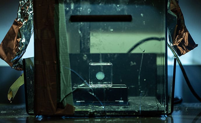
           A scaled-down 3D model of the brain, about 15% of a real brain’s size.

      O’Bryan and colleagues in UM’s College of Engineering recently published a study in the journal Materialia describing how the researchers designed a unique 3D printer ink made from a modified polymer — called a photo-crosslinkable poly(vinyl alcohol) methacrylate polymer — and then used it to print a scaled-down 3D model of the brain, about 15% of a real brain’s size. The scientists relied on a method called embedded 3D printing, crosslinking the polymer in a microgel bath, to achieve a model that mimicked the soft tissue and the structural complexities of a real human brain.

      How They Did It

      To provide a broader picture of why they conducted the study, O’Bryan described his lab’s goals. “The focus areas in our lab include the designing of soft materials, the testing of these materials, and then how to actually manufacture things out of these soft materials. And that’s especially challenging when you take into account that most soft materials are hydrogels and biopolymers which start out in a liquid phase ,” he said.

      photo of 3D brains
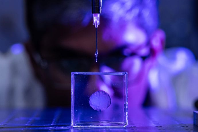
      Researchers used embedded 3D printing, crosslinking the polymer with a gel bath to mimic brain tissue.

      The researchers’ embedded 3D printing approach uses “sacrificial support baths,” jelly-like baths, which act as a supportive matrix during the printing phase. “What this allows us to do is to print materials as liquids and keep them as liquids until we’ve completely printed our structure, and then we crosslink the printed structure using heat or UV [ultraviolet] light to make it solid. Then we take it out of the gel bath, and it’s become a soft, deformable structure — meaning it can change shape when a force is applied,” O’Bryan said.

      Wednesday, March 18, 2026

      UMBRELLA: Using AI to improve stroke care from SAFE(Stroke Alliance for Europe)


       A complete fucking failure! 'Care' NOT RECOVERY!

      Really hope your comeuppance is incredibly difficult when you become the 1 in 4 per WHO that has a stroke! Not recovering 100% is going to be a real bitch since you DID NOTHING when you could have! Until 100%recovery research is out there, artificial intelligence isn't going to do much good. Wrong goal: the only goal in stroke is 100% recovery! GET THERE!

      UMBRELLA: Using AI to improve stroke care

      On Brain Awareness Week’s third day we are focusing on UMBRELLA, a European research project that uses artificial intelligence (AI) and digital tools to improve how strokes are diagnosed, treated and managed(Survivors want recovery; NOT managing their compensation! Don't you people HAVE ANY BRAINS AT ALL?). 

      A stroke occurs when blood flow to the brain is blocked or a blood vessel bursts. Quick treatment is vital, but recovery can be long and complex and many survivors remain at risk of another stroke.

      UMBRELLA aims to support doctors by using AI to help them make faster and more personalised decisions, while keeping patient information safe and private.

      The project follows a careful process: health data is securely collected, organised and analysed inside hospitals. Personal details are removed, and strict privacy rules are followed. The AI is then trained to recognise patterns and tested to make sure it works safely and truly helps patient care. 

      Led by Vall d’Hebron Research Institute and Siemens Healthineers, and supported by the EU’s Innovative Health Initiative, the project brings together hospitals, researchers, patient organisations and companies from across Europe. 

      “Stroke survivors live with many long-term challenges. Better tools to support them are urgently needed. UMBRELLA is an exciting step toward more personalised and effective stroke care, with the potential to make a real difference for patients and families across Europe,” said Arlene Wilkie, Director General of SAFE. 

      By improving diagnosis, treatment and prevention, UMBRELLA aims to transform stroke care(NOT RECOVERY!) and improve the quality of lives(This should be return them to their previous lives!) of thousands of people. 

      To learn more about the project: 

      Visit the Umbrella website https://umbrella-ihi.eu
      Or contact research@safestroke.eu 

      This project is supported by the Innovative Health Initiative Joint Undertaking (IHI JU) under grant agreement No 101172825. The JU receives support from the European Union’s Horizon Europe research and innovation programme and COCIR, EFPIA, Europa Bío, MedTech Europe and Vaccines Europe.

      Friday, March 6, 2026

      Wearable sensors may detect early disability progression in multiple sclerosis

       With ANY BRAINS AT ALL your stroke medical 'professionals' would immediately use this to objectively diagnose gait abnormalities. And then objectively monitor the recovery. But nothing will occur, we have blithering idiots in stroke!

      Wearable sensors may detect early disability progression in multiple sclerosis

      A longitudinal study of people with multiple sclerosis (MS) found that declines in daytime physical activity detected through wearable accelerometers were associated with increased risk of disability progression and brain atrophy.

      The findings, published in Neurology, suggest continuous activity monitoring may serve as a sensitive, noninvasive biomarker for early disease worsening before clinical symptoms become apparent.

      “Timely identification of patients at risk for disease progression is essential to reduce long-term disability, but the current tests for measuring MS disability are not designed to detect small changes,” said Kathryn C. Fitzgerald, Johns Hopkins University, Baltimore, Maryland. “Using a relatively inexpensive and accessible device around the wrist may help us identify early changes in the disease.”

      The study enrolled 238 adults aged ≥40 years who underwent annual brain MRI scans and wore wrist-based accelerometers 24 hours per day for 2 straight weeks every 3 months, for up to 3 years. The devices captured several activity metrics, including total activity levels, sedentary time, circadian rhythm parameters, and activity during specific two-hour daytime windows.

      Participants had been living with MS for an average of 13 years. At the start of the study, they had an average disability level of 3 on the Expanded Disability Status Scale (EDSS).

      Over an average follow-up of 2.9 years, 120 participants experienced confirmed disability progression based on the composite EDSS-plus. Overall physical activity declined by an average of about 2% per year.

      Importantly, within-person decreases in daytime activity -- particularly between 8:00 AM and 2:00 PM -- were associated with a significantly higher risk of disability progression. A one-standard deviation decrease in morning or midday activity increased the risk of confirmed progression by roughly 20% to 24%.

      MRI analyses also showed that reductions in morning activity were linked to greater brain atrophy, including loss of whole-brain, deep Gray matter, and thalamic volume. While individuals with lower average moderate-to-vigorous activity had smaller brain volumes overall, these between-person differences were not associated with disability progression.

      “More research is needed to confirm these findings, but it’s exciting to think that using easily accessible devices could help us predict who is at risk of worsening disease and potentially prevent those changes,” said Ellen M. Mowry, Johns Hopkins University. “Detecting small changes could also help us speed up research on new treatments.”

      A limitation of the study is that people who did not have MS were not included, which would help researchers understand how activity levels may change as a part of normal aging. Also, the participants were relatively older and more disabled, so the results may not apply to younger people with MS and those with less disability.

      Reference: https://www.neurology.org/doi/10.1212/WNL.0000000000214678

      SOURCE: American Academy of Neurology

      Tuesday, December 2, 2025

      Higher B12 Levels Linked to Slower Cognitive Decline

       With nothing on how to measure or what higher means: COMPLETELY FUCKING USELESS! Does no one in stroke have any brains at all?

      Higher B12 Levels Linked to Slower Cognitive Decline

      TOPLINE:

      Higher vitamin B12 status in mid-to-late life was associated with slower declines in memory, executive function, and language compared to lower status in patients without baseline dementia, new research showed. Additionally, the associations for executive function and language — but not memory — persisted regardless of folate status.

      METHODOLOGY:

      • Researchers analyzed 1994 dementia-free adults (mean age, 60 years; 54% women; 99% non-Hispanic White individuals) from the Framingham Heart Study Offspring Cohort. All had at least two assessments of vitamin B12 biomarkers and neuropsychological test scores between 1998 and 2018.
      • The participants were divided into four quartiles on the basis of the cumulative average of three vitamin B12 biomarkers: serum levels of cobalamin, methylmalonic acid, and total homocysteine, corrected for low folate levels.
      • The primary outcomes were changes in memory, executive function, and language factor scores over a mean follow-up duration of 14.2 years.
      • The analysis was adjusted for demographic, lifestyle, and medical factors, including age, education, and apolipoprotein E allele status.

      TAKEAWAY:

      • Participants with a higher cumulative vitamin B12 status showed significantly slower annual cognitive decline than those with a lower B12 status in the domains of language (P for trend = .003), memory (P for trend = .004), and executive function (P for trend = .03).
      • The highest B12 level was also associated with less cognitive decline (by 0.05-0.09 SD) over 10 years than the lowest level.
      • The results were consistent across folate levels, though the association between B12 status and memory was slightly stronger in those with elevated folate levels (≥ 20 ng/mL).

      IN PRACTICE:

      “Given that even small delays in the onset of symptoms can reduce the prevalence of cognitive impairment, efforts to manage vitamin B12 status and improve nutrition throughout the life course may help mitigate cognitive decline into older age,” the investigators wrote.

      SOURCE:

      The study was led by Francesca R. Marino, Boston University Chobanian and Avedisian School of Medicine, Boston. It was published online on October 21 in Alzheimer’s & Dementia.

      LIMITATIONS:

      The study population consisted primarily of European American participants, possibly reducing the generalizability of the findings. Neuropsychological test scores and vitamin B12 biomarkers were not measured at the same time, and different assays were used across study exams, which could introduce nondifferential error. Genetic data on polymorphisms affecting homocysteine or other B12 biomarkers were unavailable. Additionally, excluded participants were older with lower B12 or folate levels, which may have introduced selection bias.

      DISCLOSURES:

      The study was funded by the National Heart, Lung, and Blood Institute and the National Institute on Aging. Several investigators reported serving in advisory or editorial roles for several pharmaceutical and research organizations. Full details are provided in the original article.

      Tuesday, October 28, 2025

      Prognostic assessment of acute ischemic stroke by systemic immune-inflammatory index: a comprehensive meta-analysis of multidimensional outcomes

      Predicting failure to recover IS ABSOLUTELY FUCKING USELESS! Does no one in stroke have any brains at all?

       Prognostic assessment of acute ischemic stroke by systemic immune-inflammatory index: a comprehensive meta-analysis of multidimensional outcomes


      Yanhong JiangYanhong Jiang1Yifan CuiYifan Cui1Xiaojie HuXiaojie Hu2Jiaying LianJiaying Lian3Xueying QinXueying Qin4Xingchen Wang,
Xingchen Wang1,2*Xuran Ma
Xuran Ma1*
      • 1The Second Clinical Medical College, Shandong University of Traditional Chinese Medicine, Jinan, China
      • 2Department of Neurology, The Second Affiliated Hospital of Shandong University of Traditional Chinese Medicine, Jinan, China
      • 3Department of Neurology, Quzhou Traditional Chinese Medicine Hospital, Quzhou, China
      • 4The First Clinical Medical College, Beijing University of Traditional Chinese Medicine, Beijing, China

      Introduction: Our study aimed to quantify the predictive ability of the Systemic Immune-inflammatory Index (SII) for predicting the prognosis and multidimensional complications in acute ischemic stroke (AIS) patients. The primary outcome was poor prognosis, and secondary outcomes included mortality, severity, hemorrhagic transformation/symptomatic intracerebral hemorrhage, stroke-associated pneumonia/poststroke pneumonia, early neurological deterioration, post-stroke depression, progression or recurrence, and other adverse outcomes.

      Methods: We searched 15 databases from their establishment to 13 October 2024 and selected cohort or case-control analyses that analyzed the association of continuous or categorized SII as exposures with the above adverse outcomes of AIS populations.

      Results: The results showed that 78 studies with 40,682 participants were included in meta-analyses. Continuous SII values were significantly higher in poor prognosis groups than in controls (SMD = 248.13, 95% CI: 198.77 to 297.50; p = 0.000). Poor prognosis incidences rose with higher continuous SII values (OR = 1.004, 95% CI: 1.002 to 1.005; p = 0.000). More patients in High SII groups had poor prognosis (RR = 1.95, 95% CI: 1.66 to 2.28; p = 0.000). The risk of poor prognosis was higher in the high SII groups, though this was not statistically significant (OR = 1.007, 95% CI: 0.998 to 1.015; p = 0.120).

      Discussion: In conclusion, our study found that continuous SII and high SII were associated with poor prognosis of AIS and various complications. Given the accessibility and low cost of SII, integrating it into prognostic scores merits further research for better clinical choices.

      Systematic review registration: PROSPERO (CRD42024586414), https://www.crd.york.ac.uk/PROSPERO/view/CRD42024586414.

      Monday, June 16, 2025

      Stroke Patients' Knowledge, Attitudes, and Practices Regarding Home-Based Exercise and Psychological Rehabilitation Programs

      If you had 100% recovery protocols your patients would gladly do the millions of reps needed because they would be looking forward to recovery! SO, SOLVE THAT PROBLEM!

      Leaders solve problem; Are you leaders or mice?

       Stroke Patients' Knowledge, Attitudes, and Practices Regarding Home-Based Exercise and Psycholosgical Rehabilitation Programs

      Ying Zou 2 Hongbo Li 3 Wei Xu 4 2 Lezheng Wang 2 Qiyue Sun Lei Shi Shaoxuan Ma 2 Ye Liu 1* 1 Beijing Sport University, Beijing, Beijing Municipality, China 2 Jiangsu College of Nursing, Huai'an, China Jinhu County People’s Hospital, Jiangsu, China 4 Zhenjiang Hospital of Integrated Traditional Chinese and Western Medicine, Zhenjiang, Jiangsu Province, China 5 Yangzhou University, Yangzhou, Jiangsu Province, China The final, formatted version of the article will be published soon.Notify me 

      Background: 

      Stroke rehabilitation encompasses crucial components essential for the recovery process. This study aimed to investigate the knowledge, attitudes, and practices (KAP) of stroke patients regarding home-based exercise and psychological rehabilitation programs aimed at enhancing their physical and mental well-being, as well as alleviating depressive symptoms.

      Methods: 

      This study conducted a cross-sectional survey at 37 institutions from December 2023 to January 2024. The survey gathered demographic data and assessed KAP related to stroke rehabilitation through structured questionnaires. 

      Results: 

      A total of 499 valid questionnaires were included in this study. Of these participants, 312 (62.5%) were male and 252 (50.5%) were aged over 60 years. The median [25%,75%] knowledge, attitude, and practice scores were 9.03 (5.04) (possible range: 0-20), 33.19 (3.55) (possible range: 9-45), and 21.43 (4.49) (possible range: 6-30), respectively. Multivariate logistic regression showed that knowledge score (OR = 1.19, 95% CI: [1.13,1.26], P < 0.001), attitude score (OR = 1.28, 95% CI: [1.19,1.38], P < 0.001), employed (OR = 2.41, 95% CI: [1.15,5.04], P = 0.020), retired (OR = 2.20, 95% CI: [1.20,4.02], P = 0.011), and more than 3 years of duration of stroke (OR = 0.37, 95% CI: [0.16,0.85], P = 0.019) were independently associated with practice. Structural equation modeling (SEM) showed that knowledge directly affected attitude (β = 0.407, P < 0.001), knowledge (β = 0.390, P < 0.001) and attitude (β = 0.461, P < 0.001) directly affected practice, and knowledge indirectly affected practice through attitude (β = 0.188, P = 0.007).

      Conclusion: 

      The study highlights that while stroke patients generally exhibit positive attitudes and practices towards home-based exercise and psychological rehabilitation, there remains a gap in their knowledge about these crucial interventions. Clinically, it is imperative to enhance educational strategies targeting stroke survivors, focusing on improving knowledge to further strengthen their rehabilitation outcomes and adherence to prescribed regimens.(Adherence wouldn't be a problem with 100% recovery protocols! Can't you people think at all?)

      Keywords: knowledge, Attitude, Practice, Stroke, Patient, home-based exercise, psychological rehabilitation programs

      Received: 23 Mar 2025; Accepted: 13 Jun 2025.

      Copyright: © 2025 Dai, Zou, Li, Xu, Luo, Wang, Sun, Shi, Ma and Liu. This is an open-access article distributed under the terms of the Creative Commons Attribution License (CC BY). The use, distribution or reproduction in other forums is permitted, provided the original author(s) or licensor are credited and that the original publication in this journal is cited, in accordance with accepted academic practice. No use, distribution or reproduction is permitted which does not comply with these terms.

      * Correspondence: Ye Liu, Beijing Sport University, Beijing, 100084, Beijing Municipality, China

      Disclaimer: All claims expressed in this article are solely those of the authors and do not necessarily represent those of their affiliated organizations, or those of the publisher, the editors and the reviewers. Any product that may be evaluated in this article or claim that may be made by its manufacturer is not guaranteed or endorsed by the publisher.