Changing stroke rehab and research worldwide now.Time is Brain! trillions and trillions of neurons that DIE each day because there are NO effective hyperacute therapies besides tPA(only 12% effective). I have 523 posts on hyperacute therapy, enough for researchers to spend decades proving them out. These are my personal ideas and blog on stroke rehabilitation and stroke research. Do not attempt any of these without checking with your medical provider. Unless you join me in agitating, when you need these therapies they won't be there.

What this blog is for:

My blog is not to help survivors recover, it is to have the 10 million yearly stroke survivors light fires underneath their doctors, stroke hospitals and stroke researchers to get stroke solved. 100% recovery. The stroke medical world is completely failing at that goal, they don't even have it as a goal. Shortly after getting out of the hospital and getting NO information on the process or protocols of stroke rehabilitation and recovery I started searching on the internet and found that no other survivor received useful information. This is an attempt to cover all stroke rehabilitation information that should be readily available to survivors so they can talk with informed knowledge to their medical staff. It lays out what needs to be done to get stroke survivors closer to 100% recovery. It's quite disgusting that this information is not available from every stroke association and doctors group.

Showing posts with label immune system. Show all posts

Thursday, August 13, 2026

Scientists just discovered a new brain phase between 50 and 75. It may help explain cognitive decline

 Have your competent? doctor and hospital get further research going that creates protocols to address this problem! Can't do that? PURE INCOMPETENCE!

Scientists just discovered a new brain phase between 50 and 75. It may help explain cognitive decline

A single-cell study of the hippocampus suggests brain aging is not a steady decline, but a coordinated immune, vascular, and neuronal overhaul.

Scientists at the University of California, San Diego School of Medicine just discovered a new brain phase.

A single-cell study identified a new biological phase the brain may enter between 50 and 75. The research offers new, important insights about brain aging and cognitive decline.

Between the ages of 50 and 75, the cellular landscape of the hippocampus, the brain’s memory region, begins to change. Immune cells get replaced, the genome’s physical structure loosens, and gene regulation is reshaped across multiple cell types. 

The study’s findings, which were published in the peer-reviewed journal Science, suggest that aging is not so much a gradual decline than an active biological overhaul—one that may eventually be a target for treatment.

The researchers looked at individual cells from human hippocampal tissue collected at various points across the adult lifespan. They then mapped gene regulation and three-dimensional genome architecture to produce what Sci Tech Daily called “one of the most detailed accounts yet of how these features change as the human brain ages.”

A changing immune system

One of the most significant shifts the scientists discovered appeared in the microglia—the immune cells that help maintain and protect that brain. In midlife, between the ages of 50 and 75, the microglia that formed during embryonic development declined sharply. The researchers found that cells with similar traits to blood immune cells replaced the missing microglia cells.

Related video: Study identifies protein that could help protect memory as we age (WKYC-TV Cleveland)

Those replacements carried stronger inflammatory signatures, raising the possibility that they help drive the persistent inflammation tied to brain aging. At the same time, cell populations that maintain the blood-brain barrier dropped off substantially.

“Microglia are critical for maintaining brain homeostasis,” Bing Ren, PhD, a corresponding author of the study and scientific director and CEO of the New York Genome Center told Sci Tech Daily. “When these cells fail to perform their housekeeping duties, toxic materials accumulate that can trigger inflammatory processes that may contribute to neurodegenerative diseases.”

Nathan Zemke, PhD, lead author and a principal investigator at the UC San Diego Center for Epigenomics, told Inc. that the immune system shift is directly connected to memory. 

“Between roughly ages 50 and 80, many of the brain’s original immune cells appear to be replaced by immune cells that enter from the bloodstream,” he told Inc. 

Those cells are important for clearing debris and protecting the brain, he added, but the incoming ones carry stronger inflammatory signals, which may contribute to the chronic brain inflammation commonly associated with cognitive decline.

Across several brain cell types, the genome’s three-dimensional structure grew progressively less organized with age. DNA folds into a precise architecture that helps determine which genes a cell can use. As that folding erodes, so does the cell’s control over its own machinery. The researchers suggest it may be a basic feature of brain aging.

That breakdown is what makes the structural changes matter for the mind, Zemke said. “Every type of brain cell relies on a carefully organized set of genetic instructions to perform its specialized role,” he told Inc. “As we age, that organization begins to break down, which may disrupt the genes needed to maintain healthy brain function and cognition.”

Notably, the immune, vascular, and neuronal systems appeared to change together, in a coordinated way. 

“Importantly, this study reveals that aging is not simply a gradual decline, but involves coordinated and dynamic remodeling of immune, vascular, and neuronal systems,” said Xiangmin Xu, PhD, a co-corresponding author and director of the Center for Neural Circuit Mapping at UC Irvine told Sci Tech Daily.”These findings open the door to identifying new therapeutic targets aimed at preserving circuit integrity and brain function across the lifespan.”

Implications for the future of aging research

The findings should also change how professionals think about aging itself, Zemke told Inc. “Many biological changes associated with aging appear to accelerate around age 50, including increased inflammation and reduced cellular energy production,” he said. “However, the timing varies greatly from person to person. Understanding what drives that variation could help explain why some people age more successfully than others.”

The immune-cell shift may carry the greatest significance for Alzheimer’s research. The replacement of the brain’s original immune cells by blood-derived ones occurs during the same window when Alzheimer’s-related changes are thought to begin, Zemke said. The incoming cells show inflammatory features associated with the disease, suggesting the transition could be an important contributor to Alzheimer’s risk.

This post originally appeared at inc.com.

“Click here to subscribe to the Inc. newsletter: inc.com/newsletters"

Saturday, May 23, 2026

Treating the immune system to repair the brain

 Ask your competent? doctor EXACTLY HOW THIS WILL GET YOU 100% RECOVERED!

Do you prefer your doctor, hospital and board of director's incompetence NOT KNOWING? OR NOT DOING? Your choice; let them be incompetent or demand action! You do know incompetent doctors and hospitals can be fired!

Treating the immune system to repair the brain


 Science Translational Medicine
 20 May 2026 Vol 18 Issue 850 DOI: 10.1126/scitranslmed.aeb1677

Abstract

Non-neuronal brain cells and systemic immunity play a central role in Alzheimer’s disease (AD) and other brain disorders. The immune system, initially protective, becomes dysfunctional as the disease progresses. Here, we discuss next-generation therapeutic approaches aimed at treating the immune system rather than the brain to combat AD and other neurodegenerative diseases.

Access the full article

View all access options to continue reading this article.


Monday, May 4, 2026

What Happens To Your Immune System After One Sauna Session by mindbodygreen


I'm just got a personal sauna from NuRecover but will use a cold shower instead of a chiller bucket for the cold shock proteins. Still to be put together.

What Happens To Your Immune System After One Sauna Session

 There’s a reason sauna use keeps showing up in longevity research. Regular exposure has been associated with lower risks of cardiovascular disease1 ,respiratory illness2,dementia3, and even overall mortality4 .But those outcomes don’t tell us much about the immediate effects. What actually changes in the body during a single session? And how quickly do those changes happen? A new study5set out to answer that by tracking immune responses before, during, and after a 30-minute sauna session, offering a closer look at the body’s short-term reaction to heat. 

What happens to your immune system during a 30-minute sauna

Researchers studied 51 adults, both men and women, during a standard Finnish sauna session set to about 73°C (roughly 163°F). Each participant spent 30 minutes in the sauna, with blood samples taken before, immediately after, and 30 minutes post-session. The goal was to track how immune cells and inflammatory signals changed in response to heat stress. Participants were allowed to drink water throughout, which helped control for dehydration and made the setup closer to a typical real-world sauna experience. Instead of just looking at overall immune activity, the researchers zoomed in on specific white blood cells and a wide range of cytokines, which are signaling molecules involved in inflammation and immune response. This gave a more detailed picture of how the body reacts in the short term. 

Sauna’s immediate effect on immune cells

The biggest shift came down to your white blood cells, which are a key part of your immune system. After the sauna session, those levels went up, including important types like neutrophils and lymphocytes. These are the cells that help your body spot and respond to anything that doesn’t belong, whether that’s a virus or another kind of threat. What stands out is how quickly this happens. The increase shows up right after the sauna, then goes back down within about 30 minutes. So this isn’t a long-lasting spike; it’s more like a short window where your immune system is a bit more alert. This pattern mirrors what happens during exercise. When you work out, immune cells move out of tissues and into your bloodstream, where they’re more ready to respond if needed. A sauna seems to trigger a comparable response, just through heat instead of movement. 

What about inflammation & body temperature?

One thing the researchers looked at closely was inflammation, since that’s often what people think of when they hear the body is under “stress.” But in this case, there weren’t big changes across most inflammatory markers. Out of dozens of signals they measured, only a few shifted in a meaningful way. That suggests your body isn’t going into a full inflammatory response during a sauna. Instead, the response seems more about mobilization than inflammation. Your immune cells are being redistributed and activated, not necessarily pushed into an inflammatory state. Body temperature did play a role, though. On average, people’s temperature rose by about 2°C (roughly 3.6°F) during the session. And the more it increased, the more certain immune-related signals shifted alongside it. 

Adding sauna use to your routine

 So what does this mean for your day-to-day routine? It doesn’t mean a single sauna session will prevent illness or replace other foundational habits. But it does suggest that sauna use can act as a short-term stimulus for your immune system, similar to a workout.If you already use a sauna, this adds another layer of context. That post-sauna feeling isn’t just relaxation. Your body is actively responding, mobilizing immune cells in a way that may support overall immune surveillance over time. 
If you’re considering adding it in, consistency matters more than intensity. This study looked at one session, but previous research has linked regular sauna use to broader health benefits, including a lower risk of certain chronic conditions.

It’s also worth paying attention to how you personally respond. Hydration, heat tolerance, and recovery all play a role in how beneficial the experience feels.

The takeaway

This study doesn’t suggest that sauna use is a cure-all. But it does offer a clearer picture of what’s happening in the body during a session. Your immune system isn’t passive in that environment. It’s responding, adapting, and briefly shifting into a more active state.

Friday, April 24, 2026

How the immune system may influence Alzheimer’s, Parkinson’s, and related diseases

Maybe this is why saunas are helpful in preventing dementia, boosting the immune system.

How the immune system may influence Alzheimer’s, Parkinson’s, and related diseases

A sweeping review shows how the brain’s immune defenses can both fight and fuel neurodegeneration, revealing why timing, cell type, and disease context may be crucial for future therapies.

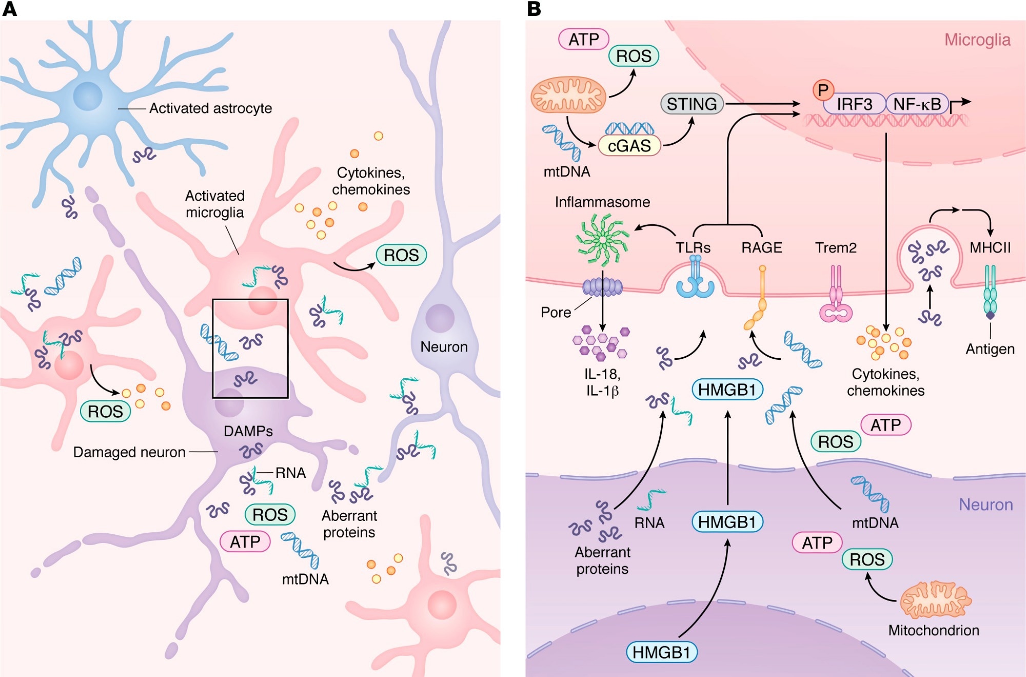
Innate immune activation and microglial transition during neurodegeneration. (A) During neurodegeneration, distressed neurons release DAMPs such as mtDNA, ROS, HMGB1, aberrant proteins, and pTau–bound RNA. These signals activate resident microglia and astrocytes, which then recruit peripheral immune cells. As shown in more detail in B, activated microglia upregulate PRRs, including TLRs, RAGE, and cGAS, enabling detection of neuron-derived DAMPs. Engagement of PRRs induces proinflammatory gene expression programs and activates inflammasome pathways that promote release of chemokines and cytokines into the CNS parenchyma. As microglia transition from homeostatic to reactive states in a TREM2-dependent manner, damage-associated microglia increase MHC-II expression to support antigen presentation and coordination of adaptive immune responses that attempt to limit neurodegeneration.

Innate immune activation and microglial transition during neurodegeneration. (A) During neurodegeneration, distressed neurons release DAMPs such as mtDNA, ROS, HMGB1, aberrant proteins, and pTau–bound RNA. These signals activate resident microglia and astrocytes, which then recruit peripheral immune cells. As shown in more detail in B, activated microglia upregulate PRRs, including TLRs, RAGE, and cGAS, enabling detection of neuron-derived DAMPs. Engagement of PRRs induces proinflammatory gene expression programs and activates inflammasome pathways that promote release of chemokines and cytokines into the CNS parenchyma. As microglia transition from homeostatic to reactive states in a TREM2-dependent manner, damage-associated microglia increase MHC-II expression to support antigen presentation and coordination of adaptive immune responses that attempt to limit neurodegeneration.

A recent review published in the Journal of Clinical Investigation synthesized current evidence on immune mechanisms in neurodegeneration. Accumulating evidence suggests that the immune system not only drives the progression of neurodegenerative diseases but also contributes to their onset through maladaptive cellular signaling and activation. The central nervous system (CNS) parenchyma depends on resident immune cells to maintain the blood-brain barrier (BBB) and respond to dysfunctional cells, pathogens, or cellular damage under steady-state conditions.

Neurons activate signaling pathways to respond to cellular dysfunction, while microglia and astrocytes alleviate danger signals and recruit peripheral immune cells. However, these responses can aggravate aberrant protein deposition during neurodegeneration, and the balance between detrimental and beneficial immune involvement is poorly defined. In the present review, researchers reviewed current knowledge of immune mechanisms in neurodegeneration.

Innate Immune Signaling in 

Neurodegenerative Disease

Aberrant proteins, including phosphorylated tau, amyloid β (Aβ), and α-synuclein, have been identified as damage-associated molecular patterns (DAMPs) that activate pattern recognition receptors in the CNS. Toll-like receptor 2 (TLR2) and TLR4 are upregulated in mouse models of Alzheimer’s disease (AD) and in the brains of AD patients.

The binding of Aβ to TLR4 or TLR2 elevates the expression of cytokines, including tumor necrosis factor (TNF)-α, interleukin (IL)-6, IL-1β, IL-17, and IL-10, in microglia and astrocytes. Pharmacological inhibition or genetic ablation of these TLRs worsens cognitive decline and increases Aβ burden in the brain by decreasing microglial activation. The review also highlighted inflammasome and DNA-sensing pathways, including NLRP3 and cGAS-STING, as additional contributors to neuroinflammation across neurodegenerative diseases.

Studies have consistently reported increased expression of the receptor for advanced glycation end products (RAGE) on neurons, astrocytes, and microglia in AD, Parkinson’s disease (PD), and amyotrophic lateral sclerosis. RAGE overexpression in mouse AD models accelerated cognitive impairment, while its deficiency decreased memory deficits.

Genetic studies in AD patients identified two single-nucleotide polymorphisms (SNPs) in the triggering receptor expressed on myeloid cells 2 (TREM2), which is highly expressed by microglia, as risk factors of a magnitude comparable to carrying the apolipoprotein E (APOE) ε4 allele. These observations indicate that microglia are key players in neurodegenerative pathogenesis.

T Cell Responses Across 

Neurodegenerative Disorders

A growing body of evidence indicates that cluster of differentiation 4 (CD4+) T cells have disease-specific functions across neurodegenerative diseases. Aβ-restricted CD4+ T cell transfusion into a transgenic AD mouse model was neuroprotective, preventing cognitive decline. In contrast, α-synuclein-specific CD4+ T cells were neurotoxic in PD models, promoting inflammation.

α-synuclein overexpression increased recruitment of CD4+ T cells producing interferon-γ, while depleting these cells decreased neuronal loss. These observations indicate that CD4+ T cells can be protective in some neurodegenerative diseases and pathogenic in others. In addition, CD8+ T cells have garnered increasing attention as contributors to CNS pathology. CD3+ and CD8+ T cells have been detected in the hippocampus in AD, with CD3+ T cell numbers correlating with tau pathology.

Similarly, increased CD8+ T cell numbers were observed in PD and correlated with neuronal death, although whether this relationship is causative or reactive remains uncertain. Further, the accumulation of neuroprotective CD8+ T cells in the brains of AD mice has been reported and shown to restrict amyloid plaque growth. Overall, studies reveal that CD8+ T cells have diverse roles in neurodegenerative diseases, shaped by antigen specificity, microglial interactions, and tissue residency. The review also noted growing interest in granzyme K-expressing CD8+ T cells as potentially specialized players in several neurodegenerative conditions.

Aging, Injury, and Viral Effects 

on CNS Immunity

Aging is a major risk factor for neurodegenerative diseases. In youth, microglia survey the CNS, astrocytes maintain the BBB, and only a small number of T cells enter the parenchyma. However, in advanced ages, chronic low-grade inflammation occurs, during which many immune pathways become dysregulated.

Further, repetitive head injury can accelerate neurodegeneration. Traumatic brain injury is linked to the development of PDAD, and chronic traumatic encephalopathy, even when it occurs in childhood. Following mild traumatic brain injury, the release of DAMPs by distressed cells triggers microglial activation, which clears debris and reinforces weakened BBB regions.

Repetitive injuries may increase reactivity and trigger microglial death, thereby allowing the entry of myelomonocytic cells. Like repetitive injuries, viral infections can alter CNS immunity and influence vulnerability to neurodegeneration. Recent studies have linked non-neurotropic and neurotropic viral infections to a higher risk of neurodegenerative diseases.

Immune Timing and 

Neurodegeneration Implications

Taken together, neurodegenerative diseases stem from a complex interplay of environmental and biological factors that govern the magnitude and timing of immune activation and the programming of T cells, microglia, and other leukocytes. Viral infection, repetitive head injury, and aging restructure the CNS immune environment. Many immune pathways involved exhibit stage-dependent and context-specific functions.

Microglial activation may initially help clear aberrant proteins, but could become maladaptive with protracted stimulation. Similarly, CD8+ T cells can promote neuronal injury or support tissue repair based on antigen specificity, local signaling cues, and effector programming. As such, appreciating the cell-intrinsic states and temporal dynamics of immune cells is crucial for understanding immune signatures and determining when immunomodulatory interventions will be beneficial.

Journal reference:
  • Latour YL, McGavern DB (2026). Immune signaling and function in neurodegeneration. Journal of Clinical Investigation, 136(8), e199850. DOI: 10.1172/JCI199850, https://www.jci.org/articles/view/199850

A single sauna session boosts immune cells within minutes

I'm getting a personal sauna from NuRecover but will use a cold shower instead of a chiller bucket for the cold shock proteins.

A single sauna session boosts immune cells within minutes

New research reveals how a brief Finnish sauna session mobilizes immune defenses within minutes, offering fresh clues into how heat exposure may influence human health.

Young couple relaxing and sweating in hot sauna wrapped in towel. Woman and man In Sauna. Interior of Finnish sauna, classic wooden steam room with hot steam. Russian bathroom. Relax in hot Bathhouse.Study: Acute Finnish sauna heat exposure induces stronger immune cell than cytokine responses. Image credit: Mr. Tempter/Shutterstock.com

Heat exposure is harmful but can sometimes have beneficial effects. Finnish sauna bathing (FSB) is an example of controlled acute heat stress that may influence immune function by altering immune cells and cytokines. A 30-minute Finnish sauna session rapidly increases circulating immune cells while leaving most cytokine levels unchanged, according to a study published in the journal Temperature.

How Finnish sauna bathing 

became a global health habit

FSB involves exposing people to relatively dry air at 70–100 °C for 10-30 minutes per session. It is part of Finnish culture and is shown to offer multiple health benefits. Existing literature reports lower risks of cardiovascular disease, dementia, psychosis, and some respiratory conditions. FSB is also associated with fewer adverse outcomes in disadvantaged socioeconomic settings and with better physical fitness.

Acute heat stress may reshape 

immune and inflammatory responses

Prior research suggests that regular FSB is associated with lower levels of systemic inflammation. The current study examined how heat stress associated with FSB manifests as acute changes in white blood cells and cytokines up to 30 minutes after the sauna.

Single sauna session 

triggers rapid immune cell mobilisation

Participants were free of known cardiovascular disease (CVD) but had one or more risk factors, such as smoking, hypertension, or obesity. The study included 51 middle-aged adults who were regular sauna users. During the 30-minute sauna session, body temperature, measured at the ear, increased steadily, reflecting the heat stress exposure, while plasma volume remained unchanged on average.

Among women, hemoglobin and hematocrit levels were lower, while platelet counts were higher than in men. In both sexes, all these factors increased with FSB. White cells also rose in women, and remained slightly higher at 30 minutes post-sauna.

Among white cells, neutrophils and lymphocytes returned to baseline at 30 minutes, unlike the persistent elevation seen with the combined MXD cell category (monocytes, eosinophils, and basophils). Overall, white cell proportions were preserved.

Only two cytokines changed significantly after FSB, and both decreased, while one additional marker showed a borderline increase. Immediately after FSB exposure, body temperature was correlated with circulating cytokine levels, though not with white cell counts. These included several interferons and interleukins, which were positively associated with raised ear temperature. Conversely, two cytokines were inversely correlated with ear temperature. These changes were independent of the frequency of sauna use across the week.

These findings suggest that white cells are mobilized in association with FSB exposure, but there was no direct correlation with increased temperature. Significant associations were observed for only a few cytokines, whereas a larger number showed correlations with temperature change in the immediate post-sauna period. The associations between temperature change and cytokines were largely unchanged after adjustment for sex or body mass index.

The authors propose that physiological challenge caused by FSB-induced heat stress may underlie these changes. The increase in hemoglobin concentration and hematocrit suggests a possible loss of plasma volume in the hot, dry FSB conditions, although average plasma volume did not change, and results were adjusted for individual variation.

The increase in neutrophil and lymphocyte counts could be due to immune cell mobilization in response to heat stress. The generalized nature of the response is suggested by the overall preservation of the differential count.

Apparently, a large temperature change is not required for such mobilization, as no correlation was found between temperature change and white cell count. Conversely, the former was associated with cytokine changes in the immediate post-sauna period, suggesting a heat-stress response.

The authors report that to the best of their knowledge, few prior studies have explored both immune cells and cytokines in relation to FSB-induced heat stress. The sauna use pattern in this study closely resembled that in real life and in some earlier research.

A few other studies have shown changes in some interleukins using different sauna protocols. This highlights the need for further research to distinguish sauna-induced responses in habitual sauna users (who are likely heat-adapted) from those in individuals who seldom or never use it.

Limited immune profiling 

leaves key cell responses unclear

The authors measured three main white cell categories (neutrophils, lymphocytes, and the combined MXD group), but did not assess monocytes, eosinophils, and basophils separately. A more detailed lymphocyte assessment could have better assessed the heat-stress response to the FSB.

The physiological effects of cytokines correlated with temperature change following FSB remain unknown. The origin of the possibly mobilized white cells is unclear. The possibility of low plasma volume remains relevant, but was not factored into the analysis. Moreover, the study measured only the acute physiological response rather than chronic resting conditions.

Future directions should include research on the relevance of intermittent heat and cold stress exposure and whether these acute responses translate into long-term health effects.

Download your PDF copy by clicking here.

Journal reference:

Monday, February 26, 2018

Discovery Reveals Way to Stop Inflammation in Alzheimer's, Arthritis, More

You probably want this to prevent your likely dementia, so ask your doctor for followup. 
https://www.rdmag.com/news/2018/02/discovery-reveals-way-stop-inflammation-alzheimers-arthritis-more?
A new discovery about the immune system may allow doctors to treat harmful inflammation that damages the brain in neurodegenerative diseases such as Alzheimer's. It might also let doctors save patients from the potentially deadly inflammation of sepsis, a full-body infection that kills a quarter-million Americans every year.
The finding "opens up a whole new research area to look at neuroinflammation in the context of Alzheimer's and Parkinson's," said lead researcher Bimal Desai, PhD, of the University of Virginia School of Medicine. "But the clinical impact will be in many, many different areas."
Neurological Treatments
Traditional treatments for neurological inflammation, such as in Alzheimer's and Parkinson's disease, are largely ineffective because biological drugs are blocked by what is known as the blood-brain barrier. That barrier protects the brain from dangers such as bacteria or toxins in the blood, but it also makes it very difficult to get drugs into the brain. "A lot of the drugs we use right now to treat inflammation, [known as] biologicals, don't work in the brain because they can't get through," explained Desai, of UVA's Department of Pharmacology and UVA's Carter Immunology Center.
His new finding, involving important immune cells known as macrophages (and microglia), could offer a way around that. He and his team have identified a specific electrical switch, known as an ion channel, within macrophages that controls the flow of calcium into the cells. Without calcium, the cells can't cause inflammation. By targeting this switch with tiny molecules, researchers could deny the macrophages calcium and prevent inflammation - even in the brain.
A Better Way to Battle Inflammation
That could let researchers develop a new and better way to stop inflammation. "Small molecules are perhaps more affordable as treatments and can hit things like this ion channel switch, TRPM7," said researcher Michael Schappe, a graduate student in Desai's lab. "We could use that to address inflammation in a bunch of contexts, but particularly in instances like neuroinflammation, where [current] treatments are particularly ineffective."
Desai noted that drug companies are already at work on drugs that could target this type of switch. And that could be good news for patients with many inflammatory diseases. "Right now, you have conditions like arthritis or IBD [inflammatory bowel disease], where inflammation plays a huge role. They do have very good drugs for them, but these drugs are extremely expensive and cannot be taken orally by the patients. They can cost as much as $20,000 a year," he said. "The reason for that is that they're biologicals. They're protein molecules that are very difficult to make and distribute. But having identified an ion channel as a target in this context allows you to use small molecules, which are ridiculously cheap compared to biologicals and can be taken orally by the patients."
The discovery of the new drug target, the researchers noted, was made possible by something very unusual about UVA. To learn more, visit the Making of Medicine blog at https://makingofmedicine.virginia.edu/2018/02/26/the-switch-that-could-shut-down-inflammation-even-in-the-brain/

Wednesday, June 7, 2017

Natural sugar may trigger immune system to treat atherosclerosis, study finds

In mice, so not to be done on your own. But you do expect your competent? doctor and hospital to get human testing going, right!

http://www.news-medical.net/news/20170607/Natural-sugarc2a0may-trigger-immune-system-to-treat-atherosclerosis-study-finds.aspx
Researchers have long sought ways to harness the body's immune system to treat disease, especially cancer. Now, scientists have found that the immune system may be triggered to treat atherosclerosis and possibly other metabolic conditions, including fatty liver disease and type 2 diabetes.
Studying mice, researchers at Washington University School of Medicine in St. Louis have shown that a natural sugar called trehalose revs up the immune system's cellular housekeeping abilities. These souped-up housecleaners then are able to reduce atherosclerotic plaque that has built up inside arteries. Such plaques are a hallmark of cardiovascular disease and lead to an increased risk of heart attack.
The study is published June 7 in Nature Communications.
"We are interested in enhancing the ability of these immune cells, called macrophages, to degrade cellular garbage -- making them super-macrophages," said senior author Babak Razani, MD, PhD, an assistant professor of medicine.
Macrophages are immune cells responsible for cleaning up many types of cellular waste, including misshapen proteins, excess fat droplets and dysfunctional organelles -- specialized structures within cells.
"In atherosclerosis, macrophages try to fix damage to the artery by cleaning up the area, but they get overwhelmed by the inflammatory nature of the plaques," Razani explained. "Their housekeeping process gets gummed up. So their friends rush in to try to clean up the bigger mess and also become part of the problem. A soup starts building up -- dying cells, more lipids. The plaque grows and grows."
In the study, Razani and his colleagues showed that mice prone to atherosclerosis had reduced plaque in their arteries after being injected with trehalose. The sizes of the plaques measured in the aortic root were variable, but on average, the plaques measured 0.35 square millimeters in control mice compared with 0.25 square millimeters in the mice receiving trehalose, which translated into a roughly 30 percent decrease in plaque size. The difference was statistically significant, according to the study.
The effect disappeared when the mice were given trehalose orally or when they were injected with other types of sugar, even those with similar structures.
Found in plants and insects, trehalose is a natural sugar that consists of two glucose molecules bound together. It is approved by the Food and Drug Administration for human consumption and often is used as an ingredient in pharmaceuticals. Past work by many research groups has shown trehalose triggers an important cellular process called autophagy, or self-eating. But just how it boosts autophagy has been unknown.
In this study, Razani and his colleagues show that trehalose operates by activating a molecule called TFEB. Activated TFEB goes into the nucleus of macrophages and binds to DNA. That binding turns on specific genes, setting off a chain of events that results in the assembly of additional housekeeping machinery -- more of the organelles that function as garbage collectors and incinerators.
"Trehalose is not just enhancing the housekeeping machinery that's already there," Razani said. "It's triggering the cell to make new machinery. This results in more autophagy -- the cell starts a degradation fest. Is this the only way that trehalose works to enhance autophagy by macrophages? We can't say that for sure -- we're still testing that. But is it a predominant process? Yes."
The researchers are continuing to study trehalose as a potential therapy for atherosclerosis, especially since it is not only safe for human consumption but is also a mild sweetener. One obstacle the scientists would like to overcome, however, is the need for injections. Trehalose likely loses its effectiveness when taken orally because of an enzyme in the digestive tract that breaks trehalose into its constituent glucose molecules. Razani said the research team is looking for ways to block that enzyme so that trehalose retains its structure, and presumably its function, when taken by mouth.

Thursday, April 20, 2017

Immune discovery points to therapies to improve stroke recovery

What is your doctor doing to solve this problem? ANYTHING AT ALL? Or just sitting on asses because SOMEONE ELSE WILL SOLVE THE PROBLEM?  
https://medicalxpress.com/news/2017-04-immune-discovery-therapies-recovery.html


Having a stroke damages immune cells as well as affecting the brain, research has found.
The findings help explain why patients have a greater risk of catching life-threatening infections, such as pneumonia, after having a stroke.
Therapies that boost survival of the affected immune cells or compensate for their damage could help improve the recovery of stroke patients, the researchers say.
The study found that patients have reduced levels of protective antibodies in their blood after having a stroke, which might explain why they are more susceptible to infections.
Tests with mice revealed those which experienced a stroke had fewer numbers of specialised immune cells called marginal zone B cells, which produce antibodies.
Affected mice were more susceptible to bacterial lung infections, the researchers found.
Loss of the B cells was caused by a chemical called noradrenaline produced by nerves activated during stroke.
Researchers, led by the University of Edinburgh's Roslin Institute, found they could protect the mice from infections using a therapy to block the effects of noradrenaline.
Noradrenaline is part of the body's fight or flight response. It helps to prepare the body for action and has a range of effects, such as raising heart rate, boosting blood supply and triggering the release of energy from stores.
Blocking noradrenaline would probably be too dangerous in stroke patients, the researchers caution. They say development of other therapies that block or bypass the damage to the immune system could offer new approaches to help cut the risk of infection after stroke.
The study could also lead to new tests to identify which stroke patients have the highest chances of developing an infection, so that they can be monitored more closely.
Around one-third of stroke patients are stricken by infections, which can lessen their chances of making a good recovery. Treatment with antibiotics does not protect patients from developing infections and new therapies are urgently needed.
The research is published in the journal Nature Communications and was funded by the Biotechnology and Biological Sciences Research Council and the Medical Research Council. The Roslin Institute receives strategic funding from the BBSRC.
Experts from The University of Manchester and Salford Royal NHS Foundation Trust also contributed to the research.
Dr Barry McColl, of The Roslin Institute at the University of Edinburgh, said: "Our work shows that stroke has damaging effects on the normal ability of the immune system to protect us from infections such as pneumonia, which are particularly life-threatening in stroke patients. This could partly explain why people who have strokes are so prone to getting infections.
"We now plan to build on our findings by developing and testing new treatments that can block or bypass these immune deficits with B cells a particular target"
Professor Craig Smith, on behalf of the stroke research group at Salford Royal NHS Foundation Trust, said: "Infections are a major complication of stroke and lead to a worse outcome for patients. This is an important study which provides new insights about how stroke affects the immune system, which we hope will lead to new approaches to preventing infections after stroke."
More information: Laura McCulloch et al, Adrenergic-mediated loss of splenic marginal zone B cells contributes to infection susceptibility after stroke, Nature Communications (2017). DOI: 10.1038/NCOMMS15051

Having a stroke damages immune cells as well as affecting the brain, research has found.
The findings help explain why patients have a greater risk of catching life-threatening infections, such as pneumonia, after having a .
Therapies that boost survival of the affected immune cells or compensate for their damage could help improve the recovery of , the researchers say.
The study found that patients have reduced levels of protective antibodies in their blood after having a stroke, which might explain why they are more susceptible to infections.
Tests with mice revealed those which experienced a stroke had fewer numbers of specialised called marginal zone B cells, which produce antibodies.
Affected mice were more susceptible to bacterial lung infections, the researchers found.
Loss of the B cells was caused by a chemical called noradrenaline produced by nerves activated during stroke.
Researchers, led by the University of Edinburgh's Roslin Institute, found they could protect the mice from infections using a therapy to block the effects of noradrenaline.
Noradrenaline is part of the body's fight or flight response. It helps to prepare the body for action and has a range of effects, such as raising heart rate, boosting blood supply and triggering the release of energy from stores.
Blocking noradrenaline would probably be too dangerous in stroke patients, the researchers caution. They say development of other therapies that block or bypass the damage to the immune system could offer new approaches to help cut the risk of after stroke.
The study could also lead to new tests to identify which stroke patients have the highest chances of developing an infection, so that they can be monitored more closely.
Around one-third of stroke patients are stricken by infections, which can lessen their chances of making a good recovery. Treatment with antibiotics does not protect patients from developing infections and new therapies are urgently needed.
The research is published in the journal Nature Communications and was funded by the Biotechnology and Biological Sciences Research Council and the Medical Research Council. The Roslin Institute receives strategic funding from the BBSRC.
Experts from The University of Manchester and Salford Royal NHS Foundation Trust also contributed to the research.
Dr Barry McColl, of The Roslin Institute at the University of Edinburgh, said: "Our work shows that stroke has damaging effects on the normal ability of the immune system to protect us from infections such as pneumonia, which are particularly life-threatening in stroke patients. This could partly explain why people who have strokes are so prone to getting infections.
"We now plan to build on our findings by developing and testing new treatments that can block or bypass these immune deficits with B a particular target"
Professor Craig Smith, on behalf of the stroke research group at Salford Royal NHS Foundation Trust, said: "Infections are a major complication of stroke and lead to a worse outcome for . This is an important study which provides new insights about how stroke affects the immune system, which we hope will lead to new approaches to preventing infections after stroke."
More information: Laura McCulloch et al, Adrenergic-mediated loss of splenic marginal zone B cells contributes to infection susceptibility after stroke, Nature Communications (2017). DOI: 10.1038/NCOMMS15051


Read more at: https://medicalxpress.com/news/2017-04-immune-discovery-therapies-recovery.html#jCp