Changing stroke rehab and research worldwide now.Time is Brain! trillions and trillions of neurons that DIE each day because there are NO effective hyperacute therapies besides tPA(only 12% effective). I have 523 posts on hyperacute therapy, enough for researchers to spend decades proving them out. These are my personal ideas and blog on stroke rehabilitation and stroke research. Do not attempt any of these without checking with your medical provider. Unless you join me in agitating, when you need these therapies they won't be there.

What this blog is for:

My blog is not to help survivors recover, it is to have the 10 million yearly stroke survivors light fires underneath their doctors, stroke hospitals and stroke researchers to get stroke solved. 100% recovery. The stroke medical world is completely failing at that goal, they don't even have it as a goal. Shortly after getting out of the hospital and getting NO information on the process or protocols of stroke rehabilitation and recovery I started searching on the internet and found that no other survivor received useful information. This is an attempt to cover all stroke rehabilitation information that should be readily available to survivors so they can talk with informed knowledge to their medical staff. It lays out what needs to be done to get stroke survivors closer to 100% recovery. It's quite disgusting that this information is not available from every stroke association and doctors group.

Showing posts with label fibrinogen. Show all posts

Monday, March 9, 2026

Higher fibrinogen levels contributes to thrombosis in intracranial atherosclerosis-related large vessel occlusion strokes

 You described a problem, but uselessly did nothing to prevent it! You're fired!

Higher fibrinogen levels contributes to thrombosis in intracranial atherosclerosis-related large vessel occlusion strokes


  • 1. Department of Neurology, Huashan Hospital, Fudan University, Shanghai, China

  • 2. State Key Laboratory of Medical Neurobiology, Fudan University, Shanghai, China

Abstract


Background: 

The mechanisms underlying large-vessel occlusion strokes (LVOS) caused by intracranial atherosclerosis (ICAS) remain incompletely understood. This study aimed to characterize the distinct features of ICAS-LVOS to elucidate its pathological basis.


Methods: 

We conducted a cross-sectional analysis of a prospective, single-center cohort of acute ischemic stroke patients (January 2017 to January 2023). Participants were classified into three groups: ICAS-LVOS, atrial fibrillation-related LVOS (AF-LVOS), and symptomatic ICAS without LVOS (sICAS). Clinical and laboratory data were compared.


Results: 

The study included 279 patients, comprising 70 ICAS-LVOS patients, 78 AF-LVOS patients, and 131 sICAS patients. Compared to AF-LVOS, ICAS-LVOS patients demonstrated associations with younger age (OR: 0.898; p = 0.007), previous stroke (OR: 6.672; p = 0.031), posterior circulation involvement (OR: 30.299; p = 0.011) and higher fibrinogen levels (OR: 3.421; p = 0.006). A ratio of fibrinogen/D-dimer ≥6 effectively identified ICAS-LVOS with high specificity. Relative to sICAS, ICAS-LVOS was associated with higher body mass index (OR: 1.176; p = 0.002), white blood cell counts (OR: 1.234; p = 0.002), and fibrinogen levels (OR: 1.600; p = 0.029). Within the ICAS-LVOS group, higher thrombus burden was correlated with hypertension (OR: 6.071; p = 0.029) and higher fibrinogen levels (OR: 2.322; p = 0.046). Notably, in patients with fibrinogen levels <3.2 g/L, intravenous thrombolysis was associated with fewer passes of thrombectomy devices.


Conclusion: 

ICAS-LVOS exhibits a unique profile distinct from AF-LVOS and sICAS. Fibrinogen appears to play a significant role in thrombogenesis and the occurrence of LVOS in ICAS, influencing thrombus characteristics and potentially modifying the efficacy of thrombolysis in specific patient subgroups.

Highlights

  • What is already known: The specific mechanisms by which intracranial atherosclerosis (ICAS) leads to large-vessel occlusion stroke (LVOS) remain poorly defined, unlike the well-established causes of cardioembolic stroke.

  • What this study adds: We identify that ICAS-LVOS has a distinct profile, including younger age and higher fibrinogen levels. A fibrinogen/D-dimer ratio ≥6 helps identify it, and fibrinogen is linked to both its occurrence and a heavier thrombus burden.

  • How this study might affect research, practice or policy: Fibrinogen could be a future therapeutic target, and the fibrinogen/D-dimer ratio may aid in early etiology diagnosis, potentially guiding acute treatment decisions.

Saturday, February 22, 2025

High Fibrinogen Levels Linked to Cognitive Decline in Ischemic Cerebrovascular Disease Patients

Your competent? doctor has been working on fibrinogen for a long time, right? Oh no, you don't have a functioning stroke doctor implementing research to get you recovered and prevent dementia! RUN AWAY!

fibrinogen (6 posts to December 2016)

High Fibrinogen Levels Linked to Cognitive Decline in Ischemic Cerebrovascular Disease Patients

Read at link.
 

Saturday, September 14, 2024

Association between fibrinogen and white matter lesions and cerebral atrophy in patients with acute ischemic stroke

 ABSOLUTELY FUCKING USELESS RESEARCH! Associations DO NOTHING to get survivors recovered!

My doctor told me I had a bunch of white matter hyperintensities but never showed me them on any scan, so I don't know the size, location or any intervention needed, because my doctor knew nothing and did nothing.

This told me nothing useful. Like how to reverse white matter hyperintensities.

Association between fibrinogen and white matter lesions and cerebral atrophy in patients with acute ischemic stroke

, , , , , , , , ,
https://doi.org/10.1016/j.jstrokecerebrovasdis.2024.108008
Get rights and content

Highlights

  • In patients with acute ischemic stroke (AIS), increased fibrinogen levels were independently associated with white matter hyperintensity.

  • Moreover, higher levels of fibrinogen were also independently associated with increased risk of cerebral atrophy in AIS.

  • Fibrinogen has the potential to serve as a biomarker for identifying cerebral small vessel disease (CSVD).

Abstract

Background

Inflammation is a potential mechanism underlying the development of white matter lesions (WMLs) and cerebral atrophy. We aimed to investigate the relationship of fibrinogen levels with WMLs and cerebral atrophy in patients with acute ischemic stroke (AIS).

Methods

A total of 701 AIS patients were enrolled. Participants were divided into four groups according to the quartiles of fibrinogen levels: Q1 < 2.58 g/L, Q2: 2.58-3.12 g/L, Q3: 3.12-3.67 g/L, Q4: ≥ 3.67 g/L. White matter hyperintensity (WMH), periventricular hyperintensity (PVH) and deep white matter hyperintensity (DWMH) were defined according to the Fazekas scale. Cerebral atrophy was defined according to global cortical atrophy scores. Univariate and multivariate logistic regression were used to explore the relationship of fibrinogen levels and WMHs, PVH, DWMH and cerebral atrophy.

Results

Among 701 AIS patients, 498 (71.0 %), 425 (60.6 %), 442 (63.1 %), and 560 (79.9 %) had WMHs, PVH, DWMH and cerebral atrophy, respectively. After adjustment for potential covariates, the highest fibrinogen quartiles were significantly associated with increased risk of WMHs (odds ratio [OR] 1.97, 95 % confidence intervals [CI] 1.10-3.50), PVH (OR 1.85, 95 % CI 1.08-3.16) and cerebral atrophy (OR 2.53, 95 % CI 1.19-5.40) but not DWMH (OR 1.37 95 % CI 0.81-2.31) compared with the lowest fibrinogen quartile. Moreover, the association between elevated fibrinogen levels and the risk of WMLs and cerebral atrophy remained significant as continuous variables.

Conclusions

Increased baseline fibrinogen levels were independently associated with WMHs, PVH and cerebral atrophy in patients with ischemic stroke. Fibrinogen could be the potential blood biomarker of WMLs and cerebral atrophy.

Saturday, September 11, 2021

Fibrinogen Level Combined With Platelet Count for Predicting Hemorrhagic Transformation in Acute Ischemic Stroke Patients Treated With Mechanical Thrombectomy


So you predicted a problem, WHAT THE FUCK IS THE SOLUTION TO PREVENT IT? My directors would never let me get away with describing a problem without having a possible solution in hand. I'd be fired in no time.

Fibrinogen Level Combined With Platelet Count for Predicting Hemorrhagic Transformation in Acute Ischemic Stroke Patients Treated With Mechanical Thrombectomy

Changchun Lin, Hui Pan, Yuan Qiao, Peisheng Huang, Jingjing Su* and Jianren Liu*
  • Department of Neurology, Shanghai Ninth People's Hospital, Shanghai Jiao Tong University School of Medicine, Shanghai, China

A serious complication of acute ischemic stroke (AIS) after mechanical thrombectomy (MT) is hemorrhagic transformation (HT), which is potentially associated with clinical deterioration. This study examined predictors of HT following MT in AIS patients. Patients with AIS due to large artery occlusion in the anterior circulation, treated with MT and successfully recanalized (modified Thrombolysis in Cerebral Infarction score 2b/3), were studied retrospectively. HT was evaluated by computed tomography (CT) 24 h after MT and was diagnosed and classified into parenchymal hematoma (PH) and hemorrhagic infarction (HI). Multivariate logistic regression models were used to determine the risk factors for HT. Receiver operating characteristic (ROC) curve analysis was performed to determine the predictive utility of risk factors for HT. We enrolled 135 patients: 49 in the HT group and 86 in the non-HT group. The two groups differed significantly in baseline fibrinogen levels (p = 0.003) and platelet counts (p = 0.006). Multivariate logistic regression analyses showed that lower fibrinogen levels [odds ratio (OR), 0.41; 95% CI, 0.23–0.72; p = 0.002] and platelet counts (OR, 0.58; 95% CI, 0.33–0.99; p = 0.048) were independently associated with a higher risk of HT. Together, the binary variates fibrinogen and platelets well-predicted HT (area under the curve, 0.703; specificity, 77.9%; sensitivity, 55.1%). The combination of fibrinogen <2.165 g/L and platelets <171.5 × 10⁹/L was the strongest predictor of HT (OR, 23.17; 95% CI, 5.75–126.80; p < 0.0001). Our study suggests that lower baseline fibrinogen levels and platelet counts may be risk factors for HT in AIS patients following MT and reperfusion. Specifically, the combination of fibrinogen level and platelet count may predict the risk of HT after MT in these patients.

Introduction

Acute ischemic stroke (AIS) is the leading cause of long-term disability in developed countries and the leading cause of mortality worldwide (1). Mechanical thrombectomy (MT) has become the standard of care for patients with acute intracranial large-vessel occlusion. With the DIFFUSE 3 and DAWN trials extending the time window to up to 24 h, more AIS patients are now eligible for MT (2, 3). Hemorrhagic transformation (HT), a common and severe complication, is usually associated with a poor functional outcome, or even death, after MT and has a reported incidence of up to 46.1% in clinical MT trials (4). Therefore, identifying risk factors for HT could help guide patient selection for MT, which will improve procedural safety and clinical outcomes.

Studies have examined possible risk factors for HT in the setting of MT in AIS patients. Li et al. (5) found that a higher National Institutes of Health Stroke Scale (NIHSS) score, increased systolic blood pressure, history of coronary heart disease, and use of intravenous thrombolysis or oral anti-platelet or anticoagulation drugs were associated with HT in patients undergoing MT. Moreover, ischemic volume, cerebral collateral circulation, baseline Alberta Stroke Program Early CT Score (ASPECTS), and delayed endovascular treatment are associated with an increased risk of HT after MT (68). However, most of these risk factors are assessed using clinical and imaging data (9) that are complex and subjective. Hence, it is necessary to identify blood biomarkers that can accurately predict HT after MT.

Studies of blood biomarkers have shown that blood glucose, lipid profiles, bilirubin, aminotransferase, alkaline phosphatase, globulin, biomarkers of disruption of the blood–brain barrier (BBB) (10), inflammation and oxidative stress (11), vasoreactivity (12), and coagulation/fibrinolysis disorder (1315) are associated with HT in AIS patients (16). These biomarkers may reflect the pathophysiology of HT. However, most of these studies are on thrombolysis treatments, and there are limited data on blood biomarkers and the clinical relevance of HT in the setting of MT.

Platelet and fibrinogen are well-known biomarkers of the coagulation system. Fibrinogen level and platelet counts are proven to be associated with HT in AIS patients after thrombolysis (14, 17, 18). However, research about biomarkers and HT after AIS in the setting of MT is relatively less. Therefore, this study examined blood biomarkers that predict HT in AIS patients after reperfusion to provide reference data facilitating patient selection for MT.

More at link.


Saturday, June 6, 2020

Blood Clotting and Alzheimer’s


You'll have to ask your doctors how they will use this to prevent your likely chance of Alzheimers/dementia.  

Your chances of getting dementia.

1. A documented 33% dementia chance post-stroke from an Australian study?   May 2012.

2. Then this study came out and seems to have a range from 17-66%. December 2013.

3. A 20% chance in this research.   July 2013.

4. Dementia Risk Doubled in Patients Following Stroke September 2018 

5. Parkinson’s Disease May Have Link to Stroke March 2017

 The latest here:

Blood Clotting and Alzheimer’s


Alzheimer’s disease (AD) is the most common form of dementia, a condition in which the individual suffers the loss of cognitive faculties such as thinking, logic, judgment, language, problem-solving, and memory.
Image Credit: Lightspring/Shutterstock.com
It is a progressive condition and is thought to be linked to a reduced number of interneuronal connections in the brain.

Fibrinogen and AD

The brain in a patient with AD is the presence of abnormal proteins within and around the neurons, called beta-amyloid plaques and tau neurofibrillary plaques. The accumulation of these proteins is correlated with synaptic degeneration and cognitive decline.
Earlier studies have demonstrated abnormalities in the complex vascular network of the brain, but scientists have not been able to clarify if the vascular deficit caused the synaptic breakdown or if it only led to inflammation in the nervous system. Both of these conditions end in cognitive deterioration.
A study from the Gladstone Institutes demonstrated that fibrinogen, a key blood coagulation component, is involved in a cascade of events at both molecular and cellular events which lead to the loss of memory storage neurons within the brain. This is the pathophysiological basis of memory loss, according to the researchers.
The fibrinogen comes from the blood within the brain’s blood vessels. It leaks out of the blood, leading to activation of the microglia, which are the brain’s immune cells. This leads to their attack on the neurons at the synapses, causing their destruction.
The study employed cutting-edge technology to generate the first 3D volume images of AD, from humans and mouse experiments. The researchers then manipulated the mice, preventing the leakage of fibrinogen, which in turn kept the brain’s immune cells quiescent. This protected the brain against memory loss.
In a healthy brain, too, the presence of fibrinogen induced the same type of changes as in AD, but without any evidence of amyloid plaques. This was surprising, as amyloid has been the target of many research efforts to find a therapy that works for AD.
These findings lend support to the hypothesis that dementia is caused by two separate mechanisms. Or in other words, the symptoms of AD and vascular dementia could be due to independently operating underlying disease processes.

Vascular and Amyloid Hypotheses

On the one hand, vascular insufficiency causes dementia, as is seen in elderly people following a stroke. On the other hand, AD occurs in people whose brains show the appearance of amyloid plaques.
Alternatively, both could be part of a linked process. Both sets of people show the deterioration of cognitive abilities and when both pathologies are present, the decline in cognition is more profound than for either alone.

How are Amyloid-Beta and Fibrinogen Linked?

The additive effect of fibrinogen in AD could be partly explained by the fact that when amyloid interacts with fibrinogen or its successor, fibrin, increased amounts of fibrin are deposited within the blood vessels of the brain.
The amyloid-fibrinogen interaction leads to the formation of clots that are resistant to the thrombolytic action of tissue plasminogen activator (tPA) when they are formed directly from thrombin.
This resistance to breakdown could be due to the formation of a tighter fibrin network when associated with amyloid, or because amyloid interferes with the binding of plasminogen to fibrin.
In short, the interaction of fibrinogen or fibrin with amyloid could initiate or aggravate the hypoperfusion and inflammation in the brain.
Fibrin build-up interferes with the normal cerebral perfusion and induces microinfarcts, which lead to neuroinflammation, disruption of the blood-brain barrier, and dysfunction of the neurons.
Simultaneously, the amyloid activates fibrinogen, causing more fibrin to be generated via the intrinsic coagulation pathway. Amyloid also activates factor XII which triggers thrombosis, besides increasing the level of inflammation and vascular permeability through other soluble inflammatory mediators like kallikrein and bradykinin.

Fibrinogen-Linked Prothrombotic State in AD

AD patients may have a higher risk of thrombosis, as shown by higher levels of activated factors VII and V, factor V Leiden (a mutant and more degradation-resistant form of factor V), von Willebrand factor, and prothrombin fragments.
Patients with AD were tested for the risk of spontaneous emboli within the cerebral circulation, compared to healthy age-matched controls. A full 40% of them showed such events, vs only 15% in the controls, and this subset showed a faster cognitive decline over the next 6 months.
Again, microbleeds from cerebral blood vessels occur more frequently in AD than with non-AD controls, perhaps due to ischemia-induced hemorrhage.
Research in mice shows that by reducing the fibrinogen levels, the incidence of cerebral amyloid angiopathy (CAA) declines. With CAA, amyloid is deposited around cerebral blood vessels, causing narrowing and ischemia, as well as blood-brain barrier damage. CAA is present in up to 90% of AD patients.
Reduction in the level of fibrinogen also reduces microglial activation and improves cognitive performance in mouse models. On the other hand, AD is associated with a prothrombotic state: there is a higher tendency to form clots, reduced fibrin degradation, increased platelet activation, and higher levels of clotting factors.
Platelet activation might deliver fibrin directly to forming clots, contributing to the formation of resistant clots.

Directions for Future Therapy in AD

The presence of fibrin in cerebrospinal fluid helps distinguish a patient with AD from mild dementia, or non-dementia patients, and is linked with a more severe disease course. These findings could change the way the condition is perceived and treated.
The use of coagulation data in AD patients could be a novel biomarker, and research suggests that the inhibition of fibrinogen effects in AD could be a new therapeutic target.
Since anticoagulant therapy is a challenge in elderly patients, and AD increases the risk of major cerebral bleeds due to the presence of CAA, novel inhibitors that act on the amyloid-fibrinogen interaction could be useful in the therapy of this devastating condition.
Image Credit: Juan Gaertner/Shutterstock.com

Source

  • Strickland, S. (2017). Impact of the Coagulation System on the Pathogenesis of Alzheimer's Disease. Blood (2017) 130 (Supplement 1): SCI-3. https://doi.org/10.1182/blood.V130.Suppl_1.SCI-3.SCI-3. ashpublications.org/.../Impact-of-the-Coagulation-System-on-the
  • Suidan, G. L., et al. (2018). Abnormal Clotting of the Intrinsic/Contact Pathway in Alzheimer Disease Patients Is Related to Cognitive Ability. Blood Advances; 2(9): 954–963. doi: 10.1182/bloodadvances.2018017798. https://www.ncbi.nlm.nih.gov/pmc/articles/PMC5942003/
  • Cortes-Cantelli, et al. (2012). Fibrinogen and Altered Hemostasis in Alzheimer's Disease. Journal of Alzheimer's disease: JAD 32(3). DOI: 10.3233/JAD-2012-120820.
  • Merlini, M. et al. (2019). Fibrinogen Induces Microglia-Mediated Spine Elimination and Cognitive Impairment in an Alzheimer’s Disease Model. Neuron. DOI: https://doi.org/10.1016/j.neuron.2019.01.014

Wednesday, February 7, 2018

Scientists Identify Gatekeeper Cells as Origins of Dementia

You might want your doctor to test for this.
https://www.rdmag.com/article/2018/02/scientists-identify-gatekeeper-cells-origins-dementia?
Researchers have pinpointed a group of cells that, if compromised, cause about half of all dementia cases.
A team from the University of Southern California have revealed how a breakdown in the brain’s vascular system predates the accumulation of toxic plaques and tangles in the brain that bring about Alzheimer’s disease.
Approximately 50 percent of all dementias, including Alzheimer’s, begin with the breakdown of the smallest blood vessels in the brain and their protective “gatekeeper cells,” causing a communications failure called small vessel disease.
Many people suffering from small vessel disease also have white matter disease—the wearing away of fatty myelin that allows neurons to transfer messages within the brain network.
The researchers found in an animal model that brain deterioration associated with dementia might start as early as 40 in humans.
For more than 25 years, scientists have known that white matter disease impedes a person's ability to learn or remember new things, slows thinking and causes people to fall more often due to balance issues. Researchers have identified a link between crippled small blood vessels in the brain and white matter disease, but did not previously know what started the process.
“Many scientists have focused their Alzheimer's disease research on the buildup of toxic amyloid and tau proteins in the brain, but this study and others from my lab show that the problem starts earlier -- with leaky blood vessels in the brain,” Berislav Zlokovic, senior author of the study and holder of the Mary Hayley and Selim Zilkha Chair in Alzheimer's Disease Research at the Keck School of Medicine, said in a statement.
“The collapse of pericytes—gatekeeper cells that surround the brain's smallest blood vessels—reduces myelin and white matter structure in the brain,” he added. “Vascular dysfunctions, including blood flow reduction and blood-brain barrier breakdown, kick off white matter disease.”
The researchers explained that pericytes play a crucial role in white matter health and disease through fibrinogen—a protein that circulates in blood and develops blood clots so wounds can heal. When gatekeeper cells are compromised, an unhealthy amount of fibrinogen slinks into the brain and causes white matter and brain structures, including axons and oligodendrocytes to die.
“We demonstrated that controlling fibrinogen levels can, in a mouse model, reverse or slow white matter disease, the harbinger to dementia,” Axel Montagne, an assistant professor of research in physiology and neuroscience at the Zilkha Neurogenetic Institute at the Keck School of Medicine and first author of the study, said in a statement.
The research team found about 50 percent fewer gatekeeper cells and three times more fibrinogen proteins in watershed white matter areas in postmortem Alzheimer’s brains of humans compared to healthy brains. They used a MRI technique on mice and found a 50 percent increase in vessel leakage in mice that were 30-to-48 weeks old—the equivalent of a 70-year old human.
The researchers also found reduced cerebral blood flow and increased accumulation of fibrinogen in the brains of mice deficient in gatekeeper cells.
At 12 to 16 weeks old, the experimental mice had 10 times more  in the corpus callosum compared to the control group, the region that is the brain’s central transmit terminal that routes motor, sensory and cognitive information to their final destinations.
To confirm that fibrinogen proteins are toxic to the brain, researchers used an enzyme known to reduce fibrinogen in the blood and brain of mice. White matter volume in mice returned to 90 percent of their normal state, and white matter connections were back to 80 percent productivity, the study found.
"Our study provides proof that targeting fibrinogen and limiting these protein deposits in the brain can reverse or slow white matter disease," Zlokovic said. "It provides a target for treatment, but more research is needed. We must figure out the right approach.
"Perhaps focusing on strengthening the blood-brain barrier integrity may be an answer because you can't eliminate fibrinogen from blood in humans. This protein is necessary in the blood. It just happens to be toxic to the brain."
According to the World Health Organization, dementia affects 50 million people worldwide and costs about $818 billion.

Tuesday, November 7, 2017

Blood Leaking Fibrinogen Is the Cause of Multiple Neurological Diseases

We  need to know how much damage there is to the myelin sheaths post-stroke so we can determine if the solution to this is required for stroke recovery.
https://www.labroots.com/trending/cardiology/7243/blood-leaking-fibrinogen-cause-multiple-neurological-diseases

WRITTEN BY: Kara Marker
3 10 412

The body has built-in mechanisms to handle brain injury, but when something disrupts the repair process, neurological diseases like multiple sclerosis (MS), stroke, and Alzheimer’s develop. With a new study published in the journal Neuron, scientists from the Gladstone Institutes discovered the role of a blood protein in blocking repair processes in the brain.
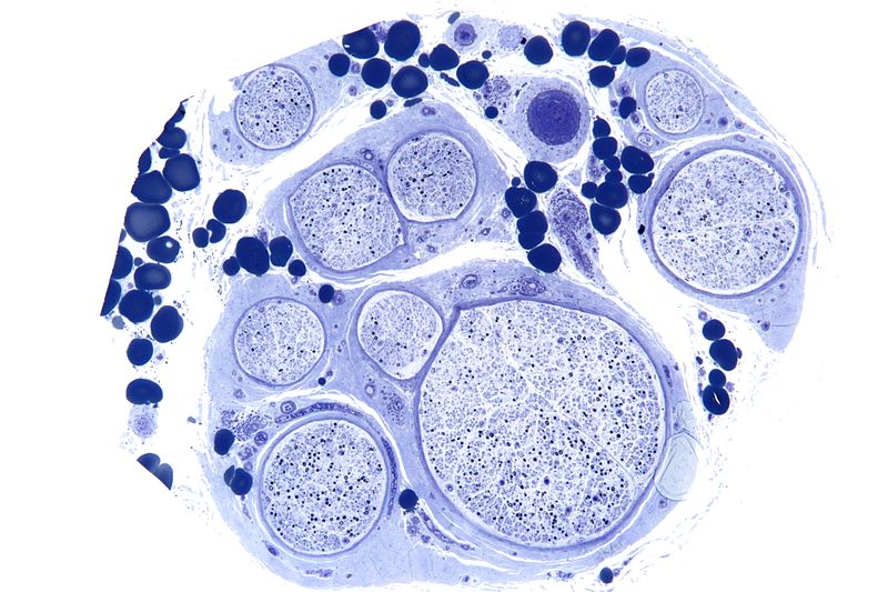
Low magnification micrograph of a vasculitic neuropathy. Credit: Wikimedia User Nephron
Without the myelin sheath, nerve fibers experience a plethora of problems centered around impaired signal transmission: cognition, sensation, movement, and more. When Gladstone researchers realized that it was the blood-clotting protein fibrinogen that was causing the repair problems, they set out to revolutionize the way scientists approach treating neurological disorders.
Fibrinogen, made in the liver, is one of 13 coagulation factors that promote blood clotting. In the right context, blood clotting can save lives by preventing excessive bleeding. In the wrong context, blood clotting can prevent the flow of blood, carrying oxygen and nutrients, to important bodily organs like the brain and heart.
They found that fibrinogen “leaks” into the central nervous system and disrupts the way nerve cells produce myelin, preventing repair. Normally, adult stem cells travel to damaged nerve sites and transform into new myelin-producing cells. However, fibrinogen prevents adult stem cells from making the reparative transition into myelin-producing cells.
"We thought it might be important to look instead at the toxic environment outside the cell, where blood proteins accumulate," explained senior investigator Katerina Akassoglou, PhD. "We realized that targeting the blood protein fibrinogen could open up the possibility for new types of therapies to promote brain repair."
Now that they know what’s stopping repair, scientists are hopeful that they can find a way to better approach treating neurological diseases like MS and Alzheimer’s.
“Repairing myelin by eliminating the toxic effects of vascular damage in the brain is a new frontier in disease therapeutics," said Gladstone’s Lennart Mucke, MD. "This study could change the way we think about how to repair the brain."
Sources: Gladstone Institutes, Healthline

Wednesday, December 14, 2016

Coagulation and fibrinolytic activity of tenecteplase and alteplase in acute ischemic stroke

For your doctor to explain because our fucking failures of stroke associations do not translate stroke research into layperson understandable terms.
 https://www.mdlinx.com/internal-medicine/medical-news-article/2015/12/09/alteplase-cerebral-hemorrhage-factor-v-prothrombin/6428892/?
Stroke, 12/09/2015
The authors compared the fibrinolytic activity of tenecteplase and alteplase in patients with acute ischemic stroke, and explored the association between hypofibrinogenaemia and intracerebral hemorrhage. In patients with acute ischemic stroke, alteplase 0.9 mg/kg caused significant disruption of the fibrinolytic system, whereas tenecteplase 0.25 mg/kg did not, consistent with the trend toward lower intracerebral hemorrhage incidence with tenecteplase in the ATTEST study.

Methods

  • Venous blood samples from a subgroup of participants in the Alteplase-Tenecteplase Trial Evaluation for Stroke Thrombolysis (ATTEST) study were obtained at pretreatment, 3 to 12 hours, and 24±3 hours post-intravenous thrombolysis for analyses of plasminogen, plasminogen activator inhibitor-1, d-dimer, factor V, fibrinogen, and fibrin(ogen) degradation products, in addition to routine coagulation assays.
  • Related sample Wilcoxon signed-rank tests were used to test the within-group changes, and independent Mann-Whitney tests for between-group differences.

Results

  • Thirty patients were included (alteplase=14 and tenecteplase=16) with similar baseline demographics.
  • Compared with baseline, alteplase caused significant hypofibrinogenaemia (P=0.002), prolonged prothrombin time (P=0.011), hypoplasminogenaemia (P=0.001), and lower factor V (P=0.002) at 3 to 12 hours after administration with persistent hypofibrinogenaemia at 24 hours (P=0.011), whereas only minor hypoplasminogenaemia (P=0.029) was seen in the tenecteplase group.
  • Tenecteplase consumed less plasminogen (P<0.001) and fibrinogen (P=0.002) compared with alteplase.
Go to PubMed Go to Abstract Print Article Summary Cat 2 CME Report