Changing stroke rehab and research worldwide now.Time is Brain! trillions and trillions of neurons that DIE each day because there are NO effective hyperacute therapies besides tPA(only 12% effective). I have 523 posts on hyperacute therapy, enough for researchers to spend decades proving them out. These are my personal ideas and blog on stroke rehabilitation and stroke research. Do not attempt any of these without checking with your medical provider. Unless you join me in agitating, when you need these therapies they won't be there.

What this blog is for:

My blog is not to help survivors recover, it is to have the 10 million yearly stroke survivors light fires underneath their doctors, stroke hospitals and stroke researchers to get stroke solved. 100% recovery. The stroke medical world is completely failing at that goal, they don't even have it as a goal. Shortly after getting out of the hospital and getting NO information on the process or protocols of stroke rehabilitation and recovery I started searching on the internet and found that no other survivor received useful information. This is an attempt to cover all stroke rehabilitation information that should be readily available to survivors so they can talk with informed knowledge to their medical staff. It lays out what needs to be done to get stroke survivors closer to 100% recovery. It's quite disgusting that this information is not available from every stroke association and doctors group.

Showing posts with label MMP. Show all posts

Sunday, March 1, 2026

NMN improves high-fat-diet-induced myocardial damage of aging mice through Sirt3/PINK1/Parkin signaling pathway

 Didn't your competent? doctor prescribe NMN years ago? NO? So fucking incompetence reigned and your doctor is still employed?

NMN improves high-fat-diet-induced myocardial damage of aging mice through Sirt3/PINK1/Parkin signaling pathway


https://doi.org/10.1016/j.tjnut.2026.101435Get rights and content

Abstract

Background

Long-term high-fat diet (HFD) consumption is associated with the development of metabolic cardiomyopathy and contributes to accelerated cardiac aging. Nicotinamide mononucleotide (NMN), a key NAD+ precursor, has shown promise in ameliorating age-related cardiac decline, but its mechanisms are not fully understood.

Methods

14-month-old male C57BL/6J mice were divided into three groups: normal-diet (ND) group, high-fat diet (HFD) group and HFD+NMN group. NMN was added to their drinking water at a dose of 400 mg/kg for 7 months. Cardiac tissues were collected and analyzed using hematoxylin and eosin (H&E) and Masson's trichrome staining, Western blotting, Quantitative Real-Time PCR, and immunohistochemistry. In vitro, H9c2 cardiomyocytes were exposed to palmitic acid (PA) to establish a lipotoxicity model, and the effects of NMN on cell viability and autophagy flux were assessed via MTT assay and mRFP-GFP-LC3 adenoviral transfection.

Results

Compared to HFD group, NMN treatment significantly reduced the heart index, ameliorated myocardial fibrosis, and decreased the expression levels of senescence-associated secretory phenotype (SASP) markers (Serpine1, MMP3, CXCL-1, CXCL-10, P16; P<0.05), as well as senescence markers (P21 and β-gal; P<0.01), pro-inflammatory cytokines (IL-1β and TNF-α; P<0.05) and apoptotic indicators (Bax/Bcl-2 ratio, cleaved caspase-3; P<0.01). Conversely, NMN treatment upregulated the levels of anti-inflammatory factor IL-10 (P<0.01) in the cardiac tissue. Furthermore, NMN treatment increased protein levels of Sirt3, PINK1 and Parkin (P<0.01), and enhanced autophagy-lysosomal markers including LC3-II/LC3-I ratio and TFEB (P<0.05), alongside decreased p62 level (P<0.01) in cardiac tissue. In H9c2 cardiomyocytes, NMN treatment significantly attenuated PA-induced cytotoxicity (P<0.01) and enhanced PA-induced the impaired autophagy flux. Notably, these protective effects were abolished by co-treatment with 3-TYP, a selective Sirt3 inhibitor.

Conclusion

NMN alleviates HFD-induced myocardial damage of aging mice by activating the Sirt3/PINK1/Parkin signaling pathway, enhancing autophagy-lysosomal function. These findings indicate that NMN is a promising therapeutic candidate for the treatment of metabolic cardiomyopathy.

Monday, March 23, 2020

Neuroinflammation: friend and foe for ischemic stroke

So it is more difficult to solve that you expected. WHAT THE FUCK IS YOUR STRATEGY TO SOLVE IT? I'm guessing burying your head in the sand because

you are waiting for SOMEONE ELSE TO SOLVE THE PROBLEM. That behavior would get you fired in my book.

Neuroinflammation: friend and foe for ischemic stroke


Abstract

Stroke, the third leading cause of death and disability worldwide, is undergoing a change in perspective with the emergence of new ideas on neurodegeneration. The concept that stroke is a disorder solely of blood vessels has been expanded to include the effects of a detrimental interaction between glia, neurons, vascular cells, and matrix components, which is collectively referred to as the neurovascular unit. Following the acute stroke, the majority of which are ischemic, there is secondary neuroinflammation that both promotes further injury, resulting in cell death, but conversely plays a beneficial role, by promoting recovery. The proinflammatory signals from immune mediators rapidly activate resident cells and influence infiltration of a wide range of inflammatory cells (neutrophils, monocytes/macrophages, different subtypes of T cells, and other inflammatory cells) into the ischemic region exacerbating brain damage. In this review, we discuss how neuroinflammation has both beneficial as well as detrimental roles and recent therapeutic strategies to combat pathological responses. Here, we also focus on time-dependent entry of immune cells to the ischemic area and the impact of other pathological mediators, including oxidative stress, excitotoxicity, matrix metalloproteinases (MMPs), high-mobility group box 1 (HMGB1), arachidonic acid metabolites, mitogen-activated protein kinase (MAPK), and post-translational modifications that could potentially perpetuate ischemic brain damage after the acute injury. Understanding the time-dependent role of inflammatory factors could help in developing new diagnostic, prognostic, and therapeutic neuroprotective strategies for post-stroke inflammation.(What is your strategy for creating these solutions?)


Friday, September 23, 2016

Mechanism of Action and Clinical Potential of Fingolimod for the Treatment of Stroke

Fingolimod already has this positive research needing more followup: Already approved for MS, so your doctor being an innovative sort will likely use this as an off-label use for stroke. That will never occur, you will just need to deal with the fact your doctor is doing nothing in the first week to save all your dying neurons.(neuronal cascade of death)

FTY720 Preserves Blood-Brain Barrier Integrity Following Subarachnoid Hemorrhage in Rats

The latest here:

Mechanism of Action and Clinical Potential of Fingolimod for the Treatment of Stroke

imageWentao Li1, imageHaoliang Xu2 and imageFernando D. Testai1*
  • 1Department of Neurology and Rehabilitation, University of Illinois College of Medicine, Chicago, IL, USA
  • 2Department of Pathology, University of Illinois College of Medicine, Chicago, IL, USA
Fingolimod (FTY720) is an orally bio-available immunomodulatory drug currently approved by the FDA for the treatment of multiple sclerosis. Currently, there is a significant interest in the potential benefits of FTY720 on stroke outcomes. FTY720 and the sphingolipid signaling pathway it modulates has a ubiquitous presence in the central nervous system and both rodent models and pilot clinical trials seem to indicate that the drug may improve overall functional recovery in different stroke subtypes. Although the precise mechanisms behind these beneficial effects are yet unclear, there is evidence that FTY720 has a role in regulating cerebrovascular responses, blood–brain barrier permeability, and cell survival in the event of cerebrovascular insult. In this article, we critically review the data obtained from the latest laboratory findings and clinical trials involving both ischemic and hemorrhagic stroke, and attempt to form a cohesive picture of FTY720’s mechanisms of action in stroke.

Introduction

Fingolimod (FTY720) is an orally bio-available immunomodulatory drug unique for its reversible leukocyte sequestration properties. In 2010, it was approved by the FDA for the treatment of multiple sclerosis (MS) (1, 2).
Given the current understanding of the role of the immune system in the pathophysiology of brain injury in cerebrovascular diseases, there is now significant interest in the potential benefits of FTY720 on stroke outcomes. Several groups have independently evaluated its effects in rodent models of brain ischemia and intracerebral hemorrhage (ICH). More recently, pilot clinical trials have been conducted demonstrating promising results, albeit in small populations (3–5). Taken together, these studies seem to indicate that administration of FTY720 results in overall improved functional recovery in different stroke subtypes.
The precise mechanisms behind these beneficial effects are still under investigation. FTY720 is a partial sphingosine-1-phosphate (S1P) agonist with immunomodulatory properties that regulates cerebrovascular responses, blood–brain barrier (BBB) permeability, and central nervous system (CNS) cell survival (6, 7). In this article, we will organize these elements and attempt to form a cohesive picture of FTY720’s mechanisms of action in stroke, and critically review the data obtained from recent clinical trials.

Role of the Immune System in Stroke Progression

Immunomodulation is a well-characterized effect of FTY720 and is thought to mediate some of the beneficial effects seen in stroke models. In order to understand the extent to which the immune system impacts the evolution of stroke outcomes, we will sketch a proposed model of the immune cascade following ischemic insult.
In the immediate aftermath of an ischemic event, complement activation, clot formation, and oxidative stress result in direct damage to local vasculature. Endothelial cells die and detach, interrupting the integrity of the BBB and exposing sub-endothelial antigens. Immune cells adhere to the vessel wall and upregulate the expression of chemoattractant and adhesion molecules that lead to the infiltration of the brain parenchyma by the innate immune system.
A combination of neutrophils, monocytes, and macrophages, this innate immune system further contributes to vascular compromise and early inflammation. One well-documented process is through the release of matrix metalloproteinases (MMPs) by the immune cells, particularly MMP-9, which contributes to the breakdown of the BBB with the resultant edema and growth of the infarcted area (8).
In the parenchyma, glial cells are also activated by the inflammation and damage-associated molecular patterns (DAMPs) released from dying neurons. These reactive astrocytes and microglia further stimulate the recruitment of leukocytes, which release their own pro-inflammatory chemokines, perpetuating a cycle of vascular damage, inflammation, and cell death (9).
The second, adaptive phase of the immune response is mediated predominantly by effector T cells, which are stimulated by DAMPS and brain-specific antigens released upon neuronal cell death (10). These T cells mobilize to the injured regions of the brain, infiltrating a compromised BBB to release pro-inflammatory cytokines, including IFN-γ, resulting in a delayed neurotoxic effect (11, 12). Of note, brain-specific antigens were identified in cervical nodes and palatine tonsils of animals with cerebral ischemia and stroke survivors. Interestingly, some of these antigens were associated with infarct volume and survival. While these studies need to be replicated in larger cohorts, they suggest the participation of peripheral lymphoid tissue in stroke-associated inflammation and outcomes.
Lastly, the inflammatory process is brought to an end via a combination of B cells and regulatory T cells. The latter acts through IL-10, which in combination with TGF-β produced by local macrophages, suppresses further helper T-cell-induced inflammation, and promotes the regeneration of remaining viable neurons (13, 14).
A reduction in various components of the innate and adaptive immune response have been associated with better stroke outcomes. Clinically, a lower ratio of CD14+ pro-inflammatory monocytes to CD16+ reparative monocytes has been correlated with better acute and long-term functional outcomes (15). Similarly, decreased complement activation, specifically the reduced expression of C3, C4, and C-reactive protein, has been associated with better recovery at 3 months post-stroke (16). Experimentally, inhibition of CD8+ and CD4+ T-cell migration into the CNS and direct disruption of CD8+ cytotoxicity has led to reduced infarct volume and post-ischemic inflammation (17). Disruption of the DAMPs-activated γδT cells have also resulted in decreased infarct size and better functional recovery in mice (18). Finally, direct delivery of B cells and IL-10 to the brain in animal models have resulted in a reduction of inflammatory cytokines produced by effector T cells and a reduction of infarct size (19, 20).

Fingolimod and Stroke-Related Mechanisms of Action

Pharmacology

Isaria sinclairii, otherwise known as “winter-insect and summer-plant,” is a fungus that has been used in traditional Chinese medicine for over 300 years. Classically prescribed as a panacea for multiple ailments, it produces an atypical amino acid myriocin (ISP-1) that blocks the synthesis of sphingolipids. This chemical compound has since been modified into fingolimod, also known as FTY720 (21). In Western Medicine, FTY720 initially showed promise in preventing ischemic–reperfusion injury following organ transplant, but failed clinical trials due to the development of acute macular edema in some patients (22, 23). In 2010, it became the first oral disease modifying drug approved for treatment of MS (6). In its base form, FTY720 is an orally bio-available, lipophilic molecule that readily crosses the BBB and steadily accumulates in the CNS white and gray matter (24). It bears a structural similarity to sphingosine and is reversibly phosphorylated primarily by Sphingosine-Kinase 2 (SphK2) and, to a lesser extent, by SphK1 (25). In its activated form, FTY720-phosphate is a sphingosine-1-phosphate (S1P) analog that binds to cell membrane G-coupled S1P receptors (S1PR) S1PR1, 3, 4, and 5, but not S1P2 (26). With the exception of S1PR4, these receptors are ubiquitously distributed in the CNS (Table 1). In addition to its action on cell surface receptors, FTY720 regulates the synthesis of different bioactive sphingolipids and, together with S1P, regulates gene expression via epigenetic mechanisms (Figure 1). The half-life of FTY720 averages ~9 days and its pharmacology is not sensibly affected by age, weight, sex, or ethnicity (27).

Wednesday, July 27, 2016

Systems Biology of Immunomodulation for Post-Stroke Neuroplasticity: Multimodal Implications of Pharmacotherapy and Neurorehabilitation

Whatever the fuck this means. Written to make sure survivors can't understand. Research like this isn't for survivors anyway, it is for our stroke doctors to implement even though they never read and analyze research for their patients. Someday 1 out of 100,00 doctors will quibble with that gross generalization.
http://journal.frontiersin.org/article/10.3389/fneur.2016.00094/full?utm_source=newsletter&
  • National Brain Research Centre, Gurgaon, India
Aims: Recent studies indicate that anti-inflammatory drugs, act as a double-edged sword, not only exacerbating secondary brain injury but also contributing to neurological recovery after stroke. Our aim is to explore whether there is a beneficial role for neuroprotection and functional recovery using anti-inflammatory drug along with neurorehabilitation therapy using transcranial direct current stimulation (tDCS) and repetitive transcranial magnetic stimulation (rTMS), so as to improve functional recovery after ischemic stroke.
Methods: We develop a computational systems biology approach from preclinical data, using ordinary differential equations, to study the behavior of both phenotypes of microglia, such as M1 type (pro-inflammatory) vis-à-vis M2 type (anti-inflammatory) under anti-inflammatory drug action (minocycline). We explore whether pharmacological treatment along with cerebral stimulation using tDCS and rTMS is beneficial or not. We utilize the systems pathway analysis of minocycline in nuclear factor kappa beta (NF-κB) signaling and neurorehabilitation therapy using tDCS and rTMS that act through brain-derived neurotrophic factor (BDNF) and tropomyosin-related kinase B (TrkB) signaling pathways.
Results: We demarcate the role of neuroinflammation and immunomodulation in post-stroke recovery, under minocycline activated-microglia and neuroprotection together with improved neurogenesis, synaptogenesis, and functional recovery under the action of rTMS or tDCS. We elucidate the feasibility of utilizing rTMS/tDCS to increase neuroprotection across the reperfusion stage during minocycline administration. We delineate that the signaling pathways of minocycline by modulation of inflammatory genes in NF-κB and proteins activated by tDCS and rTMS through BDNF, TrkB, and calmodulin kinase (CaMK) signaling. Utilizing systems biology approach, we show that the activation pathways for pharmacotherapy (minocycline) and neurorehabilitation (rTMS applied to ipsilesional cortex and tDCS) results into increased neuronal and synaptic activity that commonly occur through activation of N-methyl-d-aspartate receptors. We construe that considerable additive neuroprotection effect would be obtained and delayed reperfusion injury can be remedied, if one uses multimodal intervention of minocycline together with tDCS and rTMS.
Conclusion: Additive beneficial effect is, thus, noticed for pharmacotherapy along with neurorehabilitation therapy, by maneuvering the dynamics of immunomodulation using anti-inflammatory drug and cerebral stimulation for augmenting the functional recovery after stroke, which may engender clinical applicability for enhancing plasticity, rehabilitation, and neurorestoration.

Introduction

Recent investigations have reported that immune responses to inflammation are non-specific systemic infections associated with progression of neurodegenerative diseases via activation of macrophages (1). Minocycline is a tetracycline antibiotic having several properties, such as anti-inflammatory, anti-apoptosis, free radical scavenger, and protein misfolding (2). The therapeutic effects of minocycline in preclinical models of neurodegenerative diseases showed direct neuroprotection and reduction of microglial inflammatory responses (3). It has been reported in in vivo studies that minocycline blocks the adhesion of leukocytes to cerebrovascular endothelial cells induced by lipopolysaccharides, as well as tumor necrosis factor-α (TNF-α) production in the brain (4). In vitro studies have reported the anti-inflammatory effects of minocycline for neuroprotection (5) and in macrophages (6). Neuroprotective effects of minocycline include reduction of macrophage activation, prevention of the potentiation of ischemia-like injury to astrocytes and endothelial cells consolidating the brain tissue parenchyma (7). Although, the anti-inflammatory effects of minocycline are known to some extent, the direct effects of neuroprotection have not been well investigated in neurodegenerative diseases.
Several studies have shown that the physiological neuroprotection mechanisms that occur after stroke are targeted through various signaling pathways. Several studies suggest that the mechanisms associated with either reducing the size of infarct or enabling neurorestoration, involve the following entities: (i) anti-high mobility group box-1 activity (8); (ii) NF-κB (9); (iii) mammalian target of rapamycin (mTOR) inhibitor (10, 11); (iv) stimulation of toll-like receptors (TLR2 and TLR4) prior to brain ischemia (12, 13), (v) c-Jun N-terminal kinase (JNK) inhibitor (14); (vi) p38 mitogen-activated protein kinase (p38 MAPK) inhibitor (15); (vii) MEK1 pathway (16); (viii) MAPP/MEK/ERK inhibitor (17); and (ix) Minocycline-induced reduction of LPS-stimulated p38 MAPK activation, and stimulation of the phosphoinositide 3-kinase (PI3K)/Akt pathway (18).
Currently, little is known about endogenous counter regulatory immune mechanisms that can induce neurorestoration. The glycogen synthase kinase-3β (AKT/GSK-3β) pathway has been recognized as a protective pathway against cerebral ischemic injury. In cerebral ischemia models, it has been shown that remote limb conditioning does indeed activate and upregulate the pro-survival AKT pathway (19) and long-term protection against cerebral ischemia is afforded by limb post-conditioning that is associated with AKT, MAPK, phosphatidylinositol 3-kinase (PI3K), and protein kinase C (PKC) signaling pathways (20). NF-κB transcription factor family members, such as p50, p65/RelA in the hippocampus, are regulated by metabotropic glutamate receptor signaling and c-Rel transcription factor is responsible for the formation and maintenance of long-term memory (21). Minocycline directly inhibits matrix metalloproteinase (MMP)-9 activation through NF-κB pathway (22). In silico modeling of anti-inflammatory response has been reported for endotoxins (LPS) and corticosteroids by activating TLRs in NF-κB (23).
Taken together, the modulation of cell survival and death signaling by hypoxic/ischemic preconditioning appears to be capable of targeting multiple levels of signaling cascades. Several inhibitors targeted the point of convergence through distinct and interacting signaling pathways (crosstalk mechanism) for inflammation by activating macrophages that lead to neuroprotection. Also, cerebral stimulation-based transcranial magnetic stimulation and direct current stimulation enhances brain-derived neurotrophic factor (BDNF) and tropomyosin-related kinase B (TrkB) signaling (24, 25). In this study, we harness the convergent signaling pathways of pharmacotherapy (anti-inflammatory, immunomodulatory) and neurorehabilitation therapy (functional recovery) for efficient post-stroke neurorestoration by experimental and systems-level approach. We modeled using the systems biology approach of minocycline modulation of MMPs through NF-κB signaling pathway, a master regulator of inflammatory responses along with neurorehabilitation-based activation in BDNF and TrkB signaling.

More at link.


Thursday, October 1, 2015

Disruption of brain-blood barrier might influence progression of Alzheimer’s

This probably means that this one cause of the neuronal cascade of death should be solved first.
Inflammatory action leaking through the blood brain barrier.
 But with NO strategy survivors are fucking screwed and our doctors and hospitals are laying down on the job. What the fuck is your doctor doing about MMP to alleviate this problem. ANYTHING AT ALL?
I've written 4 posts on MMP, one post on MMP-14, and 9 posts on MMP-9.
http://www.alphagalileo.org/ViewItem.aspx?ItemId=156807&CultureCode=en
VIB - Flanders Interuniversity Institute for Biotechnology
More and more data from preclinical and clinical studies strengthen the hypothesis that immune system-mediated actions contribute to and drive pathogenesis in Alzheimer’s disease. The team of Roosmarijn Vandenbroucke in the Claude Libert Group (VIB/UGent) combined their knowledge and expertise related to inflammation with the expertise in Alzheimer’s disease present in the Bart De Strooper Group (VIB/KU Leuven). This collaboration lead to the insights that Aβ indeed induces a strong inflammatory response, thereby destroying an important but often neglected brain barrier, called the blood-cerebrospinal fluid (CSF) barrier. Disruption of this blood-CSF barrier disturbs brain homeostasis and might negatively affect disease progression. Strikingly, these effects could be blocked in the presence of a matrix metalloproteinase (MMP) inhibitor.
Roosmarijn Vandenbroucke: “Although further research is needed, these data suggest that blocking MMP activity or upstream inflammatory signalling, might have therapeutic potential to treat Alzheimer’s disease. It is important we could demonstrate the role of the blood-cerebrospinal fluid barrier, because this would be an easier target to reach in comparison with the targets of current therapies.”
The publication of Vandenbroucke et al. was picked up by Alzforum.org who combined it together with another publication about the Blood-Brain Barrier:
Barriers Between Blood and CSF, Brain Yield to Aβ—Not a Bad Thing?
The barrier between the blood and central nervous system crumbles in Alzheimer’s disease, but researchers have known little about how this happens, or what it does to brain pathology. Two new papers shed some light on how Aβ damages the cells that protect the brain parenchyma and cerebrospinal fluid. The studies examine different systems and describe distinct mechanisms, but both add to the picture of what may happen in disease.
http://www.vib.be/en/news/Pages/Disruption-of-brain-blood-barrier-might-influence-progression-of-Alzheimer%E2%80%99s.aspx

Thursday, July 2, 2015

Unbalanced metalloproteinase-9 and tissue inhibitors of metalloproteinases ratios predict hemorrhagic transformation of lesion in ischemic stroke patients treated with thrombolysis: results from the MAGIC study

Way out of my league, so you'll have to contact a genius somewhere to decipher this.
http://journal.frontiersin.org/article/10.3389/fneur.2015.00121/full?
imageBenedetta Piccardi1*, imageVanessa Palumbo2, imageMascia Nesi2, imagePatrizia Nencini2, imageAnna Maria Gori3, imageBetti Giusti3, imageGiovanni Pracucci1, imagePaolina Tonelli1, imageEleonora Innocenti1, imageAlice Sereni3, imageElena Sticchi3, imageDanilo Toni4, imagePaolo Bovi5, imageMario Guidotti6, imageMaria Rosaria Tola7, imageDomenico Consoli8, imageGiuseppe Micieli9, imageRossana Tassi10, imageGiovanni Orlandi11, imageFrancesco Perini12, imageNorina Marcello13, imageAntonia Nucera14, imageFrancesca Massaro15, imageMaria Luisa DeLodovici16, imageGiorgio Bono16, imageMaria Sessa17, imageRosanna Abbate3 and imageDomenico Inzitari1,18, On behalf of the MAGIC Study Group
  • 1Neuroscience Section, Department of Neurofarba, University of Florence, Florence, Italy
  • 2Stroke Unit, Department of Neurology, Careggi University Hospital, Florence, Italy
  • 3Department of Experimental and Clinical Medicine, Atherothrombotic Diseases Center, AOU Careggi, University of Florence, Florence, Italy
  • 4Emergency Department Stroke Unit, Department of Neurological Sciences, Sapienza University of Rome, Rome, Italy
  • 5SSO Stroke Unit, U.O. Neurologia d.O., DAI di Neuroscienze, Azienda Ospedaliera Integrata, Verona, Italy
  • 6Neurology Unit, Valduce General Hospital, Como, Italy
  • 7U.O. Neurologia, DAI Neuroscienze-Riabilitazione, Azienda Ospedaliera-Universitaria S. Anna, Ferrara, Italy
  • 8U.O. Neurologia, G. Jazzolino Hospital, Vibo Valentia, Italy
  • 9Istituto Neurologico Nazionale C. Mondino, Pavia, Italy
  • 10U.O.C. Stroke Unit, Dipartimento di Scienze Neurologiche e Neurosensoriali, Azienda Ospedaliera Universitaria Senese, Siena, Italy
  • 11Department of Neurosciences, Neurological Clinic, University of Pisa, Pisa, Italy
  • 12UOC di Neurologia e “Stroke Unit”, Ospedale San Bortolo, Vicenza, Italy
  • 13Neurology Unit, Arcispedale Santa Maria Nuova, Reggio Emilia, Italy
  • 14Department of Clinical Neurological Sciences, London Health Sciences Centre, Western University, London, ON, Canada
  • 15Neurology Unit, Misericordia e Dolce Hospital, Prato, Italy
  • 16Stroke Unit, Department of Neurology, Ospedale di Circolo e Fondazione Macchi, Varese, Italy
  • 17Department of Neurology, Istituti Ospitalieri, Cremona, Italy
  • 18Institute of Neuroscience, Italian National Research Council, Florence, Italy
Background: Experimentally, metalloproteinases (MMPs) play a detrimental role related to the severity of ischemic brain lesions. Both MMPs activity and function in tissues reflect the balance between MMPs and tissue inhibitors of metalloproteinases (TIMPs). We aimed to evaluate the role of MMPs/TIMPs balance in the setting of rtPA-treated stroke patients.
Methods: Blood was taken before and 24-h after rtPA from 327 patients (mean age 68 years, median NIHSS 11) with acute ischemic stroke. Delta median values of each MMP/TIMP ratio [(post rtPA MMP/TIMP-baseline MMP/TIMP)/(baseline MMP/TIMP)] were analyzed related to symptomatic intracranial hemorrhage (sICH) according to NINDS criteria, relevant hemorrhagic transformation (HT) defined as confluent petechiae within the infarcted area or any parenchymal hemorrhage, stroke subtypes (according to Oxfordshire Community Stroke Project) and 3-month death. The net effect of each MMP/TIMP ratio was estimated by a logistic regression model including major clinical determinants of outcomes
Results: Adjusting for major clinical determinants, only increase in MMP9/TIMP1 and MMP9/TIMP2 ratios remained significantly associated with sICH (odds ratio [95% confidence interval], 1.67 [1.17–2.38], p = 0.005; 1.74 [1.21–2.49], p = 0.003, respectively). Only relative increase in MMP9/TIMP1 ratio proved significantly associated with relevant HT (odds ratio [95% confidence interval], 1.74 [1.17–2.57], p = 0.006) with a trend toward significance for MMP9/TIMP2 ratio (p = 0.007).
Discussion: Our data add substantial clinical evidence about the role of MMPs/TIMPs balance in rtPA-treated stroke patients. These results may serve to generate hypotheses on MMPs inhibitors to be administered together with rtPA in order to counteract its deleterious effect.

Introduction

Matrix metalloproteinases (MMPs) are a family of zinc-dependent endopeptidases that are involved in extracellular matrix (ECM) degradation (1). The turnover of ECM is regulated by the balance between MMPs and a group of endogenous proteins called tissue inhibitor of metalloproteinases (TIMPs) (2). Active MMPs and some MMP proenzymes form 1:1 complexes with TIMPs and the unbalance between these two families of molecules appears implicated in a variety of diseases (3). A list of MMPs and TIMPs with their putative role in acute ischemic stroke is shown in Table S1 in Supplementary Material.
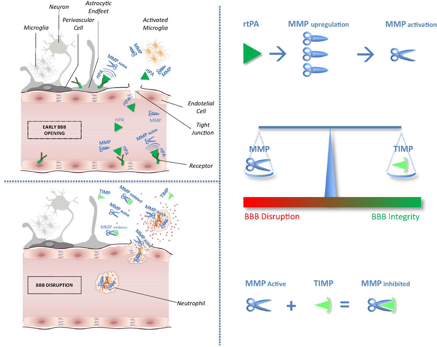
After cerebral ischemia, the general neuronal response to excitotoxic injury determines the release of pro-inflammatory cytokines that stimulate the local production of MMPs and TIMPs (4). In experimental models of brain ischemia, MMPs and MMP/TIMP unbalance play a detrimental role related to blood–brain barrier (BBB) disruption leading to hemorrhagic transformation and edema of an ischemic brain lesion (5). Circulating levels of MMP9 have been proved associated with poor outcomes in stroke patients treated with tissue plasminogen activator (rtPA) (6, 7). Furthermore, recent studies suggest that rtPA adverse effects may be mediated through MMPs upregulation and activation (2). No clinical study has hitherto considered selectively the effect of the balance between MMPs and their physiological inhibitor related to stroke outcomes after thrombolysis. Theoretical effects of rtPA on MMP/TIMP unbalance have been shown in Figure 1.
FIGURE 1
www.frontiersin.org Figure 1. Impact of tissue plasminogen activator on MMP/TIMP unbalance at the neurovascular unit level. After acute ischemic stroke, rtPA may cross blood–brain barrier (BBB), enter the brain parenchyma, and thereby damage neurovascular unit components by promoting metalloproteinase (MMPs) production and activation. Indeed, unbalance between MMPs and their natural inhibitors (tissue inhibitors of metalloproteinases, TIMPs) may exacerbate BBB disruption leading to hemorrhagic transformation and edema of an ischemic brain lesion.
The aim of this study was to evaluate the effect of MMPs/TIMPs ratio on outcomes of ischemic stroke in the same cohort of the biological markers associated with acute ischemic stroke (MAGIC) study. Because MMP inhibition is considered a possible therapeutic target for stroke patients (8), a clearer understanding of MMP/TIMP interplay, compared with the effect of MMPs only, would have important implications for acute stroke therapies.

More at link.

Saturday, May 30, 2015

Sequential Therapy with Minocycline and Candesartan Improves Long-Term Recovery After Experimental Stroke

Is this enough to start up human clinical trials? Ask your doctor and if your doctor doesn't do clinical trials you need to call the hospital president and ask why the stroke department head isn't solving stroke problems by doing clinical research.
http://link.springer.com/article/10.1007/s12975-015-0408-8

Sahar Soliman,


$39.95 / €34.95 / £29.95 *
Rent the article at a discount
Rent now
* Final gross prices may vary according to local VAT.
Get Access

Abstract

Minocycline and candesartan have both shown promise as candidate therapeutics in ischemic stroke, with multiple, and somewhat contrasting, molecular mechanisms. Minocycline is an anti-inflammatory, antioxidant, and anti-apoptotic agent and a known inhibitor of matrix metalloproteinases (MMPs). Yet, minocycline exerts antiangiogenic effects both in vivo and in vitro. Candesartan promotes angiogenesis and activates MMPs. Aligning these therapies with the dynamic processes of injury and repair after ischemia is likely to improve success of treatment. In this study, we hypothesize that opposing actions of minocycline and candesartan on angiogenesis, when administered simultaneously, will reduce the benefit of candesartan treatment. Therefore, we propose a sequential combination treatment regimen to yield a better outcome and preserve the proangiogenic potential of candesartan. In vitro angiogenesis was assessed using human brain endothelial cells. In vivo, Wistar rats subjected to 90-min middle cerebral artery occlusion (MCAO) were randomized into four groups: saline, candesartan, minocycline, and sequential combination of minocycline and candesartan. Neurobehavioral tests were performed 1, 3, 7, and 14 days after stroke. Brain tissue was collected on day 14 for assessment of infarct size and vascular density. Minocycline, when added simultaneously, decreased the proangiogenic effect of candesartan treatment in vitro. Sequential treatment, however, preserved the proangiogenic potential of candesartan both in vivo and in vitro, improved neurobehavioral outcome, and reduced infarct size. Sequential combination therapy with minocycline and candesartan improves long-term recovery and maintains candesartan’s proangiogenic potential.