Changing stroke rehab and research worldwide now.Time is Brain! trillions and trillions of neurons that DIE each day because there are NO effective hyperacute therapies besides tPA(only 12% effective). I have 523 posts on hyperacute therapy, enough for researchers to spend decades proving them out. These are my personal ideas and blog on stroke rehabilitation and stroke research. Do not attempt any of these without checking with your medical provider. Unless you join me in agitating, when you need these therapies they won't be there.

What this blog is for:

My blog is not to help survivors recover, it is to have the 10 million yearly stroke survivors light fires underneath their doctors, stroke hospitals and stroke researchers to get stroke solved. 100% recovery. The stroke medical world is completely failing at that goal, they don't even have it as a goal. Shortly after getting out of the hospital and getting NO information on the process or protocols of stroke rehabilitation and recovery I started searching on the internet and found that no other survivor received useful information. This is an attempt to cover all stroke rehabilitation information that should be readily available to survivors so they can talk with informed knowledge to their medical staff. It lays out what needs to be done to get stroke survivors closer to 100% recovery. It's quite disgusting that this information is not available from every stroke association and doctors group.

Showing posts with label blood vessels. Show all posts

Tuesday, April 4, 2023

Blood vessels – not just brain cells – can affect brain health, new report finds

Since blood vessels and stroke has been well studied, ask your doctor how that knowledge is getting you 100% recovered. No excuses allowed, your doctor if competent, should know everything about this.

Blood vessels – not just brain cells – can affect brain health, new report finds

By Laura Williamson, American Heart Association News
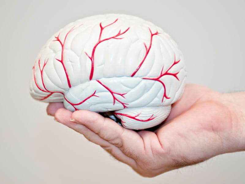
JodiJacobson/E+ via Getty Images
(JodiJacobson/E+ via Getty Images)

Lea en español

Brain cells have a complex relationship with the blood vessels around them that can greatly affect the vessels' ability to function, according to a new science report from the American Heart Association.

The scientific statement, published Monday in the AHA journal Stroke, highlights new and emerging concepts on how blood vessels affect overall brain health, including cognitive function, and where more investigation is needed.

"Brain blood vessels do a lot more than deliver blood to the brain," said writing committee chair Dr. Costantino Iadecola, director and chair of the Feil Family Brain and Mind Research Institute at Weill Cornell Medicine in New York City.

The statement encourages greater collaboration among researchers and health care professionals specializing in cardiovascular diseases and brain health, Iadecola said. As the population ages, the need to better understand brain health has grown.

"There are age-related brain problems that cannot be resolved within a singular specialty," he said. "What we need is a wide variety of specialties working in concert."

The link between blood vessel health and some brain problems, such as strokes, has been well-studied. But the association between blood vessels, Alzheimer's-related dementia and other neurodegenerative brain disorders needs further exploration, Iadecola said.

More than half a million U.S. adults are diagnosed with dementia each year, and roughly 1.5% of the population lives with the condition. While dementia is considered one of the world's biggest health problems, most research has focused on the buildup of proteins inside and between brain cells that leads to Alzheimer's disease. A growing body of research, highlighted in the new report, suggests that blood vessels also play an important role in Alzheimer's disease and dementia.

"When you think about brain health, most people think about neurons (brain cells) and how they function," said statement co-author Dr. Anusha Mishra, an assistant professor in the department of neurology at Oregon Health & Science University in Portland. "But we don't think about how those neurons are supported."

The brain needs a lot of energy to work properly. It gets that energy from oxygen and glucose delivered by blood vessels that are densely packed into brain material. But blood vessels do a lot more, the statement notes. They also help remove harmful byproducts generated by brain activity and regulate immune cell traffic in and out of the brain. When damaged, blood vessels contribute to problems with cognitive function and brain diseases.

Having a better understanding of the role blood vessels play could provide better opportunities for the prevention, recognition and treatment of brain disorders, the statement concludes.

Numerous studies have established a strong relationship between heart and brain health, finding they share many of the same risk factors, including high blood pressure. Iadecola said the new statement expands on the AHA's focus on that relationship by digging deeper into how vascular health affects brain health.

For example, stiffening of the vessels that bring blood from the heart to the brain can cause strokes, he said. But studies also show an association between hardened arteries and Alzheimer's disease. Problems with blood flow and leakage into the brain from the bloodstream also appear to occur before the appearance of Alzheimer's symptoms. But just how they're contributing to the development of Alzheimer's or dementia remains unclear.

Another area that needs further study is the role blood vessels play in helping to clear the brain of the excessive proteins that contribute to Alzheimer's disease and other types of dementia, Mishra said. Future research should ask, "What are the driving forces that are clearing these proteins, and what is the role of the vascular system in that clearance?"

"Understanding what's not going right is an important window into how dementia occurs that we are just starting to scratch the surface of," Mishra said.

The statement also addresses how blood vessels in the meninges, three layers of membranes that cover and protect the brain and spinal cord, may be affecting cognitive function. The blood-brain barrier, formed by the cells lining brain blood vessels, prevents harmful substances from entering the brain through the bloodstream, while allowing needed nutrients and oxygen to cross over. Iadecola said researchers now understand that blood vessels in the meninges, which do not have the barrier, are an important avenue through which some cells enter the brain. These cells, immune cells in particular, patrol the brain and report back to the rest of the body's immune system.

"There's traffic of immune cells in and out of the brain," Iadecola said. "This becomes relevant when the trafficking is altered and, as a result, the brain may suffer."

Iadecola said he hopes the statement will help increase awareness of the "broad array of functions" that blood vessels have on the brain.

"All of these things become more and more important as people age," he said. "Our job as neurovascular clinicians and scientists is to bring this knowledge to the general medical community."


What’s it like inside of a blood vessel? Find out with virtual reality!

 You can use this to ask your doctor to describe EXACTLY what caused your stroke and how to prevent the next one.

What’s it like inside of a blood vessel? Find out with virtual reality!

Experience immersive learning of human physiology with virtual reality 

We are excited to introduce a new virtual reality (VR) experience that is now available in our course, Introductory Human Physiology. This VR experience is accessible on a VR headset or via desktop and is designed to provide you with an immersive and engaging way to explore the human body. By using VR, you will experience what it’s like to be inside a blood vessel and how to take someone’s blood pressure. 

Introductory Human Physiology is a rigorous course for aspiring healthcare practitioners and students alike, as well as a useful refresher for healthcare professionals. Whether you are a professional, student, or curious learner, we hope this new VR experience will deepen your understanding of the subject matter as well as stimulate your curiosity to learn in new ways with emerging learning technologies like VR. 

To access this new VR experience, simply log into Coursera, enroll in Introductory Human Physiology and look for the course item with “Immersive Experience” in the title in Week 5 of the course. We believe that this new VR experience will greatly enhance your learning experience and we encourage you to give it a try.

Thank you for being a Duke learner on Coursera, and we hope you enjoy this new VR experience! Also, we encourage you to check out other digital learning opportunities with Duke University!


Wednesday, May 15, 2019

Brain Health - a small matter of the blood vessels

44 minutes that maybe your doctor might want to peruse.

Brain Health - a small matter of the blood vessels

Edinburgh Neuroscience Christmas Lecture 2018: Brain Health - a small matter of the blood vessels


Thursday, January 5, 2017

Innovative technique to examine blood vessels in 3D help unlock secrets of the brain

With this we should be able to watch and see if interventions to stop pericytes from strangling capillaries post-stroke actually work. Or even just to see how long the strangling occurs.  But with NO strategy and NO leadership, solving this part of the neuronal cascade of death will never occur.
http://www.alphagalileo.org/ViewItem.aspx?ItemId=171345&CultureCode=en

  • Blood vessels examined in 3D allow scientists to examine circulation in the brain giving greater understanding of how dementia, brain cancer and stroke may affect veins and capillaries in this important organ.
  • Technique could identify early warning signs of diseases helping to save lives.
  • Scientists previously unable to study minute blood vessels which could unlock secrets of circulation and how diseases form in brain.
A study published today in the Journal of Anatomy has made an important breakthrough in the examination of blood vessels in the brain giving scientists a clearer understanding of how dementia, brain cancer and stroke can affect veins and capillaries in this organ.
Working collaboratively researchers from the University of Surrey and the Federal University of Sao Paulo developed an innovative technique to examine and quantify blood vessels in the brain using 3D Image Analysis (Stereology) procedures.
Using experimental animal models, this technique will allow scientists to study how such diseases develop in the brain and help them identify, through examination of blood vessels, potential warning signs of illnesses before symptoms appear. These learnings can potentially be translated into humans and help reduce the number of deaths from these illnesses.
The procedure can also be used in post mortems and biopsies examinations of animal and human tissue making it easier for pathologists to determine causes of death and quickly identify alterations in the brain circulation (such as clots) or tumors.
The inexpensive technique of dissolving China Ink with gelatin creates a solution making blood vessels more visible with the use of a confocal microscope. This enables scientists and pathologists to make an accurate reading of their number, length, surface area and create 3D images which can help identify changes in their shape and size, key indicators of a number of circulation-related diseases of the brain.
This innovative method will also facilitate a greater understanding of how exercise affects the brain. Scientists will now be able to examine circulatory effects of increased or decreased heart rate, arterial pressure on the brain and the creation of new vessels (angiogenesis).
Co-author of the study Dr Augusto Coppi from the University of Surrey said: “The brain is a fascinating organ but our full understanding of its circulation is lacking. Previously we have been unable to fully sample and perform a quantification of the circulation of the brain in 3D as we simply could not see all vessels due to their minute size and sometimes due to their irregular spatial distribution.
“This new technique will allow us to sample, image and count blood vessels in 3D giving us a greater mechanistic comprehension of how the circulation of the brain works and how brain diseases such as dementia and stroke affect this organ. With an estimated 850,000 people diagnosed with dementia in England, this technique marks a significant breakthrough in the fight against this disease.”

Thursday, October 15, 2015

Vascular diseases await translation of blood vessels engineered from stem cells

We need this to support new neurons from neurogenesis and from whenever we get neuronal stem cells implanted.
http://stm.sciencemag.org/content/7/309/309rv6.abstract?
Science Translational Medicine  14 Oct 2015:
Vol. 7, Issue 309, pp. 309rv6
DOI: 10.1126/scitranslmed.aaa1805
You are currently viewing the abstract.
View Full Text

Abstract

The discovery of human induced pluripotent stem cells (hiPSCs) might pave the way toward a long-sought solution for obtaining sufficient numbers of autologous cells for tissue engineering. Several methods exist for generating endothelial cells or perivascular cells from hiPSCs in vitro for use in the building of vascular tissue. We discuss current developments in the generation of vascular progenitor cells from hiPSCs and the assessment of their functional capacity in vivo, opportunities and challenges for the clinical translation of engineered vascular tissue, and modeling of vascular diseases using hiPSC-derived vascular progenitor cells.

Thursday, May 7, 2015

New material for creating artificial blood vessels

Since we need new blood vessels to support either neurogenesis or stem cells this might become useful to our recovery. Ask your stroke association to sponsor research to figure out how to solve this for helping survivors.
http://www.alphagalileo.org/ViewItem.aspx?ItemId=152126&CultureCode=en
Blocked blood vessels can quickly become dangerous. It is often necessary to replace a blood vessel – either by another vessel taken from the body or even by artificial vascular prostheses. Together, Vienna University of Technology and Vienna Medical University have developed artificial blood vessels made from a special elastomer material, which has excellent mechanical properties. Over time, these artificial blood vessels are replaced by endogenous material. At the end of this restorative process, a natural, fully functional vessel is once again in place. The method has already been used successfully in rats.
Arteriosclerotic vascular disorders are one of the most common causes of death in industrialized countries. In this situation a bypass operation is often the only solution. Normally, blood vessels are taken from another part of the patient's body and used to replace the damaged vessel. Thanks to a joint project undertaken by TU Wien and the Medical University of Vienna, artificially manufactured vessels should be used more frequently in future.
The most important thing is to find a suitable material. The artificial materials that have been used so far are not ideally compatible with body tissue. The blood vessel can easily become blocked, especially if it is only small in diameter. 
TUW has therefore developed new polymers. "These are so-called thermoplastic polyurethanes," explains Robert Liska from the Institute of Applied Synthetic Chemistry of Vienna University of Technology. "By selecting very specific molecular building blocks we have succeeded in synthesizing a polymer with the desired properties."
A thin polymer thread spun into tubes
To produce the vascular prostheses, polymer solutions were spun in an electrical field to form very fine threads and wound onto a spool. "The wall of these artificial blood vessels is very similar to that of natural ones," says Heinz Schima of the Medical University of Vienna. The polymer fabric is slightly porous and so, initially, allows a small amount of blood to permeate through and this enriches the wall with growth factors. This encourages the migration of endogenous cells. The interaction between material and blood was studied by Martina Marchetti-Deschmann at TU Wien using spatially resolved mass spectrometry.
The new method has already proved very successful in experiments with rats. "The rats' blood vessels were examined six months after insertion of the vascular prostheses," says Helga Bergmeister of MedUni Vienna. “We did not find any aneurysms, thromboses or inflammation. Endogenous cells had colonized the vascular prostheses and turned the artificial constructs into natural body tissue." In fact, natural body tissue re-grew much faster than expected so that the degradation period of the plastic tubes can be even shorter. Further adaptations are currently being made to the material.
The project was recently awarded PRIZE prototype funding from Austria Wirtschaftsservice (AWS). A few more preclinical trials are necessary before the artificial blood vessels can be used in humans. However, based on the results so far, the research team is very confident that the new method will prove itself for use in humans in a few years time.
http://www.meduniwien.ac.at

Monday, August 11, 2014

Scientists Unlock Key to Blood Vessel Formation

Since we need new blood vessels to supply the all the neurogenesis that is occurring your doctor should be figuring out a way to handle this post-stroke. At least you hope your doctor is the one that correctly figures out how to do this out of the thousands of doctors working on it.
http://www.biosciencetechnology.com/news/2014/08/scientists-unlock-key-blood-vessel-formation?

Tuesday, November 12, 2013

Cordocytes-Stem Cells Cooperation in the Human Brain with Emphasis on Pivotal Role of Cordocytes in Perivascular Areas of Broken and Thrombosed Vessels

Your doctor can figure out how rebuilding blood vessels in the damaged area may help your recovery.
http://informahealthcare.com/doi/abs/10.3109/01913123.2013.846449
December 2013, Vol. 37, No. 6 , Pages 425-432 (doi:10.3109/01913123.2013.846449)
1Department of Neurosurgery, National Institute of Neurology and Neurovascular Diseases,
Bucharest
, Romania and
2Spectral Molecular Imaging,
Los Angeles, CA
, USA
Correspondence:
Viorel Pais, PhD, Collaborator
, Department of Neurosurgery, National Institute of Neurology and Neurovascular Diseases,
10-12 Soseaua Berceni, Sector 4, 41914 Bucharest
, Romania. Tel: (40)31-409-7790. Fax: (40)21-334-6463. E-mail:

Abstract

This study is based on data analysis by light and transmission electron microscopy of the surgical cases in cerebral tumors, cerebrovascular malformations, thromboses in the carotid system, and other injuries such as perivascular hemorrhage. We examined cortical arteries and veins, perivascular areas with old hematic masses, vasculogenic foci, and broken large vessels. We identified, characterized, and compared both undifferentiated cells and well-differentiated cordocytes within periadventitial areas where these cells cooperate very well with precursor/stem cells to perform vital functions for cerebral vasculature with immediate effect on brain parenchyma. This useful cellular cooperation was observed by serial sections pointing out the main role of cordocytes during the entire process of collateral vessel formation after thrombosis and, respectively, in vascular wall repair after ruptures. This is the first cytohistopathological study which illustrates and explains some facets of cordocytes-stem cells cooperation around the vessels of human brain with emphasis on the fundamental role of cordocytes in response to vascular injuries. Our pioneering study will be completed for both basic science and modern medical care by further studies.


Read More: http://informahealthcare.com/doi/abs/10.3109/01913123.2013.846449

Monday, February 11, 2013

How Blood Vessels Regroup After Stroke

You will need to followup with your doctor to push this to a stroke protocol ready in time for your next stroke.  Or do you think your doctor will do anything to prepare for your next stroke? 

How Blood Vessels Regroup After Stroke


By thinking of cells as programmable robots, researchers at Rice University hope to someday direct how they grow into the tiny blood vessels that feed the brain and help people regain functions lost to stroke and disease.

Rice bioengineer Amina Qutub and her colleagues simulate patterns of microvasculature cell growth and compare the results with real networks grown in their lab. Eventually, they want to develop the ability to control the way these networks develop.
The results of a long study are the focus of a new paper in the Journal of Theoretical Biology.
"We want to be able to design particular capillary structures," said Qutub, an assistant professor of bioengineering based at Rice's BioScience Research Collaborative. "In our computer model, the cells are miniature adaptive robots that respond to each other, respond to their environment and pattern into unique structures that parallel what we see in the lab."
When brain cells are deprived of oxygen -- a condition called hypoxia that can lead to strokes -- they pump out growth factor proteins that signal endothelial cells. Those cells, which line the interior of blood vessels, are prompted to branch off as capillaries in a process called angiogenesis to bring oxygen to starved neurons.
How these new vessels form networks and the shapes they take are of great interest to bioengineers who want to improve blood flow to parts of the brain by regenerating the microvasculature.
"The problem, especially as we age, is that we become less able to grow these blood vessels," Qutub said. "At the same time, we're at higher risk for strokes and neurodegenerative diseases. If we can understand how to guide the vessel structures and help them self-repair, we are a step closer to aiding treatment."
First, they need to understand how individual cells respond to stimuli. To model the process in a computer requires rules, Qutub said. In these simulations, each cell is a "state machine," a unit that goes from one "state" to the next in time based on input. In the case of these endothelial cells, the input comes from vascular endothelial growth factor and/or brain-derived neurotrophic factor, proteins that encourage angiogenesis.
"There's a memory in each of these cells that helps define how they emerge into these very elaborate vasculature structures," she said. The cells that quickly differentiate into tip and stalk cells follow particular rules to advance, grow, divide and branch, depending on input from growth factors and from neighboring cells that can dictate how far and how fast they develop.
For instance, she said, in a chain of endothelial cells, a tip cell at its maximum length can only continue to migrate if the stalk cell immediately behind grows and pushes it forward. Similarly, the team set rules for branching and changes in direction, as well as an "idle" state, all based on observation of real vessel growth.
The researchers modeled a set of endothelial cells growing from a sphere and exposed them to simulated growth factors. They let them grow for what, in real life, would be a period of 24 hours, but in the computer took fractions of a second. They ran tens of thousands of simulations to see how the cells would migrate, proliferate and branch under various conditions.
The next step was to figure out which of the computer simulations matched actual behavior. Qutub's lab cultured spheres of human umbilical vein endothelial cells in collagen scaffolds, exposed them to growth factors and took microscope images as they developed networks over several days.
They compared these images with the simulations. The closest matches -- of which there are only a few amid thousands of simulations -- became the basis for a refined set of rules.
"We know how cells are connected as a function of the growth factors, and there are very distinct patterns to the way these networks are organized," Qutub said. "So when we compare the simulations with the assays, we find parallels that let us classify what we see in the real networks. This gives us a paradigm where we can start to think of actually programming real cells with chemical signaling.
"It opens up a lot of doors," she said. "Now we can think about changing things in the cells or giving them drugs that target pathways and growth factors to induce a particular vasculature structure."
Byron Long, a postdoctoral research associate in the Qutub Group, is lead author of the paper. Co-authors are Rice undergraduates Rahul Rekhi and Jiwon Jung and Amada Abrego, an undergraduate student at the Monterrey Institute of Technology, Mexico, who took part in the Rice Summer Undergraduate Research Program in Biosciences and Bioengineering, funded by the Howard Hughes Medical Institute, and has a complimentary appointment to the Qutub Lab.

Monday, January 28, 2013

Large-vessel correlates of cerebral small-vessel disease

Definitely a question for your doctor. How does knowing this prevent your next stroke?

http://www.docguide.com/large-vessel-correlates-cerebral-small-vessel-disease
OBJECTIVE: Our aim was to investigate the relationship of carotid structure and function with MRI markers of cerebral ischemic small-vessel disease. METHODS: The study comprised 1,800 participants (aged 72.5 ± 4.1 years, 59.4% women) from the 3C-Dijon Study, a population-based, prospective cohort study, who had undergone quantitative brain MRI and carotid ultrasound. We used multivariable logistic and linear regression adjusted for age, sex, and vascular risk factors. RESULTS: Presence of carotid plaque and increasing carotid lumen diameter (but not common carotid artery intima-media thickness) were associated with higher prevalence of lacunar infarcts: odds ratio (OR) = 1.60 (95% confidence interval [CI]: 1.09-2.35), p = 0.02 and OR = 1.24 (95% CI: 1.02-1.50), p = 0.03 (by SD increase). Carotid plaque was also associated with large white matter hyperintensity volume (WMHV) (age-specific top quartile of WMHV distribution): OR = 1.32 (95% CI: 1.04-1.67), p = 0.02, independently of vascular risk factors. Increasing Young elastic modulus and higher circumferential wall stress, reflecting augmented carotid stiffness, were associated with increasing WMHV (effect estimate [β]± standard error: 0.0003 ± 0.0001, p = 0.024; β ± standard error: 0.005 ± 0.002, p = 0.008). Large WMHV was also associated with increasing Young elastic modulus (OR = 1.22 [95% CI: 1.04-1.42], p = 0.01) and with decreasing distensibility coefficient (OR = 0.83 [95% CI: 0.69-0.99], p = 0.04), independently of vascular risk factors. Associations of carotid lumen diameter with lacunar infarcts and of carotid stiffness markers with WMHV were independent of carotid plaque. CONCLUSIONS: In addition to and independently of carotid plaque, increasing carotid lumen diameter and markers of carotid stiffness were associated with increasing prevalence of lacunar infarcts and increasing WMHV, respectively.

Friday, December 28, 2012

Novel Drug May Protect Organs in Acute HF

My take on this. If it is relaxing blood vessels then maybe something like this needs to be researched if the reason it relaxes blood vessels is that pericytes let go. If so then it could be used in hyperacute therapy to open the capillaries that have been closed down by clamping pericytes.  Have your researcher get back to you with the answer.
http://www.medpagetoday.com/Cardiology/CHF/36635
The mortality reduction seen in acute heart failure with the novel blood vessel relaxer serelaxin may be a real effect from reduced end-organ damage and faster decongestion, exploratory analysis of the RELAX-AHF trial data suggested.
Safety results from that trial indicated a 37% reduction at 6 months in both cardiovascular death risk and in all-cause mortality with numbers needed to treat of less than 30.
The effect was virtually identical in combined analysis of the phase II and phase III studies with the drug (hazard ratio 0.62 for all-cause death, P=0.0076), Marco Metra, MD, of the University of Brescia, Italy, and colleagues reported online in the Journal of the American College of Cardiology.
Serelaxin treatment was associated with reduced markers of cardiac, renal, and liver damage and with less congestion at day two of the heart failure admission, which all correlated with 6-month mortality "providing a potential mechanism for the improved survival of serelaxin-treated patients."

The rest at the link


Thursday, December 27, 2012

Stroke Treatment Reveals Targets For Better Recovery

regenerating blood vessels in the brain.
http://www.webpronews.com/stroke-treatment-reveals-targets-for-better-recovery-2012-12

From 2003

The ACCESS Study

Evaluation of Acute Candesartan Cilexetil Therapy in Stroke Survivors

http://stroke.ahajournals.org/content/34/7/1699.short
-------------------------------------------------------------------------------------

The angiotensin-receptor blocker candesartan for treatment of acute stroke (SCAST): a randomised, placebo-controlled, double-blind trial

http://www.sciencedirect.com/science/article/pii/S0140673611601049

------------------------------------------------

Candesartan but not ramipril pretreatment improves outcome after stroke and stimulates neurotrophin BDNF/TrkB system in rats

http://journals.lww.com/jhypertension/Abstract/2008/03000/Candesartan_but_not_ramipril_pretreatment_improves.25.aspx 


Thursday, December 20, 2012

UGA research offers new targets for stroke treatments

We're going to need this so get your doctor pushing for clinical trials. You do expect your doctor to initiate stuff like this, don't you?
http://www.sciencecodex.com/uga_research_offers_new_targets_for_stroke_treatments-104253
New research from the University of Georgia identifies the mechanisms responsible for regenerating blood vessels in the brain.
Looking for ways to improve outcomes for stroke patients, researchers led by the UGA College of Pharmacy assistant dean for clinical programs Susan Fagan used candesartan, a commonly prescribed medication for lowering blood pressure, to identify specific growth factors in the brain responsible for recovery after a stroke.
The results were published online Dec. 4 in the Journal of Pharmacology and Experimental Therapeutics.
Although candesartan has been shown to protect the brain after a stroke, its use is generally avoided because lowering a person's blood pressure quickly after a stroke can cause problems—like decreasing much-needed oxygen to the brain—during the critical period of time following a stroke.
"The really unique thing we found is that candesartan can increase the secretion of brain derived neurotrophic factor, and the effect is separate from the blood pressure lowering effect," said study coauthor Ahmed Alhusban, who is a doctoral candidate in the College of Pharmacy. "This will support a new area for treatments of stroke and other brain injury."
Alhusban and Fagan worked with Anna Kozak, a research scientist in the college, and Adviye Ergul, a professor and director of the physiology graduate program at Georgia Health Sciences University. They are the first to show that the positive effects of candesartan on brain blood vessel growth are caused by brain derived neurotrophic factor, or BDNF.
The research shows that when candesartan blocks the angiotensin II type 1 receptor, which lowers blood pressure, it stimulates the AT2 receptor and increases the secretion of BDNF, which encourages brain repair through the growth of new blood vessels.
"BDNF is a key player in learning and memory," said Fagan, the Albert W. Jowdy Professor. "A reduction of BDNF in the brain has been associated with Alzheimer's disease and depression, so increasing this growth factor with a common medication is exciting."
AT2 is a brain receptor responsible for angiogenesis, or the growth of new blood vessels from pre-existing vessels. Angiogenesis is a normal and vital process in human growth and development—as well as in healing.
For the study, the investigators used both living rat models and human brain cells. Groups were treated with either a low or high dose of angiotensin II alone or in combination with a dose of candesartan. Candesartan promoted angiogenesis, but this effect was entirely prevented by blocking BDNF or inactivating the AT2 receptor. This method identified the involvement of the AT2 receptor in BDNF secretion.
"This target is a key to enhance recovery and reduce the subsequent disability in stroke victims," said Alhusban. "We know angiogenesis proteins are upregulated in the week after a brain injury. Stimulation of the AT2 receptor with a medication is likely to enhance this part of the brain's own recovery mechanisms."
Medications proven to kick-start BDNF will not only benefit stroke victims but could have a role in other brain injury, particularly veterans with combat-related traumatic brain injuries.
There are currently medications in development activating the AT2 receptor as a mechanism for brain protection, but drug development will take five to 10 years before such a therapy is available to the public.

Wednesday, December 19, 2012

Ve-ptp Modulates Vascular Integrity by Promoting Adherens Junction Maturation

You doctor and researcher needs to inform you if this condition is a problem for you as a bleeder stroke. And what they are doing to correct it.
http://www.plosone.org/article/info%3Adoi%2F10.1371%2Fjournal.pone.0051245

Abstract

Background

Endothelial cell junctions control blood vessel permeability. Altered permeability can be associated with vascular fragility that leads to vessel weakness and haemorrhage formation. In vivo studies on the function of genes involved in the maintenance of vascular integrity are essential to better understand the molecular basis of diseases linked to permeability defects. Ve-ptp (Vascular Endothelial-Protein Tyrosine Phosphatase) is a transmembrane protein present at endothelial adherens junctions (AJs).

Methodology/Principal Findings

We investigated the role of Ve-ptp in AJ maturation/stability and in the modulation of endothelial permeability using zebrafish (Danio rerio). Whole-mount in situ hybridizations revealed zve-ptp expression exclusively in the developing vascular system. Generation of altered zve-ptp transcripts, induced separately by two different splicing morpholinos, resulted in permeability defects closely linked to vascular wall fragility. The ultrastructural analysis revealed a statistically significant reduction of junction complexes and the presence of immature AJs in zve-ptp morphants but not in control embryos.

Conclusions/Significance

Here we show the first in vivo evidence of a potentially critical role played by Ve-ptp in AJ maturation, an important event for permeability modulation and for the development of a functional vascular system.