Changing stroke rehab and research worldwide now.Time is Brain! trillions and trillions of neurons that DIE each day because there are NO effective hyperacute therapies besides tPA(only 12% effective). I have 523 posts on hyperacute therapy, enough for researchers to spend decades proving them out. These are my personal ideas and blog on stroke rehabilitation and stroke research. Do not attempt any of these without checking with your medical provider. Unless you join me in agitating, when you need these therapies they won't be there.

What this blog is for:

My blog is not to help survivors recover, it is to have the 10 million yearly stroke survivors light fires underneath their doctors, stroke hospitals and stroke researchers to get stroke solved. 100% recovery. The stroke medical world is completely failing at that goal, they don't even have it as a goal. Shortly after getting out of the hospital and getting NO information on the process or protocols of stroke rehabilitation and recovery I started searching on the internet and found that no other survivor received useful information. This is an attempt to cover all stroke rehabilitation information that should be readily available to survivors so they can talk with informed knowledge to their medical staff. It lays out what needs to be done to get stroke survivors closer to 100% recovery. It's quite disgusting that this information is not available from every stroke association and doctors group.

Wednesday, March 31, 2021

Rebuilding Microbiome for Mitigating Traumatic Brain Injury: Importance of Restructuring the Gut-Microbiome-Brain Axis

 Nothing here told me anything useful in understandable actions I could take even though this was in TBI not stroke.

Rebuilding Microbiome for Mitigating Traumatic Brain Injury: Importance of Restructuring the Gut-Microbiome-Brain Axis

Molecular Neurobiology (2021)

Abstract

Traumatic brain injury (TBI) is a damage to the brain from an external force that results in temporary or permanent impairment in brain functions. Unfortunately, not many treatment options are available to TBI patients. Therefore, knowledge of the complex interplay between gut microbiome (GM) and brain health may shed novel insights as it is a rapidly expanding field of research around the world. Recent studies show that GM plays important roles in shaping neurogenerative processes such as blood-brain-barrier (BBB), myelination, neurogenesis, and microglial maturation. In addition, GM is also known to modulate many aspects of neurological behavior and cognition; however, not much is known about the role of GM in brain injuries. Since GM has been shown to improve cellular and molecular functions via mitigating TBI-induced pathologies such as BBB permeability, neuroinflammation, astroglia activation, and mitochondrial dysfunction, herein we discuss how a dysbiotic gut environment, which in fact, contributes to central nervous system (CNS) disorders during brain injury and how to potentially ward off these harmful effects. We further opine that a better understanding of GM-brain (GMB) axis could help assist in designing better treatment and management strategies in future for the patients who are faced with limited options.

Introduction

Traumatic brain injury (TBI) occurs due to an external force causing skull damage which could invariably affect the brain [1]. The trauma leading to brain injury can be broadly categorized as an impact or a non-impact event depending upon whether the external object had a direct contact with the head (impact) or was it a non-impact force like the blast waves or a rapid acceleration, and deceleration (non-impact) with the head [2]. In the USA, frequency of TBI occurs every 15 s (roughly about 1.7 million new TBI cases/year) and costs more than US $77 billion/year [3]. In brief, TBI events are responsible for 50,000 deaths together with 80,000 individuals that are left with permanent disabilities each year [4,5,6,7]. It is believed that the frequency of brain injury is estimated to be higher than any other type of diseases such as Parkinson’s disease, multiple sclerosis, AIDS, and breast carcinoma [3]. For example, motor-vehicle or traffic-related accidents constitute 17% cases while walking-falls are responsible for 35% of cases in USA [4,5,6,7]. As per one estimate 130,000 children in the age between 5 and 18 years suffered from sport-related concussions [8]. Besides, blast injury was the most common cause of TBI-related event that was observed among the military personnel [9]. In recent years, several experimental animal models have been developed to replicate human TBI pathophysiological aspects employing the pre-clinical settings [10] including fluid percussion, weight-drop injury, and controlled cortical impact (CCI). These animal models are routinely used in simulating TBI-related events in small animals with characteristics of mild or severe TBI. In fact, these models remain the workhorses for studying characteristic features of the primary, as well as secondary brain injuries in humans [11].

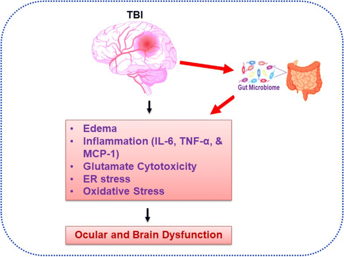
An acquired insult during TBI could potentially change various structural components of the brain resulting in temporary or even permanent brain impairment [12, 13]. Interestingly, GM and its role(s) in various system disorders has recently been the major focus area of research worldwide. For example, previous work reveals that GM plays important roles in neurogenerative processes such as formation of BBB, myelination, neurogenesis, and microglial maturation [14]. It has been shown that microbiome also modulates many aspects of our behavior since GM is involved in the modulation of cellular and molecular processes by balancing microbial eubiosis and dysbiosis condition and also involved in the progression of TBI-induced pathologies including BBB permeability, immune response to neuroinflammation, astroglial activation, and mitochondrial dysfunction (Fig. 1) [14]. Currently, efficacious treatments for TBI patients are acutely lacking [15,16,17]. Additionally, gut dysbiosis is known to exacerbate behavioral impairment as shown in studies that employed animal models of TBI and the spinal cord injury [18,19,20,21]. Furthermore, the dysbiotic milieu negatively affects the post stroke recovery [22,23,24,25]. Treatments for TBI and related disorders are severely limited, but recent research shows that microbiome transplants could mitigate CNS damage and functional impairments in spinal cord injury and stroke in animals [18]. In addition, probiotics were shown to reduce the rate of infections and time spent in intensive care units of hospitalized patients suffering from the brain trauma [18, 26, 27]. Thus, establishment of a protective, that is, eubiotic GM, is a promising therapeutic avenue since the brain injuries induce dysbiosis (Fig. 2). Reiner and colleagues 2014 reported that Novel CB2 Inverse Agonist SMM-189 reduce motor, visual, and emotional deficits after closed-head mild traumatic brain injury mouse model via mitigation of microglial inflammatory action [28]. ER stress was found to be increased early in juvenile rats exposed to TBI and that these rats developed tau oligomers over the course of 30 days and had significant short-term and spatial memory deficits following injury [29]. Treangen and colleagues suggested that acute bacterial dysbiosis within the gut microbiome was observed after TBI post-injury in mice [30]. The overall researched layout is represented in Fig. 2. However, post-TBI associated ocular and brain dysfunction via direct regulation of altered gut microbiome homeostasis is still needed to be demonstrated. This review discusses how GM alterations during post TBI contribute to CNS dysfunction and how to potentially target GM for therapeutic benefits in patients.

Fig. 1.
figure1

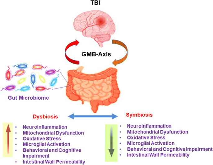
TBI induced dysbiosis via the gut microbiome brain (GMB) axis. The GMB-axis could potentially contribute, and further worsen the injury profile by promoting dysbiosis over eubiosis wherein harmful microbes in the gut can lead to an increase in neuroinflammation, mitochondrial dysfunction, oxidative stress, microglial activation, behavioral, and cognitive impairment, and intestinal wall permeability.

Fig. 2.
figure2

Resolution of the gut dysbiotic environment. Treatment with probiotics may help break the vicious dysbiotic cycle thus reducing the impact of brain injury, and hence improve substantially the TBI-related biochemical, pathological, and behavioral markers.

An Overview of Traumatic Brain Injury (TBI)

 More at link.

Exercise, healthy diet in midlife may prevent serious health conditions in senior years

This is just an excuse to NOT SOLVE all the problems in strok and allow blame the patient for everything. This way your doctor is not responsible for curing you of your stroke but allowed to say: 'Told you so, you didn't take care of yourself, you caused your problems.'

Exercise, healthy diet in midlife may prevent serious health conditions in senior years

Research Highlights:

  • Regular exercise and a healthy diet for middle-aged adults may be key to achieving optimal cardiometabolic health later in life.
  • Cardiometabolic health risk factors include the metabolic syndrome, a cluster of health conditions such as excess body fat around the waist, high blood pressure, high blood sugar, abnormal cholesterol or triglyceride levels that increase the risk of heart disease, stroke and Type 2 diabetes.
  • In an analysis of more than 2,300 U.S. adults, researchers found that following the U.S. Department of Health and Human Services’ physical activity guidelines in tandem with dietary recommendations was associated with a lower chance of developing the metabolic syndrome and other serious health conditions as people age from midlife to their senior years.

Embargoed until 4 a.m. CT/5 a.m. ET Wednesday, March 31, 2021

DALLAS, March 31, 2021 — Following a routine of regular physical activity combined with a diet including fruits, vegetables and other healthy foods may be key to middle-aged adults achieving optimal cardiometabolic health later in life, according to new research using data from the Framingham Heart Study published today in the Journal of the American Heart Association, an open access journal of the American Heart Association.

Cardiometabolic health risk factors include the metabolic syndrome, a cluster of disorders such as excess fat around the waist, insulin resistance and high blood pressure. Presence of the metabolic syndrome may increase the risk of developing heart disease, stroke and Type 2 diabetes.

Researchers noted it has been unclear whether adherence to both the U.S. Department of Health and Human Services’ 2018 Physical Activity Guidelines for Americans and their 2015-2020 Dietary Guidelines for Americans - as opposed to only one of the two – in midlife confers the most favorable cardiometabolic health outcomes later in life. The physical activity guidelines recommend that adults achieve at least 150 minutes of moderate or 75 minutes of vigorous physical activity per week, such as walking or swimming. The dietary guidelines, which were updated in January 2021, offer suggestions for healthy eating patterns, nutritional targets and dietary limits.

In an analysis of data from participants of the Framingham Heart Study, which began more than 70 years ago in Framingham, Massachusetts, investigators examined data from 2,379 adults ages 18 and older and their adherence to the two guidelines. They observed that meeting a combination of the two recommendations during midlife was associated with lower odds of metabolic syndrome and developing serious health conditions as participants aged in their senior years in 2016-2019 examinations.

“Health care professionals could use these findings to further promote and emphasize to their patients the benefits of a healthy diet and a regular exercise schedule to avoid the development of numerous chronic health conditions in the present and in later life,” said corresponding author Vanessa Xanthakis, Ph.D., FAHA, assistant professor of medicine and biostatistics in the Section of Preventive Medicine and Epidemiology at Boston University School of Medicine in Boston. “The earlier people make these lifestyle changes, the more likely they will be to lower their risk of cardiovascular-associated diseases later in life.”

Study participants were selected from the third generation of the Framingham Heart Study. Participants (average age 47, 54% women) were examined between 2008 and 2011. Researchers evaluated physical activity using a specialized device known as an omnidirectional accelerometer. The device, which tracks sedentary and physical activity, was worn on the participant’s hip for eight days. Researchers also collected dietary information from food frequency questionnaires to measure the kinds and levels of food and nutrients consumed.

In this investigation, researchers observed that among all participants, 28% met recommendations of both the physical activity and dietary guidelines, while 47% achieved the recommendations in only one of the guidelines. Researchers also observed that:

  • participants who followed the physical activity recommendations alone had 51% lower odds of metabolic syndrome;
  • participants who adhered to the dietary guidelines alone had 33% lower odds; and
  • participants who followed both guidelines had 65% lower odds of developing metabolic syndrome.

“It is noteworthy that we observed a dose-response association of adherence to diet and physical activity guidelines with risk of cardiometabolic disease later in life,” Xanthakis said. “Participants who met the physical activity guidelines had progressively lower risk of cardiometabolic disease as they increased adherence to the dietary guidelines.”

All study participants were white adults, therefore, the findings cannot be generalized to people in other racial or ethnic groups. Additional studies with a multiethnic participant sample are needed, researchers said.

Co-authors are Joowon Lee, Ph.D.; Maura E. Walker, Ph.D.; Maximillian T. Bourdillon, M.D., Nicole Spartano, Ph.D.; Gail T. Rogers, M.A.; Paul F. Jacques, D.Sc.; and Ramachandran S. Vasan, M.D.

The study was funded by the National Heart, Lung, and Blood Institute of the National Institutes of Health, the American Heart Association, the Evans Medical Foundation and Boston University School of Medicine.

Additional Resources:

Statements and conclusions of studies published in the American Heart Association’s scientific journals are solely those of the study authors and do not necessarily reflect the Association’s policy or position. The Association makes no representation or guarantee as to their accuracy or reliability. The Association receives funding primarily from individuals; foundations and corporations (including pharmaceutical, device manufacturers and other companies) also make donations and fund specific Association programs and events. The Association has strict policies to prevent these relationships from influencing the science content. Revenues from pharmaceutical and biotech companies, device manufacturers and health insurance providers are available here, and the Association’s overall financial information is available here.


Decreases in Stroke-Related Dementia Result from Smaller Memory Decrements

 So they didn't come up the reasons for this and thus can't create any stroke protocols on it. But they did suggest further research so they realize they failed at doing useful research.

Decreases in Stroke-Related Dementia Result from Smaller Memory Decrements

Declines in stroke-associated dementia may be due to a reduction in memory deficits occurring immediately after stroke onset, according to a cohort study published on March 16 in the journal Stroke.

“Consistent with recent trends observed in the Framingham Study, we observed improvements in cognitive functioning after stroke in recent years. However, we found that improvements in poststroke memory functioning were driven primarily by lessening of immediate memory deficits at the time of stroke onset, not differences in memory decline around stroke onset," wrote Chloe W. Eng, MSPH, of the University of California San Francisco, and colleagues.

The study authors found, however, that pre-stroke memory functioning for people who had a stroke had improved in recent years compared with earlier periods. And they attributed that, in part, to the fact that “determinants of stroke are less strongly associated with memory than in the past," adding, “Recent decreases in stroke-related dementia are also likely partially attributable to smaller memory decrements in the immediate aftermath of stroke."


These data add to the current understanding of stroke-associated decline by considering possible trends in the association between cognition and stroke across nearly 20 years, the researchers noted.

The researchers assessed a nationally representative cohort and looked at evidence on temporal trends in memory change associated with incident stroke.

The study authors assessed 2,434 participants with a first stroke, of which 590 were fatal, and 1,844 were nonfatal. At baseline, mean age was 66.3 years in epoch 1, 67.1 years in epoch 2,  and 66.5 years in epoch 3.

They evaluated adults age 50 years and older from the Health and Retirement Study across six-year successive epochs from 1998 to 2016. Participants were selected from six-year epochs and at baseline were stroke-free.

They used demographic-adjusted linear regression models to compare yearly rates of change in a composite z-standardized memory score.

The findings indicate that crude stroke incidence rates declined from 8.5 per 1,000 person-years in epoch 1 to 6.8 per 1,000 person-years in epoch 3. Moreover, the rates of memory change prior to and after stroke onset were similar across epochs.​

Notably, memory decrement immediately following stroke onset attenuated from −0.37 points in epoch 1 to −0.26 points in epoch 2 and −0.25 points in epoch 3 (pvalue for linear trend=0.02).

Limitations of the study include the inability to differentiate between stroke subtypes, that the study relied on self-reported stroke status, and inadequate information to assess potential mediation of secular trends by comorbidities like depression and vascular risk factors, the researchers noted.

Even though improvements in patient care for stroke survivors may account for trends in poststroke memory outcomes, future studies should focus on direct measures of quality of care and other potential mediators, the authors noted.Future studies may consider the impact of specific changes in stroke care during this study period to further address possible underlying mechanisms behind these observed trends," they concluded. 

EXPRESS: Dependence of seasonal dynamics of cardiovascular events on a climate of a region: a meta-analysis

How is this research of any use to getting survivors recovered? It just seems like padding to fulfill some requirement to publish research and reviews are easy.

EXPRESS: Dependence of seasonal dynamics of cardiovascular events on a climate of a region: a meta-analysis

First Published March 16, 2021 Research Article 

Background

Cardiovascular events (CVEs) occur more often in winter than in summer; however, the dependence of strokes on various meteorological factors remains unclear .

Aims

The purpose of this meta-analysis was to determine the dependence of the circannual dynamics of hospitalizations for hemorrhagic stroke (HS) and ischemic stroke (IS) on seasonal fluctuations in meteorological factors.

Summary of review and conclusions

For our meta-analysis, we selected 20 and 26 publications examining the seasonal dynamics of HS and IS, respectively . The meta-analysis showed that HS occurs more often in winter than in other seasons and does not depend on a region’s climate. The seasonal dynamics of IS are not clearly expressed and are determined by the characteristics of a region’s climate. In a climate without pronounced seasonal dynamics of atmospheric pressure and in wet winters, the vector of IS incidents will not be expressed or slightly shifted toward winter. Low atmospheric pressure in summer is associated with an increased likelihood of IS during this season compared to winter. There was also a relation between IS risk with high relative humidity and a significant decrease in ρO2 in summer, but there is not enough evidence regarding this association. We did not reveal dependence of the seasonal dynamics of strokes on the amplitude of annual fluctuations in air temperature.

Keywords: ischemic stroke, hemorrhagic stroke, season, meteorological factors, climate

Access Options
 

'Don't use your teeth, you need to learn to use your hands.'

 My exes statement to me on just getting home from hospital. She's a PT, I ignored her, no understanding of stroke deficits or recovery. So with the leftover Irish whiskey which has a cork top, there are two standard possibilities.

1. Pop the top with my left hand. Impossible, my fingers can't close around that small an object.

2. Hold the fat end of the bottle with the left hand. Impossible, I have never been able to get my hand around anything larger than 3 inches. Holding an object that big and opening up four fingers and one thumb just is not possible.

So the real solution goes back to old Westerns where bar patrons would grab the bottle and twist off the cork top with their teeth.


Tuesday, March 30, 2021

Multi-year study reveals decrease in stroke fatalities, improvements in systems of care

 NOT GOOD ENOUGH!  Just maybe you want to discuss with survivors all the failures in stroke.  And what will you think about your failure to get 100% recovered when you are the 1 in 4 per WHO that has a stroke?

This cherry picking of stroke statistics is appalling.                    

Multi-year study reveals decrease in stroke fatalities, improvements in systems of care

Acute stroke case fatality decreased substantially between 2003 and 2017 at the same time that discharge to home or rehabilitation increased and long-term care admissions decreased, according to a Canadian study.

The findings, which were published in Neurology, indicate “continuous improvements” to stroke systems of care over the 15-year study period.

“We developed this research project to understand whether outcomes among patients with acute stroke were changing over time in the last 2 decades. Other studies from developed countries have generally found that case fatality after acute stroke is going down with time,” Raed Joundi, a master’s student and stroke fellow at the University of Calgary Cumming School of Medicine, told Healio. “However, some studies have found that case fatality is decreasing after ischemic stroke, but not intracerebral hemorrhage. In addition, it is possible that the increased use of life-sustaining care may lower case fatality only in the short-term, and result in an increase in people surviving with severe disability. To address these unanswered questions, we used administrative data linkages in the entire population of Ontario, Canada.”

In this population-based study, Joundi and colleagues examined trends in acute stroke case fatality, discharge destination and long-term care admission from 2003 to 2017 for all ED visits and hospital admissions for first-ever ischemic stroke or intracerebral hemorrhage in Ontario, Canada. The researchers determined crude and standardized (according to age and sex) risk for death at 30 days and 1 year after the index stroke. They also examined trends in rates of discharge to home or to rehabilitation from acute care, as well as admission to long-term care, within 1 year after acute care discharge. Finally, Joundi and colleagues determined whether these outcome trends remained after adjusting for baseline characteristics, estimated stroke severity and use of life-sustaining care.

During the study period, 163,574 patients experienced an acute stroke, including intracerebral hemorrhage (12.4%) and ischemic stroke (87.6%). The researchers observed an increase in the number of patients over 60 years of age in the ischemic stroke group, as well as an increase in patients with hypertension, diabetes and those receiving intensive care or mechanical ventilation in both groups. They also noted a decrease in 30-day hazard of death for all patients every year of the study, with HRs that remained “virtually unchanged” after adjustment for demographics, comorbidity and type of care, while the number of patients discharged to the home or rehabilitation increased. Specifically, age/sex-standardized 30-day stroke case fatality decreased from 20.5% to 13.2% and from 32.2% to 22.8% at 1 year. Reductions in fatality occurred regardless of age or sex.

“One of the most striking results was the large decrease in case fatality after intracerebral hemorrhage. Thirty-day case fatality dropped from 45% to 30% over 15 years,” Joundi said. “This is interesting because, while hyperacute therapies for ischemic stroke (thrombolysis and thrombectomy) have become more widespread, there have been limited therapeutics for the treatment of acute intracerebral hemorrhage. The changes observed may be due to improvement in multidisciplinary stroke care, which benefits patients with both ischemic stroke and intracerebral hemorrhage.”

Joundi called the results “very encouraging.”

“Our findings ... show that efforts to improve care of those with stroke, which have spanned decades, have yielded improved outcomes,” he said. “This success should be celebrated but should also stimulate further initiatives to continuously refine and improve access to best care for all stroke patients.”


Effect of anodal versus cathodal transcranial direct current stimulation on stroke rehabilitation: a pilot randomized controlled trial

Well then write this up as a protocol AND DISTRIBUTE it to all stroke hospitals in the world.  This is your responsibility since our fucking failures of stroke associations are DOING NOTHING FOR SURVIVORS.

Is anyone ever going to put together a protocol on using tDCS and which type?   Otherwise all this fucking research and reviews are totally worthless.  This is why we need strong stroke leadership, to actually help stroke survivors.

Effect of anodal versus cathodal transcranial direct current stimulation on stroke rehabilitation: a pilot randomized controlled trial

2013, Neurorehabilitation and Neural Repair
 Eman M. Khedr, MD 1, 
Ola A. Shawky, MD 1, 
Dina H. El-Hammady, MD 2, 
John C. Rothwell, MD 3, 
Essam S. Darwish, MD 1, 
Omar M. Mostafa, MD 4, 
and Amal M. Tohamy, MD 1

Abstract

Objective
We compared the long-term effect of anodal versus cathodal transcranial direct current stimulation (tDCS) on motor recovery in patients after subacute stroke.
Methods
Forty patients with ischemic stroke undergoing rehabilitation were randomly assigned to 1 of 3 groups: Anodal, Cathodal (over-affected and unaffected hemisphere, respectively), and Sham. Each group received tDCS at an intensity of 2 mA for 25 minutes daily for 6 consecutive days over of the motor cortex hand area. Patients were assessed with the National Institutes of Health Stroke Scale (NIHSS), Orgogozo’s MCA scale (OMCASS), the Barthel index (BI), and the Medical Research Council (MRC) muscle strength scale at baseline, after the sixth tDCS session and then 1, 2, and 3 months later. Motor cortical excitability was measured with transcranial magnetic stimulation (TMS) at baseline and after the sixth session.
Results
By the 3-month follow-up, all groups had improved on all scales with P  values ranging from .01 to .0001. Improvement was equal in the Anodal and Cathodal groups. When these treated groups were combined and compared with Sham, significant interactions were seen for the OMCASS and BI scales of functional ability (P = .002 for each). There was increased cortical excitability of the affected hemisphere in all groups with the changes being greater in the real versus sham groups. There were borderline significant improvements in muscle strength.
Conclusion
A brief course of 2 types of tDCS stimulation is superior to sham stimulation in enhancing the effect of rehabilitation training to improve motor recovery after stroke.
 

Unique diagnostic signatures of concussion in the saliva of male athletes: the Study of Concussion in Rugby Union through MicroRNAs (SCRUM)

I can very obviously see a possible use for this in stroke and TBI, but no one in stroke leadership will do anything with this. Their motto must be hear no research, see no research, do no research!

hear no evil,see no evil,speak no evil


Unique diagnostic signatures of concussion in the saliva of male athletes: the Study of Concussion in Rugby Union through MicroRNAs (SCRUM)

  1. Valentina Di Pietro1,2,3,
  2. Patrick O'Halloran1,3,
  3. Callum N Watson1,
  4. Ghazala Begum3,
  5. Animesh Acharjee2,4,5,
  6. Kamal M Yakoub2,
  7. Conor Bentley2,
  8. David J Davies1,2,
  9. Paolo Iliceto6,
  10. Gabriella Candilera6,
  11. David K Menon7,
  12. Matthew J Cross8,9,
  13. Keith A Stokes8,10,
  14. Simon PT Kemp10,11,
  15. Antonio Belli1,2,3
  1. Correspondence to Dr Valentina Di Pietro, University of Birmingham, Institute of Inflammation and Ageing, Birmingham 9,000, UK; v.dipietro@bham.ac.uk

Abstract

Objective To investigate the role of salivary small non-coding RNAs (sncRNAs) in the diagnosis of sport-related concussion.

Methods Saliva was obtained from male professional players in the top two tiers of England’s elite rugby union competition across two seasons (2017–2019). Samples were collected preseason from 1028 players, and during standardised head injury assessments (HIAs) at three time points (in-game, post-game, and 36–48 hours post-game) from 156 of these. Samples were also collected from controls (102 uninjured players and 66 players sustaining a musculoskeletal injury). Diagnostic sncRNAs were identified with next generation sequencing and validated using quantitative PCR in 702 samples. A predictive logistic regression model was built on 2017–2018 data (training dataset) and prospectively validated the following season (test dataset).

Results The HIA process confirmed concussion in 106 players (HIA+) and excluded this in 50 (HIA−). 32 sncRNAs were significantly differentially expressed across these two groups, with let-7f-5p showing the highest area under the curve (AUC) at 36–48 hours. Additionally, a combined panel of 14 sncRNAs (let-7a-5p, miR-143-3p, miR-103a-3p, miR-34b-3p, RNU6-7, RNU6-45, Snora57, snoU13.120, tRNA18Arg-CCT, U6-168, U6-428, U6-1249, Uco22cjg1,YRNA_255) could differentiate concussed subjects from all other groups, including players who were HIA− and controls, immediately after the game (AUC 0.91, 95% CI 0.81 to 1) and 36–48 hours later (AUC 0.94, 95% CI 0.86 to 1). When prospectively tested, the panel confirmed high predictive accuracy (AUC 0.96, 95% CI 0.92 to 1 post-game and AUC 0.93, 95% CI 0.86 to 1 at 36–48 hours).

Conclusions SCRUM, a large prospective observational study of non-invasive concussion biomarkers, has identified unique signatures of concussion in saliva of male athletes diagnosed with concussion.

https://creativecommons.org/licenses/by/4.0/

This is an open access article distributed in accordance with the Creative Commons Attribution 4.0 Unported (CC BY 4.0) license, which permits others to copy, redistribute, remix, transform and build upon this work for any purpose, provided the original work is properly cited, a link to the licence is given, and indication of whether changes were made. See: https://creativecommons.org/licenses/by/4.0/.


Fluid Cognitive Abilities Are Important for Learning and Retention of a New, Explicitly Learned Walking Pattern in Individuals After Stroke

Useless. You tell us NOTHING about how to have these fluid cognitive abilities. 


Fluid Cognitive Abilities Are Important for Learning and Retention of a New, Explicitly Learned Walking Pattern in Individuals After Stroke

First Published March 23, 2021 Research Article 

There is significant variability in post stroke locomotor learning that is poorly understood and affects individual responses to rehabilitation interventions. Cognitive abilities relate to upper extremity motor learning in neurologically intact adults, but have not been studied in post stroke locomotor learning.

To understand the relationship between locomotor learning and retention and cognition after stroke.

Participants with chronic (>6 months) stroke participated in 3 testing sessions. During the first session, participants walked on a treadmill and learned a new walking pattern through visual feedback about their step length. During the second session, participants walked on a treadmill and 24-hour retention was assessed. Physical and cognitive tests, including the Fugl-Meyer-Lower Extremity (FM-LE), Fluid Cognition Composite Score (FCCS) from the NIH Toolbox -Cognition Battery, and Spatial Addition from the Wechsler Memory Scale-IV, were completed in the third session. Two sequential regression models were completed: one with learning and one with retention as the dependent variables. Age, physical impairment (ie, FM-LE), and cognitive measures (ie, FCCS and Spatial Addition) were the independent variables.

Forty-nine and 34 participants were included in the learning and retention models, respectively. After accounting for age and FM-LE, cognitive measures explained a significant portion of variability in learning (R2 = 0.17, P = .008; overall model R2 = 0.31, P = .002) and retention (ΔR2 = 0.17, P = .023; overall model R2 = 0.44, P = .002).

Cognitive abilities appear to be an important factor for understanding locomotor learning and retention after stroke. This has significant implications for incorporating locomotor learning principles into the development of personalized rehabilitation interventions after stroke.

Access Options
 

A review of technological and clinical aspects of robot- aided rehabilitation of upper-extremity after stroke

 You can see if your hospital has any of these or even tested any.  It only has been 6 years. Aren't you glad your stroke hospital is totally fucking incompetent? These are the ones reviewed, pictures, explanations and graphs at the link:

MIT-MANUS
Mirror Image Motion Enabler
GENTLE/s system
Bi-Manu-Track
Assisted Rehabilitation and Measurement (ARM) guide
REHAROB therapy system
Dampace
T-WREX
Maryland-Georgetown-Army (MGA) exoskeleton
L-EXOS system
ARMin

A review of technological and clinical aspects of robot- aided rehabilitation of upper-extremity after stroke

 
 Mahdieh Babaiasl 1, 
Seyyed Hamed Mahdioun 1, 
Poorya Jaryani 2, 
and Mojtaba Yazdani 3
 
1 School of Engineering Emerging Technologies, University of Tabriz, Tabriz, Iran,
 2 Department of Mechanical Engineering, Islamshahr Branch,Islamic Azad University, Islamshahr, Iran, and
 3 Control Department, Electronics Faculty, Semnan University, Semnan, Iran
 
 
Disabil Rehabil Assist Technol, Early Online: 1–18
!
2015 Informa UK Ltd. DOI: 10.3109/17483107.2014.1002539
  History
Received 9 July 2014
Revised 7 December 2014
Accepted 22 December 2014
Published online 20 January 2015

 Abstract

Cerebrovascular accident (CVA) or stroke is one of the leading causes of disability and loss of motor function. Millions of people around the world are effected by it each year. Stroke results in disabled arm function. Restoration of arm function is essential to regaining activities of daily living (ADL). Along with traditional rehabilitation methods, robot-aided therapy has emerged in recent years. Robot-aided rehabilitation is more intensive, of longer duration and more repetitive. Using robots, repetitive dull exercises can turn into a more challenging and motivating tasks such as games. Besides, robots can provide a quantitative measure of the rehabilitation progress. This article overviews the terms used in robot-aided upper-limb rehabilitation. It continues by investigating the requirements for rehabilitation robots. Then the most outstanding works in robot-aided upper-limb rehabilitation and their control schemes have been investigated. The clinical outcomes of the built robots are also given that demonstrates the usability of these robots in real-life applications and their acceptance.This article summarizes a review done along with a research on the design, simulation and control of a robot for use in upper-limb rehabilitation after stroke. 
 Mahdieh Babaiasl
1
, Seyyed Hamed Mahdioun
1
, Poorya Jaryani
2
, and Mojtaba Yazdani
3
1
School of Engineering Emerging Technologies, University of Tabriz, Tabriz, Iran,
 2
Department of Mechanical Engineering, Islamshahr Branch,Islamic Azad University, Islamshahr, Iran, and
 3
Control Department, Electronics Faculty, Semnan University, Semnan, Iran
 Disabil Rehabil Assist Technol, Early Online: 1–18
!
2015 Informa UK Ltd. DOI: 10.3109/17483107.2014.1002539
 Abstract
Cerebrovascular accident (CVA) or stroke is one of the leading causes of disability and loss of motor function. Millions of people around the world are effected by it each year. Stroke resultsin disabled arm function. Restoration of arm function is essential to regaining activities of dailyliving (ADL). Along with traditional rehabilitation methods, robot-aided therapy has emergedin recent years. Robot-aided rehabilitation is more intensive, of longer duration and morerepetitive. Using robots, repetitive dull exercises can turn into a more challenging andmotivating tasks such as games. Besides, robots can provide a quantitative measure of therehabilitation progress. This article overviews the terms used in robot-aided upper-limbrehabilitation. It continues by investigating the requirements for rehabilitation robots. Then themost outstanding works in robot-aided upper-limb rehabilitation and their control schemeshave been investigated. The clinical outcomes of the built robots are also given thatdemonstrates the usability of these robots in real-life applications and their acceptance.This article summarizes a review done along with a research on the design, simulation andcontrol of a robot for use in upper-limb rehabilitation after stroke.

Effects of Lower Limb Constraint Induced Movement Therapy in People With Stroke: A Systematic Review and Meta-Analysis

 I can't figure this out and I couldn't find a video on it. Good luck on your search.

Effects of Lower Limb Constraint Induced Movement Therapy in People With Stroke: A Systematic Review and Meta-Analysis

Auwal Abdullahi1,2*, Steven Truijen2, Naima A. Umar3, Ushotanefe Useh4, Victor A. Egwuonwu5, Tamaya Van Criekinge2 and Wim Saeys2
  • 1Neurological Rehabilitation Unit, Department of Physiotherapy, Bayero University Kano, Kano, Nigeria
  • 2Department of Rehabilitation Sciences and Physiotherapy, University of Antwerp, Wilrijk, Belgium
  • 3Department of Physiotherapy, Muhammad Abdullahi Wase Teaching Hospital, Kano, Nigeria
  • 4Lifestyle Diseases Research Entity, Faculty of Health Sciences, North-West University, Mmabatho, South Africa
  • 5Department of Medical Rehabilitation, Nnamdi Azikiwe University, Awka, Nigeria

Background: Constraint induced movement therapy (CIMT) is effective at improving upper limb outcomes after stroke.

Aim: The aim of this study was to carry out a systematic review and meta-analysis of the effects of lower limb CIMT studies of any design in people with stroke.

Materials/ Method: PubMED, PEDro, OTSeeker, CENTRAL, and Web of Science were searched from their earliest dates to February 2021. Lower limbs CIMT studies that measured outcomes at baseline and post-intervention were selected. Sample size, mean, and standard deviation on the outcomes of interest and the protocols of both the experimental and control groups were extracted. McMaster Critical Review Form was used to assess the methodological quality of the studies.

Result: Sixteen studies with different designs were included in this review. The result showed that lower limb CIMT improves functional, physiological and person's reported outcomes including motor function, balance, mobility, gait speed, oxygen uptake, exertion before and after commencement of activities, knee extensor spasticity, weight bearing, lower limb kinematics and quality of life in people with stroke post intervention. However, there were only significant differences in quality of life in favor of CIMT post-intervention [mean difference (MD) = 16.20, 95% CI = 3.30–29.10, p = 0.01]; and at follow-up [mean difference (MD) = 14.10, 95% CI = 2.07–26.13, p = 0.02] between CIMT and the control group. Even for the quality of life, there was significant heterogeneity in the studies post intervention (I2 = 84%, p = 0.01).

Conclusion: Lower limb CIMT improves motor function, balance, functional mobility, gait speed, oxygen uptake, weigh bearing, lower limb kinematics, and quality of life. However, it is only superior to the control at improving quality of life after stroke based on the current literature.

Introduction

Constraint Induced Movement Therapy (CIMT) is a translational motor rehabilitation technique following injury of the Central Nervous System (CNS). The technique originated many decades ago from use in primates; and was translated to humans following stroke and other neurological conditions (1). The original concept involved constraint of the unaffected limb and forced use of the affected one (2). Subsequent studies in humans involved voluntary massed tasks or shaping practices with the affected limb. Consequently, CIMT has been reported to be effective at improving real world arm use, motor function, and kinematic outcomes by inducing changes in the functions and structures of the brain (37). However, there have been many modifications over the years of the original protocol of CIMT, including but not limited to the length of time for the tasks practice, the constraint, and the use of a transfer package (79).

The effects of CIMT on the recovery of motor function of the upper limb have been well-investigated (7, 10). The practicability of the protocol for upper limbs could be because of the unilateral nature of the use of these limbs in most of our activities of daily living (ADL). For the lower limbs, this may seem difficult since humans are bipedal, and this requires them to use the two limbs simultaneously for ADL especially during walking. However, the positive results in the recovery of motor function of the upper limb following CIMT persuaded the neuroscientific community to consider translating the technique to the lower limbs. Consequently, a lower limb CIMT protocol was designed to comprise mainly of intensive practice with the affected limb, shaping activities, transfer package, and encouraging the increased use of the affected limb (11). So far, there are several small sample size studies that have investigated the effects of lower limb CIMT on gait parameters, balance, and motor function using different study designs such as case reports, experimental studies, quasi-experimental studies, and randomized controlled trials (RCTs) (12). These studies reported that lower limbs CIMT improved gait speed, step length, motor function, functional mobility, balance, and kinematic outcomes. However, small sample size studies may overestimate the effect of an intervention (1315). Second, the only difference in the protocols of the CIMT and control groups was the use of a constraint in the CIMT group, with no difference in the types of tasks used in most of these studies, including the intensity. According to Abdullahi, task practice is the most important component of CIMT (16, 17). Therefore, it is possible that the effects of lower limb CIMT reported in those studies were overestimated.

In addition, in upper limb CIMT constraint is used to immobilize the unaffected limb to prevent movement at joints essential for the functioning of the limb. This is to done to maximize the use of the affected limb, and to help recover function. However, for lower limb CIMT, the types of constraints used include encouraging weight bearing on the affected limb, the use of an insole in the affected limb, the use of knee braces or a splint, and attaching weight to the ankle of the affected limb (12). Constraining one of the limbs may cause asymmetry which could negatively affect normal functions such as walking, especially since humans are bipedal. The aim of this study was to therefore carry out a systematic review and meta-analysis on the effects of lower limb CIMT on outcomes after stroke such as gait parameters, balance, motor function, functional mobility, and quality of life. This review sought to answer this question: What are the effects of lower limb CIMT on this information is important as, to date, there does not seem to be any review and/ or meta-analysis on the effects of lower limb CIMT following stroke.

More at link.