Changing stroke rehab and research worldwide now.Time is Brain! trillions and trillions of neurons that DIE each day because there are NO effective hyperacute therapies besides tPA(only 12% effective). I have 523 posts on hyperacute therapy, enough for researchers to spend decades proving them out. These are my personal ideas and blog on stroke rehabilitation and stroke research. Do not attempt any of these without checking with your medical provider. Unless you join me in agitating, when you need these therapies they won't be there.

What this blog is for:

My blog is not to help survivors recover, it is to have the 10 million yearly stroke survivors light fires underneath their doctors, stroke hospitals and stroke researchers to get stroke solved. 100% recovery. The stroke medical world is completely failing at that goal, they don't even have it as a goal. Shortly after getting out of the hospital and getting NO information on the process or protocols of stroke rehabilitation and recovery I started searching on the internet and found that no other survivor received useful information. This is an attempt to cover all stroke rehabilitation information that should be readily available to survivors so they can talk with informed knowledge to their medical staff. It lays out what needs to be done to get stroke survivors closer to 100% recovery. It's quite disgusting that this information is not available from every stroke association and doctors group.

Showing posts with label dental pulp. Show all posts

Tuesday, April 8, 2025

Human dental stem cells can be transformed into excitable neuronal cells, study shows

 Didn't your fuckingly incompetent hospital get research going on this a decade ago?

NO? So, you DON'T have a functioning stroke hospital, do you? Why hasn't the board of directors reconstituted the whole hospital and get it into competent hands?

Human dental stem cells can be transformed into excitable neuronal cells, study shows

The cells with electrophysiological activity obtained at the University of the Basque Country (UPV/EHU) are opening up new avenues for the study of neurodegenerative diseases and the development of future autologous transplants.

A UPV/EHU study published in the prestigious journal Stem Cell Research & Therapy has proven that stem cells extracted from human dental pulp can be transformed into excitable neuronal cells and has highlighted the potential of these easily accessible cells for nerve tissue engineering. This finding will enable the furthering of advances in cell therapy for treating various neurodegenerative diseases, such as Huntington's disease and epilepsy.

"An adult neuron cannot be divided. When you lose it, you lose it for ever. And unlike other organs, the brain has a comparatively low capacity for natural regeneration due to its low presence of stem cells," said Gaskon Ibarretxe, a researcher in the UPV/EHU's Signaling Lab research group. The scientific community is looking for a way to obtain functional neurons that can be transplanted to restore impairments in neurodegenerative pathologies, brain lesions, strokes, etc. "But if the cells transplanted into the brain are to be able to integrate into a damaged brain circuit and replace the lost neurons, they need to be able to produce electrical impulses," added the researcher José Ramón Pineda, co-author of the study and a researcher in the group.

Stem cells have the capacity to divide and differentiate into different types of specialised cells. The UPV/EHU research group has obtained cells very similar to neurons; they "manage to produce electrical impulses like those of neurons by means of the differentiation of stem cells in human dental pulp, the soft tissue located inside the tooth," they said. The main milestone of this study was "the obtaining of cells that display functional excitability and which synthesise a type of neurotransmitter that regulates neuronal activity, without having been genetically modified; the primary dental cells were simply cultured with differentiation factors and were subjected to precise stimuli to generate cells with neuronal electrophysiological activity", said the researchers of the Signaling Lab at the UPV/EHU. This had never been achieved before."

Resembling inhibitory neurons

Neurotransmitters are substances released by neurons that can send excitatory or inhibitory signals to make neurons generate an electrical impulse or not. "The cells that we managed to differentiate are capable of synthesising a neurotransmitter known as GABA," explained the researchers. "It is a type of inhibitory signalling, in other words, it controls whether the neuron that receives it fires electrical impulses or not. And that is very important because there are neurodegenerative diseases such as Huntington's disease or conditions such as epilepsy in which there is a selective death of those types of cells in specific areas of the brain, and a resulting hyper-excitability of the brain circuitry".

Ibarretxe and Pineda, lecturers in the UPV/EHU's Department of Cell Biology and Histology, expressed great optimism when referring to the new avenues that can be explored on the basis of this finding: "We believe that these cells could be integrated into a damaged brain circuit and replace lost neurons, and thus reconnect with existing neurons and eventually regenerate the entire lost area functionally. This finding suggests a different approach to traditional cell therapy applied to the nervous system, which until now has been based, above all, on reducing inflammation, on neuroprotecting what remained alive, but not on replacing what has been lost. This opens a new door to the future of personalised medicine."

That is in fact the next step in this research: "to transplant these cells into living animals and see if they are integrated into the brain circuit and reconnect with the host's neurons. We obtained cells that generate electrical impulses characteristic of neurons that are not yet fully mature, but we are aware that they must generate trains of electrical impulses and be correctly integrated into a neuronal circuit. We haven't achieved that yet". The UPV/EHU researchers admitted that "there is a long road ahead, but we know it's going to be very promising. We believe that these cells have a great chance of being implemented in the clinical setting. The fact that they are transplanted at a relatively immature stage could even encourage their plasticity and integration into already developed brain circuits". The researchers added that these cells in fact offer an inherent advantage: "They are cells that do not have a tendency to generate tumours; on the contrary, it has been proven that they are very stable cells and differentiate better than other types of human stem cells into neurons," they concluded.

Source:
Journal reference:

Pardo-Rodríguez, B., et al. (2025). Functional differentiation of human dental pulp stem cells into neuron-like cells exhibiting electrophysiological activity. Stem Cell Research & Therapy. doi.org/10.1186/s13287-025-04134-7.

Saturday, July 27, 2024

Dental pulp stem cells regenerate neural tissue in degenerative disorders and stroke rehabilitation: A scope systematic review

I still prefer handing your competent? doctor a pee cup and asking for stem cells in return.  It has only been 11+ years, where the fuck is your doctor's protocol on turning urine into stem cells?

Turning urine into brain cells could help fight Alzheimer’s, Parkinson’s

December 2012

The latest here:

Dental pulp stem cells regenerate neural tissue in degenerative disorders and stroke rehabilitation: A scope systematic review

Open AccessPublished:July 24, 2024DOI:https://doi.org/10.1016/j.heliyon.2024.e35080
>

Abstract

Background

Dental Pulp Stem Cells (DPSCs) possess a remarkable ability for tissue differentiation, making them highly efficient in tissue regeneration and inflammation regulation. This systematic study proposes to find an answer to the question, "Do DPSCs have the ability to regenerate and rehabilitate nerve tissue?"

Methods

This systematic review was conducted based on Preferred Reporting Items for Systematic Reviews and Meta-Analyses (PRISMA) criteria, and the principle of non-bias was respected. All the articles from 2014-2024 were extracted from the Web of Science, PubMed, and Scopus databases. This study extracted the antigens and pro-inflammatory factors associated with DPSCs' involvement and how they affect the CNS's neural tissue regeneration.

Results

Two persons of researchers searched the database. After screening the full texts, they included 11 articles in their study. DPSCs control the following antigens: CD73, CD34, CD90, CD105, CD14, CD45, CD19Oct-4, CD73, CD31, CD34CD29CD44. Even though hematopoietic markers did not change much, OCT-4 and CD-73 were increased by DPSCs. DPSC-derived exosomes suppressed the expression of IL-6, IL-1β, TNF-α, and TGF, key mediators of nerve tissue inflammation. Additionally, DPSCs show high Vascular Endothelial Growth Factor (VEGF) expression in mice brain tissue cultures. DPSCs reduce Subarachnoid Hemorrhage (SAH), a condition in which blood collects in the subarachnoid space and causes ischemia.

Discussion

DPSCs showed the ability to regenerate nerve tissue and brain ganglia, stimulating angiogenesis by expressing cell markers and controlling growth factors in mice, and high therapeutic potential in neurodegenerative disorders. The present study invites further research in neurological disorders, specifically strokes, to prescribe these stem cells to the human population.

Saturday, June 25, 2022

Periodontal ligament stem cells as a promising therapeutic target for neural damage

 Just why was this research needed? There has been research on dental pulp stem cells since October 2011. Are your mentors and senior researchers that incompetent about not knowing previous research?  With a decent database of stroke research and protocols this problem wouldn't occur. But since we have fucking failures of stroke associations  no one is going to solve the database problem.

Periodontal ligament stem cells as a promising therapeutic target for neural damage

Abstract

Background

The damaged neuronal cells of adult mammalian lack the regenerative ability to replace the neuronal connections. Periodontal ligament stem cells (PDLSCs) are the promising source for neuroregenerative applications that can improve the injured microenvironment of the damaged neural system. They provide neuronal progenitors and neurotrophic, anti-apoptotic and anti-inflammatory factors. In this study, we aimed to comprehensively explore the various neuronal differentiation potentials of PDLSCs for application in neural regeneration therapy.

Main text

PDLSCs have superior potential to differentiate into various neural-like cells through a dedifferentiation stage followed by differentiation process without need for cell division. Diverse combination of nutritional factors can be used to induce the PDLSCs toward neural lineage. PDLSCs when coupled with biomaterials could have significant implications for neural tissue repair. PDLSCs can be a new clinical research target for Alzheimer's disease treatment, multiple sclerosis and cerebral ischemia. Moreover, PDLSCs have beneficial effects on retinal ganglion cell regeneration and photoreceptor survival. PDLSCs can be a great source for the repair of injured peripheral nerve through the expression of several neural growth factors and differentiation into Schwann cells.

Conclusion

In conclusion, these cells are an appealing source for utilizing in clinical treatment of the neuropathological disorders. Although significant in vitro and in vivo investigations were carried out in order for neural differentiation evaluation of these cells into diverse types of neurons, more preclinical and clinical studies are needed to elucidate their therapeutic potential for neural diseases.

Introduction

The central nervous system (CNS) and the peripheral nervous system (PNS) are parts of the nervous system. The brain and spinal cord make up the CNS, whereas cranial and spinal nerves, as well as their associated ganglia, constitute the PNS. The PNS has a built-in ability to regenerate and repair itself, whereas the CNS is essentially incapable of self-repair. Furthermore, depending on the characteristics and type of damage, the inherent regenerating ability is limited through injury itself [1].

In neural injuries, the damaged neural cells such as neurons and glial cells of adult mammalian lack the regenerative ability to replace the neuronal connections. This is due to the limited ability of neuronal progenitors to regenerate functional neuronal cells and inhibition of neural regeneration by the local injured microenvironment, especially in the glial scar [2].

One of the main formidable reasons for the limited success of pharmacotherapeutic strategies in neural damages is the microenvironment of the injury site with many molecular growth inhibitors that are hostile to any neuroregenerative therapy and function restoring of nerve fibers. It leads to the incapability of the damaged nerves to regrow and develop new synaptic connections. Targeting these inhibitors could be an efficient approach to overcome the permanent stopping of nerve growth [3].

The development of more precise therapies focused at specific molecular targets linked with a specific disease or injury of the nervous system has resulted from advances in neuroregenerative research. Due to several pathological injury processes and mechanisms, any neuroregenerative approach that focuses on just one of the events or mechanisms will not probably lead to a considerable therapeutic effect on neural injuries. The reasons for the limited therapeutic options are mainly because of both the extracellular and intracellular components of the nervous system that inhibits regeneration. To bridge the short-term requirements and revive immediate function of the nervous system, changes in plasticity and neuroregeneration firstly occur at the regional level. The lengthy and more permanent process of restoring function occurs at cellular level and promotes one or more of the restorative mechanisms which may improve neurological damages [4, 5].

Neuroregenerative medicine (NRM) is a growing field with the goal of neurogenesis, angiogenesis and synaptic plasticity [5] through replacement of lost cells and tissues and restoration of normal function [2]. Scientists are optimistic about the potentials of NRM to lead to providing novel approaches for the treatment of neural diseases and answer the ethical questions about their clinical applications [6].

NRM uses stem cells as a promising tool that make up for the scarcity of cell alternatives. Transplanted stem cells can improve the microenvironment in the injured site of the neural system and provide neuronal progenitors [7]; then, they help to slow or repair the deterioration related to degenerative or traumatic neural diseases and trigger a great effort in the field of preclinical and clinical neural research [8].

Mesenchymal stem cells (MSCs) have potential to integrate into host neuronal networks and renew functional neural connections. They can restore synaptic transmitter secretion, modulate the plasticity of damaged host tissues as well as release growth and neurotrophic factors with ability to promote cell survival [9]. In addition, MSCs have been found to diminish inflammation in vivo by suppressing pro-inflammatory cytokines and increasing anti-inflammatory cytokines and antigen-specific T-regulatory cells [10]. Researchers suggested that MSCs can cross the blood–brain barrier (BBB) [11], and this ability is the main reason for the treatment potential in neural diseases like cerebral ischemic diseases or spinal cord injuries [12, 13].

The oral cavity as an available source of MSCs includes two kinds of cells. Nondental oral MSCs which comprise periodontal ligament stem cells (PDLSCs), gingival MSCs (GMSCs), and dental follicle stem cells (DFSC) and the dental MSCs which consist of stem cells from apical papilla (SCAP), dental pulp stem cells (DPSCs) and stem cells from exfoliated deciduous teeth (SHED) [7].

Oral stem cells are rather accessible and show broad differentiation potential and high plasticity; hence, they can make autologous cell transplantation possible [8]. Moreover, they have advantages such as a higher proliferation rate and potential of immunosuppression [14, 15]; therefore, they are an excellent cell source in order for allogeneic transplantation.

They originate from cranial neural crest-derived ectomesenchymal cells (CNCCs); thus, they are capable of differentiation into neural cells in order for the reconstruction of central nervous system tissues. These cells express neural progenitors markers, including nestin, Pax6, Tuj1 and p75/NGFR, and have a more favorable neurotrophic secretome [16].

More at link.

Tuesday, August 6, 2019

Dental Pulp Stem Cells: An Attractive Alternative for Cell Therapy in Ischemic Stroke

You mean your mentors and senior researchers are so fucking incompetent they missed this from May 2016? My god, is there a need for a smidgen of stroke leadership.

TOOTH (The Open study Of dental pulp stem cell Therapy in Humans): Study protocol for evaluating safety and feasibility of autologous human adult dental pulp stem cell therapy in patients with chronic disability after stroke May 2016


Dental Pulp Stem Cells: An Attractive Alternative for Cell Therapy in Ischemic Stroke

  • 1Department of Neurology, Dalian Municipal Central Hospital Affiliated to Dalian Medical University, Dalian, China
  • 2Department of Pharmacy, Dalian Municipal Central Hospital Affiliated to Dalian Medical University, Dalian, China
  • 3School of Life Sciences, University of Warwick, Coventry, United Kingdom
Ischemic stroke is a major cause of disability and mortality worldwide, but effective restorative treatments are very limited at present. Regenerative medicine research revealed that stem cells are promising therapeutic options. Dental pulp stem cells (DPSCs) are autologously applicable cells that origin from the neural crest and exhibit neuro-ectodermal features next to multilineage differentiation potentials. DPSCs are of increasing interest since they are relatively easy to obtain, exhibit a strong proliferation ability, and can be cryopreserved for a long time without losing their multi-directional differentiation capacity. Besides, use of DPSCs can avoid fundamental problems such as immune rejection, ethical controversy, and teratogenicity. Therefore, DPSCs provide a tempting prospect for stroke treatment.
The past decade has witnessed intense advancement and tremendous therapeutic achievements in the ability to diagnose and treat stroke, a cerebrovascular disease of which 87% is ischemic in nature. Nevertheless, stroke remains a major cause of disability, morbidity, and mortality worldwide, and constitutes a major socioeconomic problem (1, 2). Ischemic stroke, due to partially or completely blocked blood flow in a cerebral artery, causes ischemic necrosis of brain tissue seriously impairing the health of affected individuals. The main therapeutic strategy for ischemic stroke is timely recanalization. This can either be achieved by tissue-type plasminogen activator application or mechanical thrombectomy. Particularly the latter can be applied up to 24 h after stroke in patients exhibiting a penumbra, and has revolutionized acute stroke treatment. However, the absolute number of patients qualifying for recanalization remains very low (3, 4). Hence, additional treatment approaches being effective beyond the first hours after stroke onset are urgently required.
Stem cell transplantation is a promising strategy to restore neurological function after stroke (5). Experimental stem cell transplantation in animals showed that numerous cell populations can improve functional recovery by a broad spectrum of mechanisms (68). Several kinds of stem cells are currently considered for therapy. These include embryonic stem cells (ESCs), fetal stem/progenitor cells, induced pluripotent stem cells (iPSCs), and adult stem cells. While embryonic or induced pluripotent stem cells exhibit a tremendous differentiation potential, they may also inherit a risk for tumor formation (9). The use of embryonic stem cells or fetal stem/progenitor cells raises ethical concerns. Adult stem cells show a limited proliferation and differentiation potential, but can still be beneficial after stroke due numerous mechanisms beyond tissue restoration. They are further believed to be safer in clinical application and their use is ethically less challenging (913).
Recent systematic reviews and meta-analyses on the most prominent adult stem cell therapy candidates, mesenchymal stem cells (MSCs), presented evidence that MSCs improve the outcome after stroke in animals (14) and patients, and confirmed the safety and feasibility of the approach (15). Nevertheless, there is still a lack of adult (stem) cells that can be derived from an autologous source, and may exhibit therapeutic abilities beyond those of MSCs.

Dental Pulp Stem Cells (DPSCs): A New Source of Adult Stem Cells

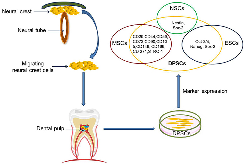
The dental pulp is a soft tissue located in the center of teeth. It comprises blood vessels, neural fibers, and connective tissue. The dental pulp contains both mesenchymal and ectodermal tissue as well as neural crest cells (16). Limited dentinal repair in the postnatal organism relies on specialized precursor cell populations residing in the dental pulp tissue. Gronthos et al. first reported the isolation and characterization of stem cells from dental pulp tissue of the third molar in 2000 (17). DPSCs are ectoderm-derived stem cells, originating from migrating neural crest cells (Figure 1). They are a subpopulation among dental pulp cells (DPCs) which possess MSC properties, such as a fibroblast-like morphology, adherence to a plastic surface, as well as surface marker expression, proliferation and colony forming behavior similar to that of MSCs (18, 19). It is not clear whether or not DPSCs are a kind of MSC population. Given their differentiation abilities as reviewed below, it might be assumed that DPSCs are a more naïve stem cell population that also, but not exclusively, exhibits MSC properties. A major benefit of DPSCs is that they can be isolated during routine dental procedures such as the eruption of deciduous teeth or extraction of impacted wisdom teeth (20) in simple and autologous fashion without ethical concerns. Another primary advantage of DPSCs is their potential for cell banking. Several studies have demonstrated that DPSCs retain their stem cell properties after long cryopreservation (21, 22). This is essential as cryopreservation can impact therapeutic capacities of other adult stem cell-containing populations in stroke (23). In addition, DPSC cultures can be established from extracted human molars with high efficiency, even after the whole tooth has been cryopreserved for up to 1 month (24). DPSCs also exhibit a multilineage differentiation potential into chondrocytes, adipocytes, odontoblasts, and potentially even neural-like cells (2528).
FIGURE 1
www.frontiersin.org Figure 1. DPSCs origin, isolation, and marker expression. DPSCs originate from migrating neural crest cells, coming to rest in dental pulp, and express markers overlapping with MSCs, ESCs, and NCSs.
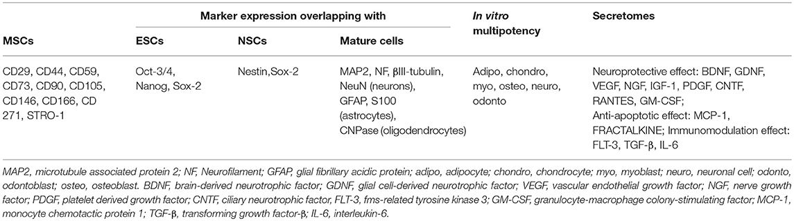
Currently, there are no specific markers that uniquely define DPSCs. In general, DPSCs, as a heterogeneous population, express a variety of markers similar to MSCs (Table 1) (Figure 1), and do not express hematopoietic markers such as CD14, CD19, CD34, and CD45 (18, 26, 2932). DPSCs isolated by their high proliferative potential tend to include a large population of cells expressing CD44+, CD90+, and CD166+. However, DPSCs also express stemness-related markers similar to ESCs such as Oct-3/4, Nanog, and Sox-2, as well as the cytoskeleton-related markers nestin and vimentin (Figure 1) (3335). They further express insulin-like growth factor 1 receptor (IGF1R) which is regarded as a pluripotency marker in ESCs. DPSC-secreted IGF1 interacts with IGF1R through an autocrine signaling pathway to maintain self-renewal and proliferation potential (36).
TABLE 1
www.frontiersin.org Table 1. Characteristics of DPSCs.
In addition, DPSCs (as neural crest-derived stem cells) not only express a number of neural stem cell (NSC) associated markers including nestin (26, 37) and Sox2 (38) (Figure 1), but also express low basal levels of markers associated with mature central nervous system cell types, including the neuronal markers βIII-tubulin, microtubule-associated protein 2 (MAP2), neurofilaments (NF) (33, 39), NeuN (40), the astrocytic marker glial fibrillary acidic protein (GFAP) (26, 33), and oligodendrocyte-associated CNPase (33). Taken together, this suggests that DPSCs can indeed differentiate into neuron-like cells under appropriate conditions, and differentiated cells even exhibit typical electrophysiological properties after neuronal differentiation (41, 42).

DPSCs as a Potential Candidate for Therapy of Neurological Diseases

Brain-derived NSCs are considered a promising population for stroke treatment due to their ability to self-renew and to differentiate into neural cells types (neurons, astrocytes, oligodendrocytes) (43). However, autologous harvest of adult human NSCs requires neurosurgical procedures due to their brain parenchymal residence (44), while allogeneic or even xenogenic NSCs grafting imposes the risk of graft rejection and additional immunological damage. Only a limited number of clinical trials currently explore the potential of NSCs for stroke treatment because of these limitations.
Adult stem cells or stem cell-containing populations are more frequently applied in translational research. As stated above, DPSCs share many biological characteristics with MSCs including bone marrow MSCs (BM-MSCs), adipose tissue-derived stem cells (ADSCs) and umbilical cord MSCs (UC-MSCs) but there are some variations in their proliferation potential (17, 27, 45), differentiation potential (17, 27, 46), immunomodulatory activity (27), secretome characteristics, and secretory capacity (4749). Specifically, DPSCs have a higher proliferation rate and a greater clonogenic potential than MSCs (17, 45). Next to DPSCs, the DPC population also contains a higher number of stem/progenitor cells as compared to bone marrow (50). This may be attributed to the developmental state of the respective tissues. All teeth, even the permanent molars, are generated early in individual development and rest in the jar until they erupt. Abilities and capacities of stem cells may be much better preserved in tissue with a slow turnover such as the dental pulp when compared to BM, which exhibits a tremendous turn-over throughout life apart from some niches. DPSCs maintain their high rate of proliferation even after extensive subculturing.
Like MSCs, DPSCs can differentiate into cells of mesenchymal and non-mesenchymal tissues in vitro and in vivo. However, DPSCs exhibit stronger odontogenesis and neurogenesis capabilities, in turn being not as potent to produce adipogeneic, osteogeneic and chondrogeneic tissue than BM-MSCs (51) and ADSCs (46). Besides, DPSCs also have immunomodulatory capacities exceeding those of BM-MSCs, for example a higher suppression rate of T lymphocyte growth (17, 27).
DPSCs exhibit superior neuroprotective and neuro-supportive properties in neurological injuries and pathologies as compared with BM-MSCs and ADSCs (52). This might be related to a higher expression of trophic factors including brain derived neurotrophic factor (BDNF), glial cell-derived neurotrophic factor (GDNF), nerve growth factor (NGF), vascular endothelial growth factor (VEGF), and platelet derived growth factor (PDGF) in DPSCs as compared to BM-MSCs (47, 48), although the spectrum of growth and trophic factors secretion is similar (53). DPSCs also express higher quantities of CXCL14 and monocyte chemoattractant protein 1 (MCP-1) than ADSCs (49). Besides, the DPSC secretome contains higher concentrations of RANTES, FRACTALKINE, fms-related tyrosine kinase 3 (FLT-3), granulocyte-macrophage colony-stimulating factor (GM-CSF), and MCP-1 than the BM-MSCs secretome (54). DPSCs show higher angiogenic and neurogenic potentials in ectopic transplantation models compared to BM-MSCs and ADSCs, and exhibit the highest migration capacity. Transplantation of DPSCs in a mouse hindlimb ischemia model produced higher blood flow and capillary density than transplantation of BM-MSCs and ADSCs, which being associated with superior recovery of limb movement abilities and reduction of ischemic hindlimb damage (55). DPSCs also mediate stronger anti-apoptotic effects in a microenvironment challenged by oxidative and serum deprivation than BM-MSCs, ADSCs and UC-MSCs (45).
Cell size and diameter are important for safety after intravascular delivery as they are the major, but not the only, determinants of vascular obstruction and complications (56) (Table 2). Previously reported studies showed that the cell diameter of human DPSCs is around 15–16 μm (59), which is comparable to NSCs (57, 58) but slightly smaller than for most MSC populations (57, 60) including human BM-MSCs (61). Still this means that one has to expect a considerable pulmonary passage filtering effect after intravenous delivery, as well as a risk for microembolism after intraarterial administration (62). Hence, thorough investigations identifying the optimal route of DPSC administration by considering safety and efficacy aspects are recommended in DPSC translational research.

Saturday, May 11, 2019

Using Dental Pulp Stem Cells for Stroke Therapy

I still prefer handing your doctor a pee cup and asking for stem cells in return. 

Turning urine into brain cells could help fight Alzheimer’s, Parkinson’s

December 2012


Did this from August 2016 provide any answers?  Did you even know about it? 

TOOTH (The Open study Of dental pulp stem cell Therapy in Humans): Study protocol for evaluating safety and feasibility of autologous human adult dental pulp stem cell therapy in patients with chronic disability after stroke

The latest here:

Using Dental Pulp Stem Cells for Stroke Therapy


Maria R. Gancheva1*, Karlea L. Kremer1, Stan Gronthos2,3 and Simon A. Koblar1,3,4
  • 1Stroke Research Programme Laboratory, Adelaide Medical School, Faculty of Health and Medical Sciences, University of Adelaide, Adelaide, SA, Australia
  •  3South Australian Health and Medical Research Institute, Adelaide, SA, Australia
  • 4Central Adelaide Local Health Network, Adelaide, SA, Australia
Stroke is a leading cause of permanent disability world-wide, but aside from rehabilitation, there is currently no clinically-proven pharmaceutical or biological agent to improve neurological disability. Cell-based therapies using stem cells, such as dental pulp stem cells, are a promising alternative for treatment of neurological diseases, including stroke. The ischaemic environment in stroke affects multiple cell populations, thus stem cells, which act through cellular and molecular mechanisms, are promising candidates. The most common stem cell population studied in the neurological setting has been mesenchymal stem cells due to their accessibility. However, it is believed that neural stem cells, the resident stem cell of the adult brain, would be most appropriate for brain repair. Using reprogramming strategies, alternative sources of neural stem and progenitor cells have been explored. We postulate that a cell of closer origin to the neural lineage would be a promising candidate for reprogramming and modification towards a neural stem or progenitor cell. One such candidate population is dental pulp stem cells, which reside in the root canal of teeth. This review will focus on the neural potential of dental pulp stem cells and their investigations in the stroke setting to date, and include an overview on the use of different sources of neural stem cells in preclinical studies and clinical trials of stroke.

Introduction

The central nervous system (CNS) functions through complex molecular and cellular interactions, and disruption by severe injury or disease leads to irreversible neuronal loss and associated functional deficits. This results in highly debilitating pathologies associated with significant health and economic burden for patients, their families, carers, and the health systems.
Stroke is a global health care problem and a leading cause of acquired adult neurological disability (1). With an aging population, the incidence and prevalence of stroke is predicted to rise. A stroke is characterised by reduced and insufficient blood supply to part of the brain. Inadequate oxygen and nutrients lead to tissue infarction, resulting in disability due to loss-of-function associated with the damaged area of the brain.
There are two main types of stroke; haemorrhagic and ischaemic. Haemorrhagic strokes, accounting for 13 percent of strokes (1), result from bleeding when a blood vessel is ruptured. Ischaemic stroke is the most common presentation of stroke at 87 percent of all cases (1), and is due to an obstruction in the blood supply, which could be formed locally (thrombosis) or formed elsewhere in the body (embolism).
During an ischaemic stroke, a complex chain of events takes place at the molecular and cellular levels, which results in cell necrosis at the site of the vascular insult (the ischaemic core), while the region surrounding the core (the ischaemic penumbra) remains viable for some time due to collateral blood supply and can thus be salvaged. A strong inflammatory response is initiated within hours of stroke onset, characterised by reactive astrogliosis, microglial activation, disruption to the blood-brain barrier (BBB), and infiltration of neutrophils and monocytes/macrophages (2). Growth factors and inflammatory mediators, from local glial and inflammatory cells, alter the reaction of endogenous neural stem and progenitor cells. Over time, reorganisation of the neural network around the core takes place. If untreated, the penumbra will transform into ischaemic tissue, expanding the irreversibly damaged area of brain. There is an opportunity to save the penumbral tissue via acute recanalisation therapies.
The currently available therapeutic interventions, such as thrombectomy and thrombolysis, are limited to a narrow therapeutic window and eligibility criteria, and though they have a significant impact on stroke outcome, disability remains after any intervention. Thrombectomy refers to the mechanical removal of a blood clot, which has been effective when performed within 24 h post-stroke (3). The more common intervention is thrombolysis by intravenously administered recombinant tissue plasminogen activator, to breakdown the clot. This is currently the only approved pharmacological agent that shows significant benefits in acute ischaemic stroke, but is only applicable within a short time frame of 4.5 h from symptomatic onset (4). Unfortunately, many patients are ineligible for these reperfusion therapies. In addition, poor patient outcomes can still be observed. Once a stroke patient is stabilised, rehabilitation interventions are relied upon to promote neuroplasticity, as patients adapt to residual disability. Improvements are most significant in the first several months following a stroke (5). There is currently no therapy that can restore damaged neural tissue and its associated functions.
Cell-based therapies have the potential to promote functional recovery in patients affected by stroke and other neurological diseases. Stem cells are promising candidates, as they can act through multiple cellular and molecular mechanisms to provide support for endogenous cells, stimulate endogenous processes, and act as a source of cell replacement. Neural stem cells (NSC), which reside in specific areas of the CNS, are the most appropriate stem cells for brain repair. Research is focused on two therapeutic paradigms; enhancing and manipulating endogenous NSC, and implanting exogenous NSC. Reprogramming strategies are being applied to develop NSC from more easily accessible and abundant cell types (6). Dental pulp stem cells (DPSC) are adult stem cells obtained from the dental pulp tissue in the tooth chamber (7). These cells are easily sourced and have neurogenic potential. They are being investigated as an alternative source of neural cells and in preclinical models of neurological diseases, including stroke. This review will focus on the potential use of human DPSC for stroke therapy and will include an overview of different types of NSC being studied.

Monday, August 22, 2016

TOOTH (The Open study Of dental pulp stem cell Therapy in Humans): Study protocol for evaluating safety and feasibility of autologous human adult dental pulp stem cell therapy in patients with chronic disability after stroke

You will notice that there is no mention of even measuring if the stem cells survived. Totally worthless research, stroke leaders should be reaming out researchers who attempt this crap. Does no one in the stroke world even know how to design research?
http://wso.sagepub.com/content/11/5/575.abstract
  1. Anjali Nagpal1
  2. Karlea L Kremer1
  3. Monica A Hamilton-Bruce2,3
  4. Xenia Kaidonis1
  5. Austin G Milton2
  6. Christopher Levi4
  7. Songtao Shi5
  8. Leeanne Carey6,7
  9. Susan Hillier8
  10. Miranda Rose7
  11. Andrew Zacest9
  12. Parabjit Takhar10
  13. Simon A Koblar3,11
  1. 1School of Medicine, The University of Adelaide, South Australian Health and Medical Research Institute (SAHMRI), Adelaide, South Australia
  2. 2Neurology Department, The Queen Elizabeth Hospital, Woodville, South Australia
  3. 3School of Medicine, University of Adelaide, Adelaide, South Australia
  4. 4Hunter Medical Research Institute, University of Newcastle, New South Wales, Australia
  5. 5School of Dental Medicine, University of Pennsylvania, Philadelphia, USA
  6. 6Neurorehabilitation and Recovery research group, Stroke Division, Florey Institute of Neuroscience and Mental Health La Trobe University, Melbourne, Victoria, Australia
  7. 7School of Allied Health, La Trobe University, Melbourne, Australia
  8. 8Health Sciences Divisional Office School of Health Sciences, University of South Australia, Adelaide, South Australia
  9. 9Department of Neurosurgery, Royal Adelaide Hospital, Adelaide, South Australia
  10. 10Molecular Imaging and Therapy Research Unit, South Australian Health and Medical Research Institute, Adelaide, South Australia
  11. 11SAHMRI & Basil Hetzel Institute, The Queen Elizabeth Hospital, Woodville, South Australia
  1. Simon A Koblar, Stroke Research Programme, SAHMRI & School of Medicine, University of Adelaide, Adelaide South Australia, South Australia. Email: simon.koblar@adelaide.edu.au

Abstract

Rationale Stroke represents a significant global disease burden. As of 2015, there is no chemical or biological therapy proven to actively enhance neurological recovery during the chronic phase post-stroke. Globally, cell-based therapy in stroke is at the stage of clinical translation and may improve neurological function through various mechanisms such as neural replacement, neuroprotection, angiogenesis, immuno-modulation, and neuroplasticity. Preclinical evidence in a rodent model of middle cerebral artery ischemic stroke as reported in four independent studies indicates improvement in neurobehavioral function with adult human dental pulp stem cell therapy. Human adult dental pulp stem cells present an exciting potential therapeutic option for improving post-stroke disability.
Aims TOOTH (The Open study Of dental pulp stem cell Therapy in Humans) will investigate the use of autologous stem cell therapy for stroke survivors with chronic disability, with the following objectives: (a) determine the maximum tolerable dose of autologous dental pulp stem cell therapy; (b) define that dental pulp stem cell therapy at the maximum tolerable dose is safe and feasible in chronic stroke; and (c) estimate the parameters of efficacy required to design a future Phase 2/3 clinical trial.
Methods and design TOOTH is a Phase 1, open-label, single-blinded clinical trial with a pragmatic design that comprises three stages: Stage 1 will involve the selection of 27 participants with middle cerebral artery ischemic stroke and the commencement of autologous dental pulp stem cell isolation, growth, and testing in sequential cohorts (n = 3). Stage 2 will involve the transplantation of dental pulp stem cell in each cohort of participants with an ascending dose and subsequent observation for a 6-month period for any dental pulp stem cell-related adverse events. Stage 3 will investigate the neurosurgical intervention of the maximum tolerable dose of autologous dental pulp stem cell followed by 9 weeks of intensive task-specific rehabilitation. Advanced magnetic resonance and positron emission tomography neuro-imaging, and clinical assessment will be employed to probe any change afforded by stem cell therapy in combination with rehabilitation.
Sample size estimates Nine participants will step-wise progress in Stage 2 to a dose of up to 10 million dental pulp stem cell, employing a cumulative 3 + 3 statistical design with low starting stem cell dose and subsequent dose escalation, assuming that an acceptable probability of dose-limiting complications is between 1 in 6 (17%) and 1 in 3 (33%) of patients. In Stage 3, another 18 participants will receive an intracranial injection with the maximum tolerable dose of dental pulp stem cell.
Outcomes The primary outcomes to be measured are safety and feasibility of intracranial administration of autologous human adult DPSC in patients with chronic stroke and determination of the maximum tolerable dose in human subjects. Secondary outcomes include estimation of the measures of effectiveness required to design a future Phase 2/3 clinical trial.

Tuesday, May 31, 2016

Adult human dental pulp stem cells promote blood–brain barrier permeability through vascular endothelial growth factor-a expression

Now all we have to do if find an intervention that promotes brain recovery that needs to get thru the blood brain barrier. That a job for 'super stroke leader', but NO leadership is around! 

Adult human dental pulp stem cells promote blood–brain barrier permeability through vascular endothelial growth factor-a expression


  1. Joshua N Winderlich1,2
  2. Karlea L Kremer1,2
  3. Simon A Koblar1,2,3
  1. 1Stroke Research Programme, School of Medicine, University of Adelaide, Adelaide, Australia
  2. 2Centre for Stem Cell Research, Robinson Institute, Adelaide, Australia
  3. 3Department of Neurology, Queen Elizabeth Hospital, Woodville, Australia
  1. Simon A Koblar, School of Medicine, University of Adelaide, Adelaide, SA 5005, Australia. Email: simon.koblar@adelaide.edu.au

Abstract

Stem cell therapy is a promising new treatment option for stroke. Intravascular administration of stem cells is a valid approach as stem cells have been shown to transmigrate the blood–brain barrier. The mechanism that causes this effect has not yet been elucidated. We hypothesized that stem cells would mediate localized discontinuities in the blood–brain barrier, which would allow passage into the brain parenchyma. Here, we demonstrate that adult human dental pulp stem cells express a soluble factor that increases permeability across an in vitro model of the blood–brain barrier. This effect was shown to be the result of vascular endothelial growth factor-a. The effect could be amplified by exposing dental pulp stem cell to stromal-derived factor 1, which stimulates vascular endothelial growth factor-a expression. These findings support the use of dental pulp stem cell in therapy for stroke.(No, it doesn't, getting thru the barrier is not a therapy in itself)

Friday, May 27, 2016

TOOTH (The Open study Of dental pulp stem cell Therapy in Humans): Study protocol for evaluating safety and feasibility of autologous human adult dental pulp stem cell therapy in patients with chronic disability after stroke

Whom is following all these other stem cell  possibilities? NO ONE IS, because we have NO stroke strategy or leadership. You want your children and grandchildren to have the same pathetic stroke rehab that you did? Then just let our fucking failures of stroke associations continue to fail survivors.  The status quo allows them to keep their jobs. The goal should be to not need stroke associations in 5 years because we have stroke protocols that get all survivors to 100% recovery

Are they using this to help the stem cells?

Hydrogel Matrix to Support Stem Cell Survival After Brain Transplantation in Stroke

Are they tracking the stem cells? 

Stem Cells Transplanted, Followed in Brain

Tracking of Administered Progenitor Cells in Brain Injury and Stroke by Magnetic Resonance Imaging

So many places to get stem cells from;

1.  brain biopsies 

2.  Blood

3.  Bone marrow stem cells

4.  Skin cells 

5.  Re-programme other cells to become nerve cells, directly in the brain. 

6.  UK scientists use 3D printer to print human stem cells

7.  Reprogramming Adult Schwann Cells to Stem Cell-like Cells by Leprosy Bacilli Promotes Dissemination of Infection

8.  Fat cells 

9.  Urine 

10.  Bone marrow

11.  Placenta

12.  Umbilical cord blood

13.  hair follicles

14.  menstrual blood

15.   liver cells

16.  Dental pulp

17.  Stem Cell Scaffolding

18.  Converting fibroblasts into functional neurons 

19.  Cells grown from the lining of their nose.


adult dental pulp stem cell therapy

  1. Anjali Nagpal1
  2. Karlea L Kremer1
  3. Monica A Hamilton-Bruce2,3
  4. Xenia Kaidonis1
  5. Austin G Milton2
  6. Christopher Levi4
  7. Songtao Shi5
  8. Leeanne Carey6,7
  9. Susan Hillier8
  10. Miranda Rose7
  11. Andrew Zacest9
  12. Parabjit Takhar10
  13. Simon A Koblar3,11
  1. 1School of Medicine, The University of Adelaide, South Australian Health and Medical Research Institute (SAHMRI), Adelaide, South Australia
  2. 2Neurology Department, The Queen Elizabeth Hospital, Woodville, South Australia
  3. 3School of Medicine, University of Adelaide, Adelaide, South Australia
  4. 4Hunter Medical Research Institute, University of Newcastle, New South Wales, Australia
  5. 5School of Dental Medicine, University of Pennsylvania, Philadelphia, USA
  6. 6Neurorehabilitation and Recovery research group, Stroke Division, Florey Institute of Neuroscience and Mental Health La Trobe University, Melbourne, Victoria, Australia
  7. 7School of Allied Health, La Trobe University, Melbourne, Australia
  8. 8Health Sciences Divisional Office School of Health Sciences, University of South Australia, Adelaide, South Australia
  9. 9Department of Neurosurgery, Royal Adelaide Hospital, Adelaide, South Australia
  10. 10Molecular Imaging and Therapy Research Unit, South Australian Health and Medical Research Institute, Adelaide, South Australia
  11. 11SAHMRI & Basil Hetzel Institute, The Queen Elizabeth Hospital, Woodville, South Australia
  1. Simon A Koblar, Stroke Research Programme, SAHMRI & School of Medicine, University of Adelaide, Adelaide South Australia, South Australia. Email: simon.koblar@adelaide.edu.au

Abstract

Rationale Stroke represents a significant global disease burden. As of 2015, there is no chemical or biological therapy proven to actively enhance neurological recovery during the chronic phase post-stroke. Globally, cell-based therapy in stroke is at the stage of clinical translation and may improve neurological function through various mechanisms such as neural replacement, neuroprotection, angiogenesis, immuno-modulation, and neuroplasticity. Preclinical evidence in a rodent model of middle cerebral artery ischemic stroke as reported in four independent studies indicates improvement in neurobehavioral function with adult human dental pulp stem cell therapy. Human adult dental pulp stem cells present an exciting potential therapeutic option for improving post-stroke disability.
Aims TOOTH (The Open study Of dental pulp stem cell Therapy in Humans) will investigate the use of autologous stem cell therapy for stroke survivors with chronic disability, with the following objectives: (a) determine the maximum tolerable dose of autologous dental pulp stem cell therapy; (b) define that dental pulp stem cell therapy at the maximum tolerable dose is safe and feasible in chronic stroke; and (c) estimate the parameters of efficacy required to design a future Phase 2/3 clinical trial.
Methods and design TOOTH is a Phase 1, open-label, single-blinded clinical trial with a pragmatic design that comprises three stages: Stage 1 will involve the selection of 27 participants with middle cerebral artery ischemic stroke and the commencement of autologous dental pulp stem cell isolation, growth, and testing in sequential cohorts (n = 3). Stage 2 will involve the transplantation of dental pulp stem cell in each cohort of participants with an ascending dose and subsequent observation for a 6-month period for any dental pulp stem cell-related adverse events. Stage 3 will investigate the neurosurgical intervention of the maximum tolerable dose of autologous dental pulp stem cell followed by 9 weeks of intensive task-specific rehabilitation. Advanced magnetic resonance and positron emission tomography neuro-imaging, and clinical assessment will be employed to probe any change afforded by stem cell therapy in combination with rehabilitation.
Sample size estimates Nine participants will step-wise progress in Stage 2 to a dose of up to 10 million dental pulp stem cell, employing a cumulative 3 + 3 statistical design with low starting stem cell dose and subsequent dose escalation, assuming that an acceptable probability of dose-limiting complications is between 1 in 6 (17%) and 1 in 3 (33%) of patients. In Stage 3, another 18 participants will receive an intracranial injection with the maximum tolerable dose of dental pulp stem cell.
Outcomes The primary outcomes to be measured are safety and feasibility of intracranial administration of autologous human adult DPSC in patients with chronic stroke and determination of the maximum tolerable dose in human subjects. Secondary outcomes include estimation of the measures of effectiveness required to design a future Phase 2/3 clinical trial. 

Nowhere in here do they even determine if the stem cells survived. Nothing will be learned from this research.