Use the labels in the right column to find what you want. Or you can go thru them one by one, there are only 33,991 posts. Searching is done in the search box in upper left corner. I blog on anything to do with stroke. DO NOT DO ANYTHING SUGGESTED HERE AS I AM NOT MEDICALLY TRAINED, YOUR DOCTOR IS, LISTEN TO THEM. BUT I BET THEY DON'T KNOW HOW TO GET YOU 100% RECOVERED. I DON'T EITHER BUT HAVE PLENTY OF QUESTIONS FOR YOUR DOCTOR TO ANSWER.
Changing stroke rehab and research worldwide now.Time is Brain!trillions and trillions of neuronsthatDIEeach day because there areNOeffective hyperacute therapies besides tPA(only 12% effective). I have 523 posts on hyperacute therapy, enough for researchers to spend decades proving them out. These are my personal ideas and blog on stroke rehabilitation and stroke research. Do not attempt any of these without checking with your medical provider. Unless you join me in agitating, when you need these therapies they won't be there.
What this blog is for:
My blog is not to help survivors recover, it is to have the 10 million yearly stroke survivors light fires underneath their doctors, stroke hospitals and stroke researchers to get stroke solved. 100% recovery. The stroke medical world is completely failing at that goal, they don't even have it as a goal. Shortly after getting out of the hospital and getting NO information on the process or protocols of stroke rehabilitation and recovery I started searching on the internet and found that no other survivor received useful information. This is an attempt to cover all stroke rehabilitation information that should be readily available to survivors so they can talk with informed knowledge to their medical staff. It lays out what needs to be done to get stroke survivors closer to 100% recovery. It's quite disgusting that this information is not available from every stroke association and doctors group.
None of these applied to me at all. Had my stroke at age 50 from something on a whitewater canoe trip down the Dog River in Ontario dropping 1150 feet in 23 miles. Some hairy whitewater in fully loaded solo canoes. Swam a few of the rapids. Tore the plaque lining my right carotid artery, clotted and let go when I got home. My doctors did nothing to reduce my future risk of more plaque tearing. I don't consider the warfarin use an effective enough prevention since that doesn't address plaque tearing at all.
Early warning signs may show up long before a stroke strikes, offering a crucial window for prevention. New research suggests cognitive changes and daily task struggles can signal risk years in advance.
Rapid mental decline predicts stroke
A long-term cohort of 5 810 middle-aged adults in the Whitehall II study had reasoning, memory, vocabulary and verbal fluency assessed three times over 10 years¹. Researchers found that those in the highest midlife stroke-risk quartile experienced accelerated decline in global cognition up to ten years before their first stroke.
Daily tasks become challenging
Up to three years before a stroke, participants began reporting greater difficulty with routine activities—dressing, showering and meal preparation all became markedly harder². Such early functional impairments may be overlooked, but they offer a clear snapshot of rising stroke risk.
Women at higher risk
Globally, women account for approximately 56 % of all stroke cases³, highlighting a gender disparity in stroke vulnerability. Carriers of the APOE ε4 allele—best known for its association with Alzheimer’s disease—may also face elevated stroke risk⁴.
Did you know? Women’s lifetime risk of stroke is roughly 1 in 5, compared with about 1 in 6 for men³.
Prevention through lifestyle changes
Public health bodies emphasise that around 90 % of strokes are attributable to modifiable risk factors⁵. Experts recommend key lifestyle changes:
Engage in at least 150 minutes of moderate exercise per week (e.g. brisk walking)
Follow a diet rich in fruits, vegetables and whole grains, limiting salt intake
Monitor and manage blood pressure, cholesterol and blood sugar with regular checks and medical advice
By spotting subtle warning signs and adopting these habits early, individuals can significantly reduce their stroke risk and protect long-term brain health.
If they have done nothing you need to fire them all starting with the board of directors. 9 years of incompetence is way too long. There is no acceptable excuse for your hospital DOING NOTHING.
Sweet, juicy, and delicious, melons make a great addition to any
meal. Although melons often appear on the table during summer, you can
eat them year-round. And you should if you want their many health
benefits.
For instance, eating watermelon before or after a
workout could improve your performance. The same fruit may prevent heart
disease and soothe your skin. To learn all the health benefits of
watermelon, read on. And if you’re not a watermelon fan, don’t worry;
many other melons offer health benefits, too.
Can watermelon prevent cognitive decline? Read on to find out.
1. Watermelon Helps You Hydrate
As
its name implies, watermelon is 92% water. According to the USDA’s
National Nutrient Database, one cup of watermelon serves over half a cup
of water, which is more than most other fruits. It’s not empty
calories, either. Watermelon supplies fiber, vitamin C, vitamin A, and
magnesium.
Because
of its high water content, watermelon has few calories per serving.
Research has linked watermelon’s low-calorie density to lower weight.
During a 2014 study published in Advances in Nutrition, researchers found that foods like watermelon keep you fuller for longer. The result is less weight gain and more hydration.
2. Watermelon Lowers Inflammation
Chronic diseases
occur through inflammation in the body. Fortunately, watermelon has two
antioxidants, vitamin C and lycopene, which fight inflammation. In 2018,
British researchers added vitamin C to a lycopene-rich juice and fed it
to participants. The juice reduced inflammation, increased
antioxidants, and lowered the participants’ risk of chronic disease.
The
American Cancer Society credits lycopene with having at least twice the
antioxidant power of beta carotene. While other carotenoids are
converted to vitamin A in the body, which weakens their
anti-inflammatory properties, lycopene is not.
3. Watermelon Protects The Brain
There is some evidence that watermelon and its nutrients improve brain health. During an animal study published in Toxicology Reports,
researchers found that watermelon juice protected rats’ brains and
livers. Specifically, the juice guarded against oxidative stress caused
by toxins in ethanol.
According
to Penelope Perkins-Veazie, a professor of horticulture at North
Carolina University, watermelon’s nutrients also help the brain.
Lycopene and citrulline support blood flow, which feeds the brain with
essential amino acids. Its antioxidants, such as beta-carotene, also
slow down cognitive decline.
4. If You Have Sore Muscles, Eat Watermelon
Citrulline,
an amino acid in watermelon, is sold as a supplement to relieve muscle
soreness. But the body doesn’t absorb the supplement as well as it
absorbs watermelon’s citrulline. According to 2013 research in the Journal of Agricultural and Food Chemistry, watermelon juice helps muscles relax more than citrulline alone.
The
study’s authors credit watermelon’s healthy compounds and
anti-inflammatory properties. So far, this small study is the only one
that relates watermelon to muscle soreness. Further research is being
conducted on citrulline’s effects on exercise.
5. It Soothes And Protects The Skin
In Korea, people
used to rub watermelon rinds on rashes or sunburns to heal them faster.
This herbal remedy has some scientific backing. Watermelon contains
several minerals that support skin health. “The vitamins A and C found
in watermelon can help restore damaged skin after it’s exposed to sun,
wind, or even stress,” says dermatologist Anne Guanche.
Remember the antioxidant lycopene? There’s some evidence that it helps your skin, too. In 2012, research in Molecular Nutrition & Food Research noted that beta-carotene and lycopene supplements guard the skin against sunburns.
6. Watermelon Is Good For Your Heart
Your diet is a
primary contributor to cardiovascular disease, and watermelon fits into a
heart-healthy diet. In 2012, scientists from Purdue University and the
University of Kentucky conducted an animal study on watermelon juice.
They found that the fruit reduced plaque in the arteries and lowered
cholesterol.
The
researchers were building on previous studies that connected citrulline
to lower blood pressure. As recently as 2019, studies have shown that
watermelon’s compound citrulline may decrease blood pressure, which
significantly reduces your risk of heart disease.
7. Eat More Melon, Work Out Harder
Because of its ability to alleviate muscle soreness, watermelon has been a favorite among athletes. But 2013 research in the Journal of Agricultural and Food Chemistry found further benefits from watermelon. According to the study, watermelon eaters work out more intensely and recover faster.
The
study’s authors credit L-citrulline, an essential compound that widens
the blood vessels. Athletes who ate watermelon experienced better heart
and muscle recovery 24 hours after their workout. Plus, watermelon
reduced muscle fatigue and hydrated the athletes. You can’t go wrong
pairing watermelon with a healthy workout.
8. It May Keep Your Eyes Sharp
There is some
evidence that watermelon guards against age-related macular degeneration
(AMD), which worsens eyesight with age. According to research in the Archives of Ophthalmology,
those who have low levels of lycopene are twice as likely to have AMD
as they age. Later research in 2005 showed that lycopene is the only
carotenoid that affects AMD.
The
National Watermelon Promotion Board reports that watermelon has 15 to
20 mg of lycopene per two cups. That’s more than most other fruits. So
if you want to protect your eyes against age-related blindness, eat more
watermelon.
9. Keep Your Gut Healthy With Watermelon’s Fiber
An
average wedge of watermelon contains just over one gram of fiber. While
it may not sound like much, remember that most people eat more than one
slice in one serving. It adds up. A registered dietitian at Health, Cynthia Sass, says that watermelon’s dietary fiber improves digestive health.
Although
watermelon helps your gut during the day, it may not do so at night.
Nutritionist Dr. Shilpa Arora does not recommend watermelon after 7 p.m.
because it is acidic. It may delay your digestion overnight, leading to
some stomach upset in the morning.
10. It Increases Libido
All the reasons your doctor should be prescribing you lots of sex in this post.
Although watermelon usually
appears in summer barbecues, some studies suggest that it might be more
appropriate for Valentine’s Day. In 2008, researchers from Texas A&N
University reported that watermelon has “Viagra-like effects.” Its
amino acids relax blood vessels as Viagra does, but without any side
effects.
This
effect has many other benefits besides increasing libido. Improving
your blood circulation also increases immunity, removes toxins from the
body, and reduces blood pressure, according to the study. The
researchers believe that watermelon could potentially help erectile
dysfunction as well.
11. It Has Cancer-Fighting Nutrients
Although
watermelon has not been directly related to fighting cancer, its many
nutrients have. Most notably, lycopene has displayed anti-cancer
properties. For instance, a 2014 scientific review of lycopene concluded
that the nutrient might prevent cancer in the digestive system. Colon,
prostate, breast, and ovarian cancers have been observed to shy away
from lycopene.
How does lycopene work? According to the Frontiers in Endocrinology,
it interrupts insulin-like growth factors (IGF). IGF causes
inflammation and cell division that, over time, may contribute to
cancer. Fortunately, watermelon has higher lycopene levels than most
other fruit.
12. Watermelon May Help Wounds Heal
Watermelons contain pantothenic acid, which has been shown to accelerate wound healing. In 2018, a study in the Turkish Journal of Biology
reported that bitter melon extract healed wounds in rats more quickly. A
year earlier, scientists found that this extract also cured diabetic
wounds more quickly.
Watermelon’s
amino acid citrulline also comes into play here. According to a 2007
scientific report from the USDA, citrulline helps cells divide quickly,
which contributes to wound healing. Research participants who drank
watermelon juice absorbed more citrulline in their bodies than those who
took supplements.
This
study aims to investigate the association between the characteristics
of atherosclerotic plaques of middle cerebral artery and recurrent
ischemic stroke using magnetic resonance vessel wall imaging.
Methods—
One
hundred and five patients with ischemic stroke attributed to middle
cerebral artery plaque underwent high-resolution black-blood magnetic
resonance vessel wall imaging. They were divided into group 1, with the
first episode of acute stroke (imaging within 4 weeks of stroke, n=44);
group 2, with recurrent acute stroke (n=29); and group 3, with chronic
stroke (imaging after 3 months of stroke, n=32). Plaque characteristics
including plaque area, plaque burden, contrast-enhancement ratio,
eccentricity, and degree of stenosis were measured and compared across 3
groups. Association between plaque characteristics and recurrent
strokes was investigated by multivariate analysis.
Results—
Plaque
burden was significantly greater in recurrent stroke group than the
other 2 groups (median: group 2, 82.7%, versus group 1, 76.3%, and group
3, 73.4%; P=0.001). Patients with acute stroke had higher
enhancement ratio than patients with chronic stroke (median: group 1,
1.59, and group 2, 1.90, versus group 3, 1.33; P=0.014).
Comparing to first-onset acute stroke patients, recurrent stroke
patients were older, more likely with female sex and hypertension, and
had higher plaque burden. After adjustment of clinical factors, plaque
burden was the only independent imaging feature associated with
recurrent stroke (odds ratio, 2.26, per 10% increase [95% CI,
1.03–4.96]; P=0.042).
Conclusions—
Higher
plaque burden of middle cerebral artery identified on magnetic
resonance vessel wall imaging is independently associated with recurrent
ischemic stroke.
Correspondence
to Jingliang Cheng, MD, PhD, First Affiliated Hospital of Zhengzhou
University, 1st Construction of E Rd, Two Seven District, Zhengzhou
450052, China, Email fccchengjl@zzu.edu.cn
Yuting
Wang, MD, University of Electronic Science and Technology of China, No.
2006 Xiyuan Ave, Gaoxin District (W), Chengdu 611731, China, Email wangyuting_330@163.com
Large-vessel
atherosclerotic disease is an important pathogenesis of deep-perforator
infarction (DPI). However, altered vessel walls of intracranial large
arteries and distribution of small arteries in DPI are unclear because
of the limited resolution of current imaging techniques. In this study
the intracranial plaque burden and lenticulostriate artery (LSA)
distribution in patients with recent DPI and non-DPI using whole-brain
vessel-wall imaging (WB-VWI) were investigated.
Methods:
A
total of 44 patients with recent DPI (23 patients) or non-DPI (21
patients) due to intracranial atherosclerotic disease were prospectively
enrolled. WB-VWI was performed in all the patients using a
three-dimensional T1-weighted vessel-wall magnetic resonance technique.
Hemispheres with DPI and non-DPI were considered as the DPI group and
non-DPI group, respectively. Hemispheres without a history of stroke
were the control group. The intracranial plaque burden was compared
between the DPI and non-DPI groups. The number and length of visualized
LSA branches among DPI, non-DPI, and control groups were also evaluated.
Results:
A
total of 77 hemispheres were analyzed (23 in the DPI group, 21 in the
non-DPI group, and 33 in the control group). Plaque burden was lower (p = 0.047) in the DPI group (82.0 ± 45.9 mm3) compared with the non-DPI group (130.9 ± 90.3 mm3). There was a significant reduction (p
= 0.002) in length of visualized LSA branches in the DPI group (74.1 ±
21.7 mm) compared with the control group (104.6 ± 33.3 mm).
Conclusions:
WB-VWI
enables the combination of vessel-wall and LSA imaging in one image
setting, which can provide information about plaque burden and LSA
distribution.
Well shit, the whole problem is inflammation grabbing cholesterol out of the bloodstream and packing it into plaque. Cholesterol has never been the problem. So these interventions have never been reducing the primary cause of the problem, just a tackling a secondary issue. When will your doctors acknowledge this fact?
CHICAGO – Agents that
decrease LDL cholesterol levels also reduce the risk for major adverse
CV events, though they do not limit inflammation that may lead to these
events, according to findings presented at American Heart Association
Scientific Sessions.
“Statins, PCSK9 inhibitors and ezetimibe have all been shown to
reduce major adverse CV events, owing to [a] reduction in LDL
cholesterol,” the researchers wrote. “There has been an increasing
interest [in] targeting inflammation to reduce major adverse CV events
in patients with known or increased risk [for] coronary artery disease.”
The aim of the current study was “to determine whether contemporary
lipid-lowering therapy in the form of statins or PCSK9 inhibition is
associated with a discernible reduction in inflammation as measured by
the levels of high-reactivity C-reactive protein.” Haris Riaz, MD, a
fellow at the Cleveland Clinic, and colleagues searched the PubMed,
Embase and CENTRAL databases to identify randomized controlled clinical
trials that compared statins and PCSK9 inhibitors with placebo or active
control groups. All trials selected by the researchers reported rates
of major adverse CV events and high-reactivity CRP (as mg/L).
Thirteen randomized controlled trials, including nine with statins
and four with PCSK9 inhibitors, were selected from an initial review of
1,362 records. Meta analyses did not reveal substantial differences in
high-reactivity CRP levels with high-dose statins when compared with
placebo or low-dose statins (standard mean difference, –0.016; 95% CI,
–0.099 to 0.066), regardless of a considerable decrease in major adverse
CV events across all stratified high-reactivity CRP levels (HR = 0.81;
95% CI, 0.72-0.91). There was also no difference observed in
high-reactivity CRP levels with treatment among patients in the PCSK9
trials (standard mean difference, 0; 95% CI, –0.017 to 0.017) in spite
of a decrease, again, in major adverse CV events (HR = 0.86; 95% CI,
0.81-0.92).
“LDL cholesterol-lowering therapies reduce relative risk of major adverse CV events
across high-reactivity CRP strata,” the researchers wrote. “However,
contemporary lipid-lowering therapies do not mitigate the inflammatory
risk.” - by Julia Ernst, MS Reference:
Riaz H, et al. Abstract Su1229/1229. Presented at: American Heart Association Scientific Sessions; Nov. 10-12, 2018; Chicago. Disclosures: The researchers report no relevant financial disclosures.
Lots of big words used but nothing that I could make sense out of to prevent my next stroke. Damn it all, stroke research should be understandable enough that laypersons can bring it to their doctor and figure out what can be done. This is needed because stroke researchers are completely failing at writing research into publicly available stroke protocols. Until survivors run stroke associations such incompetence will continue to occur. https://journals.heart.org/bloggingstroke/2018/08/24/hyperintense-plaque-biomarker-for-symptomatic-carotid-disease/?platform=hootsuite
Hatim Attar, MD
Wu
F, Song H, Ma Q, Xiao J, Jiang T, Huang X, et al. Hyperintense Plaque
on Intracranial Vessel Wall Magnetic Resonance Imaging as a Predictor of
Artery-to-Artery Embolic Infarction. Stroke. 2018
In recent years, a lot of interest has been generated on
understanding plaque morphology and identifying plaques that are truly
symptomatic. Various newer imaging modalities and techniques coupled
with knowledge of basic histopathology of plaques has placed researchers
in a position to answer these questions. Analyzing plaques, and being
able to determine which ones are actually the problem, is a focus not
only of vascular neurology, but also of cardiology. Thus, several joint
efforts with pooling of carotid and coronary plaques have provided a
rapid growth in literature in the last few years. This study has looked
specifically into intracranial Artery to Artery (A to A) subtype of
embolic strokes and used Whole Brain High Resolution MRI (WB- HRMRI) to
investigate plaque characteristics.
74 patients were prospectively recruited. Inclusion criteria included
first-time MCA strokes confirmed with MR diffusion sequences along with
intracranial ICA or MCA stenosis. There were several exclusion criteria
like concomitant high grade ipsilateral extracranial stenosis, history
of ipsilateral vessel occlusion, coexisting vulnerable plaque or
evidence of cardio-embolism, which would confound the etiology of A to A
embolism. All patients were subject to a standardized contrasted WB
HRMRI. Patients were then divided into two groups based on imaging. The
first is the A to A group, of 36 patients, which was defined as 2 or
more hyperintense areas on DW or internal border-zone MCA territory. The
second group of 38 patients, representing non A to A infarcts, based on
infarcts in deep structures corresponding to small perforator vessels.
The imaging was reviewed by two experienced neuro-radiologists, and a
culprit lesion was defined as a stenotic lesion in the vascular
territory of the stroke or the most stenotic lesion when multiple
plaques were visualized. Hyper-Intense Plaque (HIP) was defined as the
brightest spot of a plaque when compared to the reference wall. HIP were
categorized based on location and degree of plaque enhancement.
Of the 36 patients in the A to A embolism group, 30 culprit lesions
were in the MCA and 6 in the intracranial ICA. In the non A to A group,
all 38 were in the MCA. There was no difference in degree of stenosis
between the two groups. However, the occurrence of HIP was significantly
higher in the A to A group (75.0% versus 21.1%; P<0.001). 18 of the
27 HIPs (66.7%) showed the hyperintense area located adjacent to the
lumen versus 9 HIPs (33.3%) within the plaque. A higher prevalence of
plaque surface irregularity was also observed in A-to-A. These two
markers were found to be statistically significant and independent
factors for A to A embolism after multivariate analysis was completed.
Other parameters like remodeling index, degree of plaque enhancement
were not found to be statistically significant.
The authors point out that there have been some studies which
associate HIP and plaque rupture. Yet, others have not used WB HRMRI to
look specifically at intracranial plaque. They reckon that HIP can be
used as a new imaging biomarker for prediction of recurrent stroke and
assist in individualizing treatment plans. Next, two thirds of the HIPs
noted in this study were located on the plaque surface as compared to
the third located within the plaque. This suggests a probability of
intraluminal thrombus, another feature strongly associated with
thromboembolism.
This study does a good service to the literature on advanced imaging
on vascular lesions and identifying vulnerable plaque. HIP is indicative
of Intra-Plaque Hemorrhage (IPH) — a T1 hyperintensity on MRI is
subacute blood or methemoglobin. And IPH is an established marker of
plaque instability. In fact, the HR MRIs can also identify other
features of plaques, such as neovascularization, lipid rich necrotic
center, fibrous cap aside from IPH, which provide a histological
understanding of the disease. All prior clinical trials have always
focused only on degree of stenosis, but these recent studies introduce a
paradigm shift where the focus is also on the plaque morphology,
histology, progression rate. We need additional studies on advanced
imaging to justify its clinical significance and routine use balancing
our need to practice cost effective medicine.
I assume this would be the same for cerebral arteries. Go ask your doctor about this and what researcher they are working with to explore drug development opportunities and followup. You do expect your doctor to be actively working to solve your stroke recovery problems? Don't you? Or are you OK with your doctor being a bump on the log?
The buildup of plaque in the heart's arteries is an unfortunate part of aging. But by studying the genetic makeup of people who maintain clear arteries into old age, researchers led by UNC's Jonathan Schisler, PhD, have identified a possible genetic basis for coronary artery disease (CAD), as well as potential new opportunities to prevent it.
According to research published in the American Journal of Pathology, the protein CXCL5 is found in much higher levels in older adults with much clearer heart arteries.
"CXCL5 looks to be protective against CAD, and the more CXCL5 you have, the healthier your coronary arteries are," said Schisler, assistant professor of pharmacology and member of the UNC McAllister Heart Institute. "Our findings suggest that there may be a genetic basis to CAD and that CXCL5 may be of therapeutic interest to combat the disease."
Schisler and his colleagues analyzed blood samples and heart scans from 143 people over age 65 who were referred to the UNC Medical Center in Chapel Hill for cardiovascular screening. The analysis revealed that people with clear arteries had markedly higher levels of CXCL5, as well as genetic variants near the CXCL5 gene, compared with people with more plaque.
CAD is the most common cause of heart attacks and the leading cause of death in the United States. Despite increased awareness of its risk factors and a variety of available treatment options, CAD has remained a persistent public health challenge.
Previous studies linked CXCL5 to inflammation, leading some researchers to assume the protein was harmful. But recent research in mice suggested the protein could help limit plaque buildup by changing the composition of fat and cholesterol deposits in the arteries. Schisler's finding offers the first evidence that CXCL5 could play a protective role in people, at least in the context of CAD.
In addition to offering clues about how CAD develops, the study opens new possibilities for prevention and treatment. For example, it may be possible to develop a drug that mimics the effects of CXCL5 or that increases the body's natural CXCL5 production to help prevent CAD in people at high risk. The protein could even potentially be leveraged to develop a new, nonsurgical approach to help clear clogged arteries.
"Another potential application of our findings is in the use of CXCL5 as a biomarker for CAD," Schisler said. "Although our goal was not to discover biomarkers that may have diagnostic or prognostic applications, it's possible and worth exploring."
One limitation of the study is that because all participants were referred for a heart scan, researchers did not include healthy patients. Further research is needed to confirm the role of CXCL5 in CAD and explore drug development opportunities.
Schisler said that for him, although the research is in its early stages, homing in on CXCL5 provides him and his team hope in a battle worth fighting.
"I lost both of my grandfathers to cardiovascular disease - one so early I do not even have any memories of him," he said. "This has been a driving force for me to not only understand heart disease, but also find treatments that allow people to live healthy, longer lives."
You'll have to wait decades before any followup is done. It mentions coronary not cerebral so our stroke medical professionals will never put two and two together and decide this might help prevent stroke also. https://www.medpagetoday.com/cardiology/atherosclerosis/69027
Regression observed after initial dos
by MedPage Today Staff
Atherosclerotic plaque in mice began to "melt away" after a single dose of the investigational drug trodusquemine, CNBC reported.
Researchers at the University of Aberdeen in Scotland studied mice
that had established coronary atherosclerosis. The animals received
either trodusquemine or standard treatment, and the amount of
atherosclerotic plaque was measured before and after treatment. Plaque
regressed significantly after the initial dose of trodusquemine, a
protein tyrosine phosphatase 1B inhibitor also evaluated as a treatment
for cancer and diabetes.
"These
have only been tested at the preclinical level, in mice, so far, but
the results were quite impressive and showed that just a single dose of
this drug seemed to completely reverse the effects of atherosclerosis,"
said principal investigators Mirela Delibegovic, PhD, adding that
investigators plan to test the drug in humans.
Middle-aged adults who routinely skip breakfast are more likely to have clogged heart arteries than those who enjoy a big morning meal, a new study finds.
The findings are the latest to link breakfast to better heart health.
They suggest that people who eat breakfast -- especially a hearty one -- are less likely to harbor plaques in their arteries.
Plaques
are deposits of fat, calcium and other substances that can build up in
arteries, causing them to harden and narrow -- a condition called
atherosclerosis. Atherosclerosis can lead to heart attacks, strokes and other complications.
The new study does not prove that skipping breakfast directly harms people's arteries.
"It's
not that you skip breakfast, you get plaques," said senior researcher
Jose Penalvo, of Tufts University's Friedman School of Nutrition Science
and Policy in Boston.
But, he said, there are several reasons that forgoing the morning meal could contribute to the risk of atherosclerosis.
Could eating breakfast boost heart health?
For many people, skipping breakfast is part of a "cluster" of
bad habits, said Penalvo. These people tend to eat out a lot, and opt
for nutritionally dubious convenience foods, for instance.
On top
of that, Penalvo said, skipping breakfast may have negative effects on
appetite-regulating hormones, blood sugar and insulin (a hormone that
regulates blood sugar).
Prior studies have shown that breakfast fans are less likely to be obese or have diabetes or heart disease.
But
the current study actually used objective tests, Penalvo said. The
researchers used ultrasound to screen middle-aged adults for
"subclinical" atherosclerosis -- early plaque buildup that is not
causing any symptoms.
The study included more than 4,000 adults
ages 40 to 54 from Spain. Three percent were chronic breakfast-skippers,
while 27 percent regularly had a big breakfast. That meant they ate
more than 20 percent of their daily calories at their morning meal.
Most people -- 70 percent -- ate a relatively low-calorie breakfast.
It turned out that those three groups also differed in their odds of subclinical atherosclerosis.
Nearly
75 percent of breakfast-skippers showed such plaque buildup. That
compared with 57 percent of people who ate a big breakfast, and 64
percent of those who favored a light one.
Breakfast fans were
healthier in many ways, the study found. They generally ate more fruits
and vegetables, seafood and lean meat, for instance. They were also less
likely to be obese or have high blood pressure, diabetes or unhealthy
cholesterol levels.
But even with all those factors weighed, breakfast-skipping, itself, was still tied to a higher risk of atherosclerosis.
Kim Larson is a registered dietitian and a spokesperson for the Academy of Nutrition and Dietetics.
She said the findings are important, in part, because many adults -- around 30 percent -- routinely skip breakfast.
And some people, she noted, purposely cut out breakfast when they're trying to lose weight.
That's a bad idea, Larson explained, because of the effects on appetite and eating habits the rest of the day.
"People
who skip breakfast generally make up for it later in the day," said
Larson, who wasn't involved in the study. In the end, she said, they
typically down more calories over the course of the day, versus people
who eat breakfast.
The current study did not dig into the
nutritional quality of people's breakfast choices. But when it comes to
preventing disease, Larson said, "quality matters."
A bagel and
coffee is "better than nothing," she noted. However, Larson recommended
that breakfast include a healthy mix of protein, carbohydrates and fat.
She acknowledged that time is an obstacle. Many people are rushed in the morning and end up eating a muffin in the car.
But
breakfast does not need to be extravagant to be healthy, Larson said.
Some of her suggestions: oatmeal with nuts and fruit; whole-grain toast
with nut butter; granola mixed with yogurt and fruit; and apple slices
with peanut butter.
Penalvo encouraged people to look at it this way: Eating a healthy breakfast is actually an enjoyable way to potentially curb your heart disease risk.
"This is a really positive message," he said.
Of
course, breakfast is not a stand-alone solution. Penalvo said it should
be part of a generally healthy diet and other good habits such as
regular exercise.
The findings were published Oct. 2 in the Journal of the American College of Cardiology.
What is your doctor doing to determine your CVD risk due to plaque? Mine did nothing, never did find my 80% blocked right carotid artery. You'll have to demand answers since your doctor probably is just 'winging it' with no protocols to follow for anything to do with your stroke recovery or prevention of your next stroke or heart attack.
A 3-D vascular ultrasound detected elevated plaque burden in
middle-aged men, especially within the femoral territory, according to a
study published in the Journal of the American College of Cardiology.
The technology “is a feasible, reproducible and novel imaging
technique for quantifying early carotid and femoral atherosclerotic
burden in large populations,” Valentin Fuster, MD, PhD,
director of Mount Sinai Heart and physician-in-chief of The Mount Sinai
Hospital, said in a press release. “This novel method is valid for
imaging superficial peripheral atherosclerosis burden from early to
advanced stages of disease and can be applied to identification of
individuals at risk, targeting or monitoring treatment.”
Ultrasound in low-risk participants
The PESA study was an ongoing observational prospective cohort study
in which researchers analyzed data from 3,860 participants (mean age, 46
years; 63% men) without prior CVD.
Most participants (79.4%) were low risk. A baseline visit included 3-D
vascular ultrasound (Philips iU22) of the femoral and carotid
territories, 12-lead ECG and other tests. Participants were followed up
at 3 and 6 years.
A random sample (n = 32) was selected from the cohort to assess reproducibility of volume measurement and plaque detection with 3-D vascular ultrasound.
Global plaque burden was more evident in men (63.4 mm3; interquartile range [IQR], 23.8-144.8) vs. women (25.7 mm3; IQR, 11.5-61.6; P < .001). Plaque burden was more common in the femoral territory (64 mm3; IQR, 27.6-140.5) compared with the carotid territory (23.1 mm3; IQR, 9.9-48.7; P < .001), and it increased with age (P < .001).
Similar results were found for plaque presence. Plaque burden
Femoral disease burden had a stronger link to sex, age, dyslipidemia and smoking vs. carotid disease burden. Hypertension and diabetes did not show significant territorial differences.
Plaque burden was associated with higher CV risk independent of territories affected or number of plaques (P for trend < .01).
“Our results suggest that the ability of global plaque burden to
predict cardiovascular risk is likely to be improved by adding
evaluation of the femoral arteries at early stages of cardiovascular
disease,” the researchers wrote.
“[A] question is how important it will be to include femoral plaque measurement in addition to carotid measurement,” J. David Spence, MD, MBA,
professor of neurology and clinical pharmacology and director of the
Stroke Prevention and Atherosclerosis Research Centre at the Robarts
Research Institute at Western University in London, Ontario, wrote in a
related editorial. “Our experience has been that whereas carotid imaging
is very comfortable for both the patient and the technologist, it is
more difficult to deal with the disrobing (and sometimes the hygiene
issues) involved in assessing the femoral arteries.” – by Darlene Dobkowski Disclosures:The study was funded by the Fundación Centro Nacional
de Investigaciones Cardiovasculares Carlos III and Banco Santander. The
authors report no relevant financial disclosures. Spence reports serving
as an officer for Vascularis Inc.
July 13, 2017
-- A new CT technique can measure blood vessel inflammation, allowing
doctors to identify and treat patients at risk for cardiovascular
disease, according to a U.K. study published online July 12 in Science Translational Medicine.
Cardiovascular disease accounts
for one in every four deaths in the U.S., but the standard of care for
assessing it -- coronary calcium scoring -- identifies hardened arteries
when the damage has already become irreversible, and it can't
distinguish which blood vessel plaques are prone to rupture.
A team led by Dr. Alexios Antonopoulos, PhD, of the University of
Oxford created a method to identify blood vessel inflammation from
measurements of fat tissue around arteries using CT. The technique uses a
"fat attenuation index" (FAI), which is based on changes in fat cell
size that correlated with markers of blood vessel inflammation in
samples from 453 patients undergoing cardiac surgery. Among these
patients, increased FAI correlated with blood vessel inflammation in a
subset of 40 patients who were also evaluated with PET imaging.
Antonopoulos' group went on to quantify FAI in 273 additional
subjects. They found that the marker changed around suspicious lesions
in five patients who experienced heart attacks.
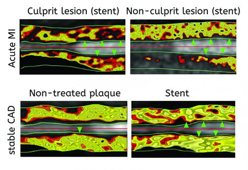
A new imaging methodology called FAI identifies inflamed artery lesions
that pose the most risk to patients. Image courtesy of Antonopoulos et
al and Science Translational Medicine.
If ongoing follow-up studies confirm the prognostic value of FAI, the
metric could transform risk stratification and clinical management for
heart disease, according to the researchers.