Changing stroke rehab and research worldwide now.Time is Brain! trillions and trillions of neurons that DIE each day because there are NO effective hyperacute therapies besides tPA(only 12% effective). I have 523 posts on hyperacute therapy, enough for researchers to spend decades proving them out. These are my personal ideas and blog on stroke rehabilitation and stroke research. Do not attempt any of these without checking with your medical provider. Unless you join me in agitating, when you need these therapies they won't be there.

What this blog is for:

My blog is not to help survivors recover, it is to have the 10 million yearly stroke survivors light fires underneath their doctors, stroke hospitals and stroke researchers to get stroke solved. 100% recovery. The stroke medical world is completely failing at that goal, they don't even have it as a goal. Shortly after getting out of the hospital and getting NO information on the process or protocols of stroke rehabilitation and recovery I started searching on the internet and found that no other survivor received useful information. This is an attempt to cover all stroke rehabilitation information that should be readily available to survivors so they can talk with informed knowledge to their medical staff. It lays out what needs to be done to get stroke survivors closer to 100% recovery. It's quite disgusting that this information is not available from every stroke association and doctors group.

Showing posts with label musical training. Show all posts

Thursday, February 23, 2023

Moderating variables of music training-induced neuroplasticity: a review and discussion

Since your doctors and therapists KNOW NOTHING on how to make neuroplasticity repeatable on demand, it is your responsibility to have musical training as a kid before you have your stroke. 

Moderating variables of music training-induced neuroplasticity: a review and discussion

  • 1Melbourne School of Psychological Sciences, The University of Melbourne, Melbourne, VIC, Australia
  • 2Department of Psychology, Université de Montréal, Montréal, QC, Canada

A large body of literature now exists to substantiate the long-held idea that musicians' brains differ structurally and functionally from non-musicians' brains. These differences include changes in volume, morphology, density, connectivity, and function across many regions of the brain. In addition to the extensive literature that investigates these differences cross-sectionally by comparing musicians and non-musicians, longitudinal studies have demonstrated the causal influence of music training on the brain across the lifespan. However, there is a large degree of inconsistency in the findings, with discordance between studies, laboratories, and techniques. A review of this literature highlights a number of variables that appear to moderate the relationship between music training and brain structure and function. These include age at commencement of training, sex, absolute pitch (AP), type of training, and instrument of training. These moderating variables may account for previously unexplained discrepancies in the existing literature, and we propose that future studies carefully consider research designs and methodologies that control for these variables.

In the last few decades, a considerable body of research has accrued on differences in the brains of musicians and non-musicians, as well as the changes created in the brain when becoming a musician. The findings of over 100 neuroimaging studies have been variously reviewed in a number of previous publications (Schlaug, 2001; Münte et al., 2002; Johansson, 2006; Altenmüller, 2008; Stewart, 2008; Habib and Besson, 2009; Jäncke, 2009; Tervaniemi, 2009; Kraus and Chandrasekaran, 2010; Wan and Schlaug, 2010; Herholz and Zatorre, 2012; Merrett and Wilson, 2012) and demonstrate convincingly that music has a significant impact on brain structure and function. Musicians and non-musicians' brains appear to have differences in volume, morphology, density, connectivity, and functional activity across a range of brain regions and structures. In addition to numerous cross-sectional studies, longitudinal music training studies in both children and adults have provided the most powerful evidence of music-induced neuroplasticity.

However, when the literature is sampled extensively, it becomes apparent that there are a number of contradictory findings. For example, a number of studies have looked at differences between musicians and non-musicians in the size or latency of electrical or magnetic potentials evoked in response to a variety of auditory stimuli. For some waveform components, such as the N1(m), there appear to be as many studies that have not found differences as those that have reported musician-non-musician differences (Pantev et al., 1998, 2001; Schneider et al., 2002; Schulz et al., 2003; Shahin et al., 2003, 2005; Kuriki et al., 2006; Lütkenhöner et al., 2006; Baumann et al., 2008). Another example can be seen in diffusion tensor imaging (DTI) studies of the corticospinal tract. Two studies found that fractional anisotropy (FA) in this tract was greater in musicians (Bengtsson et al., 2005; Han et al., 2009), while two other studies found that it was greater in non-musicians (Schmithorst and Wilke, 2002; Imfeld et al., 2009). Furthermore, there has been suprisingly little concordance between the results of whole-brain voxel-based morphometry (VBM) studies comparing musicians and non-musicians. Apart from the inferior frontal gyrus in the left hemisphere, no other brain regions were consistently found to be different between musicians and non-musicians across the five known VBM studies (Sluming et al., 2002; Gaser and Schlaug, 2003; Bermudez and Zatorre, 2005; Bermudez et al., 2009; Han et al., 2009). Although each study showed differences, the nature and location of these differences varied across studies. Even in the inferior frontal gyrus, which was significantly different in each of the VBM studies, the differences varied in their location on this gyrus across the anterior-posterior dimension.

These types of discrepancies have seldom been discussed in the literature to date. This could be due to the fact that until recently, many researchers were sceptical that music training would lead to differences in brain structure and function and/or were cautious in attributing causality to cross-sectional and correlational research. Previous studies and reviews have focused primarily on collating sufficient evidence that musicians' brains are different from non-musicians' brains, and that this reflects their experiences and not just their genetics. Because of the need to establish beyond doubt the very existence of music-induced neuroplasticity, these reviews have not focused on critically evaluating the concordance of the evidence. We would suggest that the field has matured to the point that this type of critical analysis is necessary to advance our understanding of music-induced neuroplasticity and to drive future research. It is clear that music training does induce changes in the brain, but there are numerous factors that influence when, where, and how neuroplasticity occurs in response to music training.

A number of variables that moderate the relationship between music training and neuroplasticity have been proposed in the literature. Our aim is to review these putative moderating variables and to discuss whether they could account for some of the discrepancies in the results of existing studies, including the examples given above. In addition, we will look at the implications of these moderators for future research designs and methodologies.

Age at Commencement of Training

In 1995b, Schlaug et al., published a highly influential paper that showed that the anterior half of the corpus callosum was larger in musicians than in non-musicians, but only for those musicians who commenced music training prior to 7 years of age. Since that time, a number of additional studies have reported similar findings in the corpus callosum for early trained musicians (Öztürk et al., 2002; Lee et al., 2003). Musician-specific effects in other motor regions, such as the sensorimotor cortices and pyramidal tracts, have also been correlated with age at commencement of training (Elbert et al., 1995; Amunts et al., 1997; Li et al., 2010) or practice hours in childhood (Bengtsson et al., 2005). This fits well with the behavioral literature that shows that motor skill attainment in musicians is negatively correlated with the age at which they started training. For example, an earlier age of commencement is associated with less asymmetry in hand tapping speed (Jäncke et al., 1997). Even when total years of study are accounted for, early-trained musicians outperform later-trained musicians on motor tasks (Watanabe et al., 2007).

These findings suggest that age at commencement of training is an important moderating variable of music-induced neuroplasticity. While neuroplasticity can occur throughout the lifespan, the evidence suggests that there is a sensitive period for motor learning that music training may interact with (Penhune, 2011). Based on the findings in the literature, training commenced before age seven has become a marker for early training. Those musicians who begin training prior to age seven may show greater capacity for neuroplastic changes than those who take up an instrument later in childhood or in adulthood. However, it should be noted that these types of correlations have not been found consistently across all brain regions related to motor function or related to other sensory modalities known to be influenced by music training. Although the cerebellum is a key part of the motor system, and differences between musicians and non-musicians have been found in cerebellar volume, there does not appear to be a relationship between age at commencement of training and volume in the cerebellum (Hutchinson et al., 2003). Similarly, the results in the auditory domain are mixed, with some studies finding a correlation between age of commencement of training and measures of auditory function (Pantev et al., 1998, 2001; Trainor et al., 1999; Wong et al., 2007; Musacchia et al., 2008), while another study looking at the structure of auditory cortex showed no difference (Keenan et al., 2001).

Given that this variable has not been accounted for in many studies comparing musicians and non-musicians, its impact is not fully understood. However, it could possibly account for some of the discordant findings in the literature. For example, in studies looking at FA of the corpus callosum, one study that used early-trained musicians found musician-non-musicians differences (Schmithorst and Wilke, 2002), while other studies that included musicians who commenced training after aged seven did not (Han et al., 2009; Imfeld et al., 2009). While this is a highly plausible explanation in light of the existing literature, other explanations could include differences in imaging acquisition and analysis, as the specific techniques used in these studies can have a significant impact on the results (Jones, 2010). Thus, while accounting for age at commencement of training may not solve all the existing discrepancies, it is clear that it has a significant effect on outcomes and should be reported and analyzed in future research. It might also interact with other moderating variables, such as sex and absolute pitch (AP) ability, which are discussed in more detail below.

More at link.

Monday, October 11, 2021

Therapeutic Instrumental Music Training and Motor Imagery in Post-Stroke Upper-Extremity Rehabilitation: A Randomized-Controlled Pilot Study

Why was this research needed?  Much earlier research already exists. 

Therapeutic Instrumental Music Training and Motor Imagery in Post-Stroke Upper-Extremity Rehabilitation: A Randomized-Controlled Pilot Study

https://doi.org/10.1016/j.arrct.2021.100162Get rights and content
Under a Creative Commons license
open access

Abstract

Objective

To investigate the potential benefits of three Therapeutic Instrumental Music Performance (TIMP)-based interventions in rehabilitation of the affected upper-extremity [UE] for adults with chronic post-stroke hemiparesis.

Design

Randomized-controlled pilot study

Setting

University research facility

Participants

Thirty community-dwelling volunteers [16 male/14 female; ages 33-76; mean age =55.9] began and completed the protocol. All participants had sustained a unilateral stroke > 6 months prior to enrollment [mean time post-stroke =66.9 months].

Interventions

Two baseline assessments, a minimum of one week apart; nine intervention sessions (3x/wk for 3 wks), in which rhythmically-cued, functional arm movements were mapped onto musical instruments; one post-test following the final intervention. Participants were block-randomized to one of three conditions: Group 1 - 45 minutes TIMP; Group 2 - 30 minutes TIMP, 15 minutes metronome-cued motor imagery (TIMP+cMI); Group 3 - 30 minutes TIMP, 15 minutes motor imagery without cues (TIMP+MI). Assessors and investigators were blinded to group assignment.

Main Outcome Measures

Fugl-Meyer Upper-Extremity (FM-UE); Wolf Motor Function Test- Functional Ability Scale (WMFT-FAS)

Secondary Measures

Motor Activity Log (MAL) – Amount of Use Scale; Trunk Impairment Scale.

Results

All groups made statistically significant gains on the FM-UE (TIMP, p=.005, r=.63; TIMP+cMI, p=.007, r=.63; TIMP+MI, p=.007, r=.61) and the WMFT-FAS (TIMP, p=.024, r=.53; TIMP+cMI, p=.008, r=.60; TIMP+MI, p=.008, r=.63). Comparing between-group percent change differences, on the FM-UE, TIMP scored significantly higher than TIMP+cMI (p=.032, r=.57), but not TIMP+MI. There were no differences in improvement on WMFT-FAS across conditions. On the MAL, gains were significant for TIMP (p=.030, r=.54) and TIMP+MI (p =.007, r=.63).

Conclusion

TIMP-based techniques, with and without motor imagery, led to significant improvements in paretic arm control on primary outcomes. Replacing a physical training segment with imagery-based training resulted in similar improvements; however, synchronizing internal and external cues during auditory-cued motor imagery may pose additional sensorimotor integration challenges.


Friday, May 25, 2018

New Parts of the Brain Become Active After Students Learn Physics - Study

Would learning physics be more important than learning a new musical instrument for dementia prevention and recovery from your stroke? Informed stroke survivors want to know. I'd suggest 'The Physics of Superheroes' by James Kakalios, who is a professor in the School of Physics and Astronomy at the University of Minnesota.
12 posts on music training back to June 2014.

New Parts of the Brain Become Active After Students Learn Physics - Study 


May 24, 2018


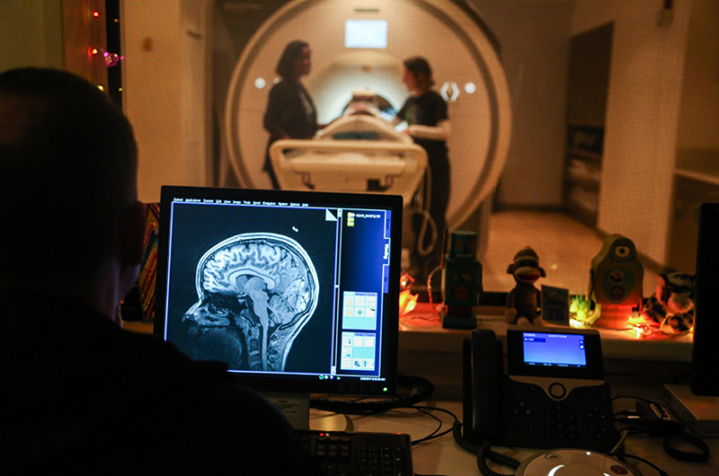
A person looking at a brain image on a monitor while someone else is helped into an MRI in the background.
Scanning using fMRI determined that physics learning activated new parts of the brain. Photo courtesy of Florida International University.

Parts of the brain not traditionally associated with learning science become active when people are confronted with solving physics problems, a new study shows.

The researchers, led by Eric Brewe, PhD, an associate professor in Drexel University’s College of Arts and Sciences, say this shows that the brain’s activity can be modified by different forms of instruction.

Using fMRI (functional magnetic resonance imaging) to measure blood flow in the brain, the researchers looked to map what areas become active when completing a physics reasoning task, both before a course on the concepts and after.

“The neurobiological processes that underpin learning are complex and not always directly connected to what we think it means to learn,” Brewe said of the findings, which were published in Frontiers in ICT.

More than 50 volunteer students took part in the study in which they were taught a physics course that utilized “Modeling Instruction,” a style of teaching which encourages students to be active participants in their learning. 

Before they participated in the class, the students answered questions from an abridged version of the Force Concept Inventory while undergoing fMRI. The Force Concept Inventory is a test that assesses knowledge of physics concepts commonly taught in early college physics classes. 

After the volunteer students completed their physics course, they again took the Force Concept Inventory, once more monitored by fMRI.

In the pre-instruction scans, parts of the brain associated with attention, working memory and problem solving — the lateral prefrontal cortex and parietal cortex, sometimes called the brain’s “central executive network” — showed activity.

“One of the keys seemed to be an area of the brain, the dorsal lateral prefrontal cortex, that generates mental simulations,” Brewe said. “This suggests that learning physics is an imaginative process, which is not typically how people think of it.”

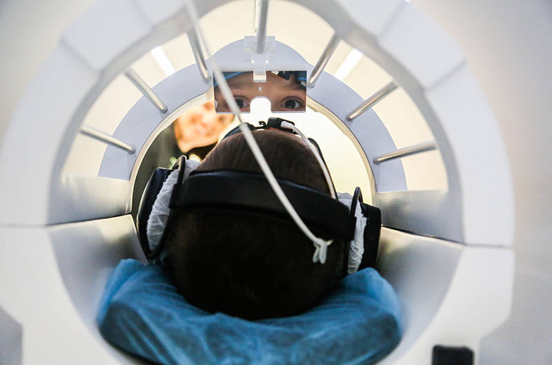
A person looking up at a mirror in an MRI machine
A person during fMRI scanning on their brain as a part of the study. Photo courtesy of Florida International University.

After the subjects had completed their class, comparison of the pre- and post-learning scans revealed increased activity in the frontal poles, which was to be expected since they’ve been linked to learning. But there was another area that also became active: the posterior cingulate cortex, which is linked to episodic memory and self-referential thought.

“These changes in brain activity may be related to more complex behavioral changes in how students reason through physics questions post- relative to pre-instruction,” Brewe and his co-authors wrote about the study. “These might include shifts in strategy or an increased access to physics knowledge and problem-solving resources.”

One of the aims of the study was to further explore how the form of teaching used, Modeling Instruction, encourages students to use their own mental models to understand new concepts. 

“The idea of mental models is something that people who research learning love to talk about, but have no evidence of what is happening inside brains other than what people say or do,” Brewe said. “We are actually looking for evidence from inside the brain.”

As such, Brewe and his fellow researchers think their study provides a good look at what might be typical when these “mental models” take hold.

But why physics? What makes this the ideal subject to study mental modeling in the brain?

Brewe said that there has been some research on the brain networks associated with learning math and reading. But mental modeling especially lends itself to physics, which has not gotten as much attention.

“Physics is a really good place to understand learning for two reasons,” Brewe said. “First, it deals with things that people have direct experience with, making formal classroom learning and informal understanding both relevant and sometimes aligned — and sometimes contrasted.”

“Second, physics is based in laws, so there are absolutes that govern the way the body works,” Brewe finished.

Moving forward, Brewe is excited by what this study opens up in his quest to improve physics learning in the United States and beyond.

“I would like to follow up on the question of mental simulations in physics, to see where that shows up at different levels of physics learning and with different populations,” he said. “But this whole study opens up many new areas of investigations and I’m pretty excited about how it will play out.”

Sunday, February 25, 2018

Playing a musical instrument could help restore brain health, research suggests

Very late to the knowledge, why the hell was this research even needed? 11 posts on music training back to June 2014.  46 posts on just listening to music back to October 2014. Does no one ever actually do anything in stroke? Like write stroke rehab protocols and get them distributed worldwide? We must have the laziest bunch of bastards in the world, with the F-bomb stroke associations doing nothing to solve any of these problems in stroke.
http://www.kurzweilai.net/playing-a-musical-instrument-could-help-restore-brain-health-research-suggests
A study by neuroscientists at Toronto-based Baycrest Rotman Research Institute and Stanford University involving playing a musical instrument suggests ways to improve brain rehabilitation methods.
In the study, published in the Journal of Neuroscience on May 24, 2017, the researchers asked young adults to listen to sounds from an unfamiliar musical instrument (a Tibetan singing bowl). Half of the subjects (the experimental group) were then asked to recreate the same sounds and rhythm by striking the bowl; the other half (the control group) were instead asked to recreate the sound by simply pressing a key on a computer keypad.
After listening to the sounds they created, subjects in the experimental group showed increased auditory-evoked P2 (P200) brain waves. This was significant because the P2 increase “occurred immediately, while in previous learning-by-listening studies, P2 increases occurred on a later day,” the researchers explained in the paper. The experimental group also had increased responsiveness of brain beta-wave oscillations and enhanced connectivity between auditory and sensorimotor cortices (areas) in the brain.
The brain changes were measured using magnetoencephalographic (MEG) recording, which is similar to EEG, but uses highly sensitive magnetic sensors.
Immediate beneficial effects on the brain
“The results … provide a neurophysiological basis for the application of music making in motor rehabilitation [increasing the ability to move arms and legs] training,” the authors state in the paper. The findings support Ross’ research in using musical training to help stroke survivors rehabilitate motor movement in their upper bodies. Baycrest scientists also have a history of breakthroughs in understanding how a person’s musical background impacts their listening abilities and cognitive function as they age.
“This study was the first time we saw direct changes in the brain after one session, demonstrating that the action of creating music leads to a strong change in brain activity,” said Bernhard Ross, PhD., senior scientist at Rotman Research Institute and senior author on the study.
“Music has been known to have beneficial effects on the brain, but there has been limited understanding into what about music makes a difference,” he added. “This is the first study demonstrating that learning the fine movement needed to reproduce a sound on an instrument changes the brain’s perception of sound in a way that is not seen when listening to music.”
The study’s next steps involve analyzing recovery by stroke patients with musical training compared to physiotherapy, and the impact of musical training on the brains of older adults. With additional funding, the study could explore developing musical training rehabilitation programs for other conditions that impact motor function, such as traumatic brain injury, and lead to hearing aids of the future, the researchers say.
The study received support from the Canadian Institutes of Health Research.

Abstract of Sound-making actions lead to immediate plastic changes of neuromagnetic evoked responses and induced beta-band oscillations during perception

Auditory and sensorimotor brain areas interact during the action-perception cycle of sound making. Neurophysiological evidence of a feedforward model of the action and its outcome has been associated with attenuation of the N1 wave of auditory evoked responses elicited by self-generated sounds, such as vocalization or playing a musical instrument. Moreover, neural oscillations at beta-band frequencies have been related to predicting the sound outcome after action initiation. We hypothesized that a newly learned action-perception association would immediately modify interpretation of the sound during subsequent listening. Nineteen healthy young adults (seven female, twelve male) participated in three magnetoencephalography (MEG) recordings while first passively listening to recorded sounds of a bell ringing, then actively playing the bell with a mallet, and then again listening to recorded sounds. Auditory cortex activity showed characteristic P1-N1-P2 waves. The N1 was attenuated during sound making, while P2 responses were unchanged. In contrast, P2 became larger when listening after sound making compared to the initial naïve listening. The P2 increase occurred immediately, while in previous learning-by-listening studies P2 increases occurred on a later day. Also, reactivity of beta-band oscillations as well as theta coherence between auditory and sensorimotor cortices was stronger in the second listening block. These changes were significantly larger than those observed in control participants (eight female, five male), who triggered recorded sounds by a keypress. We propose that P2 characterizes familiarity with sound objects, whereas beta-band oscillation signifies involvement of the action-perception cycle, and both measures objectively indicate functional neuroplasticity in auditory perceptual learning.

Thursday, July 13, 2017

Thought-controlled musical instrument may help stroke patients

But is it better than just figuring out what musical instrument the survivor can play with the deficits they have?

Thought-controlled musical instrument may help stroke patients

Neurologists have created a hands-free, thought-controlled musical instrument they hope will help empower and rehabilitate patients with motor disabilities such as those from stroke, spinal cord injury or amputation.

"There is great potential for the Encephalophone to hopefully improve rehabilitation of stroke patients and those with motor disabilities," Deuel says.
 "The Encephalophone is a musical instrument that you control with your thoughts, without movement," explained Thomas Deuel, a neuroscientist at the University of Washington, and first author of a study detailed in the journal Frontiers in Human Neuroscience.

"There is great potential for the Encephalophone to hopefully improve rehabilitation of stroke patients and those with motor disabilities," Deuel said.
 The Encephalophone collects brain signals through a cap that transforms specific signals into musical notes.

The invention is coupled with a synthesiser, allowing the user to create music using a wide variety of instrumental sounds.

In an experiment, the researchers found that participants were able to use the instrument to correctly recreate musical tones, with no prior training.
 The Encephalophone can be controlled via two independent types of brain signals -- either those associated with the visual cortex (closing one's eyes), or those associated with thinking about movement.

The researchers found that control by eye closing was more accurate than control by imagining movements.

Control by thinking about movement may be the most useful for disabled patients, and Deuel plans to continue researching this application.
 The Encephalophone is based on brain-computer interfaces using an old method, called electroencephalography, which measures electrical signals in the brain.
Neurologists have created a hands-free, thought-controlled musical instrument they hope will help empower and rehabilitate patients with motor disabilities such as those from stroke, spinal cord injury or amputation.

"There is great potential for the Encephalophone to hopefully improve rehabilitation of stroke patients and those with motor disabilities," Deuel says.

"The Encephalophone is a musical instrument that you control with your thoughts, without movement," expla ..

Tuesday, November 22, 2016

Musical training creates new brain connections in children

I'm assuming that with all the research out there that 65 posts on music and 9 posts on music training
and 31 posts on music therapy for helping survivors recover that new connections are still being made in adults. How fucking stupid does your doctor have to be to not have a music protocol for your recovery?
http://medicalxpress.com/news/2016-11-musical-brain-children.html
Taking music lessons increases brain fiber connections in children and may be useful in treating autism and Attention Deficit Hyperactivity Disorder (ADHD), according to a study being presented next week at the annual meeting of the Radiological Society of North America (RSNA).
"It's been known that musical instruction benefits children with these disorders," said Pilar Dies-Suarez, M.D., chief radiologist at the Hospital Infantil de México Federico Gómez in Mexico City, "but this study has given us a better understanding of exactly how the brain changes and where these new fiber connections are occurring."
The researchers studied 23 healthy children between the ages of five and six years old. All of the children were right handed and had no history of sensory, perception or neurological disorders. None of the children had been trained in any artistic discipline in the past.
The study participants underwent pre- and post-musical-training evaluation with (DTI) of the brain. DTI is an advanced MRI technique, which identifies microstructural changes in the brain's white matter.
"Experiencing music at an early age can contribute to better brain development, optimizing the creation and establishment of neural networks, and stimulating the existing brain tracts," Dr. Dies-Suarez said.
The brain's white matter is composed of millions of nerve fibers called axons that act like communication cables connecting various regions of the brain. Diffusion tensor imaging produces a measurement, called fractional anisotropy (FA), of the movement of extracellular water molecules along axons. In healthy white matter, the direction of extracellular water molecules is fairly uniform and measures high in fractional anisotropy. When water movement is more random, FA values decrease, suggesting abnormalities.
Over the course of life, the maturation of brain tracts and connections between motor, auditory and other areas allow the development of numerous cognitive abilities, including musical skills. Previous studies have linked autism spectrum and ADHD with decreases in volume, fiber connections and FA in the minor and lower forceps, tracts located in the of the brain. This suggests that low connectivity in the frontal cortex, an area of the brain involved in complex cognitive processes, is a biomarker of these disorders.
After the children in the study completed nine months of musical instruction using Boomwhackers—percussion tubes cut to the exact length to create pitches in a diatonic scale, DTI results showed an increase in FA and axon fiber length in different areas of the brain, most notably in the minor forceps.
"When a child receives musical instruction, their brains are asked to complete certain tasks," Dr. Dies-Suarez said. "These tasks involve hearing, motor, cognition, emotion and social skills, which seem to activate these different brain areas. These results may have occurred because of the need to create more connections between the two hemispheres of the ."
The researchers believe that the results of this study could aid in creating targeted strategies for intervention in treating disorders like autism and ADHD.
Provided by: Radiological Society of North America search and more info