Changing stroke rehab and research worldwide now.Time is Brain! trillions and trillions of neurons that DIE each day because there are NO effective hyperacute therapies besides tPA(only 12% effective). I have 523 posts on hyperacute therapy, enough for researchers to spend decades proving them out. These are my personal ideas and blog on stroke rehabilitation and stroke research. Do not attempt any of these without checking with your medical provider. Unless you join me in agitating, when you need these therapies they won't be there.

What this blog is for:

My blog is not to help survivors recover, it is to have the 10 million yearly stroke survivors light fires underneath their doctors, stroke hospitals and stroke researchers to get stroke solved. 100% recovery. The stroke medical world is completely failing at that goal, they don't even have it as a goal. Shortly after getting out of the hospital and getting NO information on the process or protocols of stroke rehabilitation and recovery I started searching on the internet and found that no other survivor received useful information. This is an attempt to cover all stroke rehabilitation information that should be readily available to survivors so they can talk with informed knowledge to their medical staff. It lays out what needs to be done to get stroke survivors closer to 100% recovery. It's quite disgusting that this information is not available from every stroke association and doctors group.

Thursday, August 29, 2019

Assessment and Rehabilitation Using Virtual Reality after Stroke: A Literature Review

A double fucking useless piece of shit. A review of assessments. How far away can you get from actual stroke rehab protocols? Maybe a review of guidelines of assessments. If someone finds one, send it to me

Assessment and Rehabilitation Using Virtual Reality after Stroke: A Literature Review


  • Pierre NolinEmail author
  • Jérémy Besnard
  • Philippe Allain
  • Frédéric Banville
  • Pierre Nolin
    • 1
    Email author
  • Jérémy Besnard
    • 2
  • Philippe Allain
    • 2
  • Frédéric Banville
    • 3
  1. 1.Département de psychologieUniversité du Québec à Trois-RivièresQuébecCanada
  2. 2.LUNAM, Laboratoire de Psychologie des Pays de la Loire (EA 4638)Université d’AngersAngersFrance
  3. 3.Département des sciences infirmièresUniversité du Québec à RimouskiRimouskiCanada
Chapter
Part of the Virtual Reality Technologies for Health and Clinical Applications book series (VRTHCA)

Abstract

This chapter presents the studies that have used virtual reality as an assessment or rehabilitation tool of cognitive functions following a stroke. To be part of this review, publications must have made a collection of data from individuals who have suffered a stroke and must have been published between 1980 and 2017. A total of 50 publications were selected out of a possible 143 that were identified in the following databases: Academic Search Complete, CINAHL, MEDLINE, PsychINFO, Psychological and Behavioural Sciences Collection. Overall, we find that most of the studies that have used virtual reality with stroke patients focused on attention, spatial neglect, and executive functions/multitasking. Some studies have focused on route representation, episodic memory, and prospective memory. Virtual reality has been used for training of cognitive functions with stroke patients, but also for their assessment. Overall, the studies support the value and relevance of virtual reality as an assessment and rehabilitation tool with people who have suffered a stroke. Virtual reality seems indeed an interesting way to better describe the functioning of the person in everyday life. Virtual reality also sometimes seems to be more sensitive than traditional approaches for detecting deficits in stroke people. However, it is important to pursue work in this emergent field in clinical neuropsychology.

Immediate and long-term memory and their relation to crystallized and fluid intelligence

Do we need this crystallized and fluid intelligence? And what is your doctor doing to ensure you have the correct amount to recover from your stroke?

Immediate and long-term memory and their relation to crystallized and fluid intelligence

Highlights

Immediate and long term memory factors predict fluid intelligence (Gf).
The same variables account for variance in crystallized intelligence (Gc).
Exploratory results suggest the memory factors account for variance in Gc due to Gf.

Abstract

The present study was conducted to investigate associations between Gc and Gf and several memory factors—namely short-term memory (STM) span, working memory capacity (WMC), and declarative long-term memory (dLTM) ability. Two hundred and thirty-six individuals completed a number of tasks assessing the above named constructs and structural equation models were fitted. In line with prior research, both WMC and dLTM ability directly accounted for variance in Gf. Notably, these same factors also accounted for variance in Gc. Importantly, however, the results of two exploratory analyses suggest that WMC and dLTM ability explain variance in Gc that is due to the investment of Gf. These exploratory analyses raise doubts about the purported role of dLTM ability in Gf and Gc performance. Ultimately, it is argued that there is need for further research investigating the cognitive components of Gc as well as dLTM ability.

Why a glass of red wine is good for your gut

Don't worry your doctor will never tell you that any alcohol is good for you. They will refer to this instead:  

Safest level of alcohol consumption is none, worldwide study shows

I ignore that, but then I'm not medically trained so I'm not biased against alcohol.  

 Why a glass of red wine is good for your gut

Tim Spector, Professor of Genetic Epidemiology, King's College London and Caroline Le Roy, Research Associate in Human Gut Microbiome, King's College London,The Conversation Wed, Aug 28 4:45 AM PDT


'To gut microbes.' View Apart/Shutterstock
Alcohol consumption guidelines vary widely between countries. In the UK and Netherlands, no more than one glass of wine or a pint of beer a day is recommended. In the US it is double these levels, and in Mediterranean countries and Chile it’s even more relaxed when it comes to drinking wine.
Though there is generally a consensus that everyone should drink less and levels of alcohol use are reducing in most countries, especially in young adults, more than 3m (or one in 20) deaths globally are attributed to alcohol consumption – making it 100 times more harmful than cannabis, cocaine and heroin.
Drinking any amount of alcohol is said to increase the risk of many diseases, including cancers, and liver disease. Yet a number of studies also seem to suggest there might be health benefits to a low intake of red wine.

Red wine and the gut

Our new research also adds support to the idea that a small glass of red wine a day might actually be beneficial to your health – specifically to your gut bacteria.
This community of trillions of microbe inhabiting our lower intestines is known as the gut microbiota. Research shows that our gut microbiota can affect multiple aspects of our general health and play a role in many illnesses but also dictate how the food we eat or the drugs we take affect us. This is partly due to the fact that gut microbes are responsible for producing thousands of chemical metabolites, that have effects on our brain, metabolism and immune systems.

Read more: Moving to another country could mess with your gut bacteria

Previous research in small studies in humans and in artificial gut models has suggested that red wine could impact our gut bacteria. And in our recent study we investigated this relationship on a large population scale in different countries to understand how drinking red wine may impact gut health compared to other alcoholic drinks.
We looked at food and drink questionnaire responses and gut bacteria diversity (that is recognised as a marker of gut health) in almost a thousand female twins in the UK, and then checked our results against two other studies of similar size in the US (the American Gut project) and the Belgium (Flemish Gut Project).


Looks moderate to me. Kinga
We found that drinking red wine (even if combined with other alcohols) is linked with an increase in gut bacteria diversity in all three countries. And as a check on other possible genetic or family biases, we also found that twins who drank more red wine than their co-twin also had more diverse gut bacteria. White wine drinkers who should be socially and culturally similar, had no significant differences in diversity, as did drinkers of other types of alcohol, like beer and spirits.
There were other associated benefits of drinking red wine too. Twins who drank red wine had lower levels of obesity and “bad” cholesterol, which we also think is partly because of the associated changes in the gut bacteria.

Precious polyphenols

Our study adds to the growing body of evidence that red wine can, when drunk in moderation, have positive effects on health. The benefits of red wine likely boil down to one key agent: polyphenols.


Guts love the polyphenols. Marako85
These molecules are natural defence chemicals found in nuts and seeds as well as many brightly coloured vegetables and fruits, including grapes. In grape, polyphenols are mostly found in the skins that are in much longer contact in the making of red wine than white. They include the tannins that have a drying effect on your tongue or resveratrol that promotes good health in people, and they also act as a fuel for our gut bacteria. This probably explains why red wine has a much stronger effect on gut bacteria than white wine. Although non-alcoholic grape juice also contains polyphenols, the fermented version contains more.
While our results are very consistent, as an observational study – where we see if factors are associated more than by chance – we cannot prove causality. To show this we’d ideally need some form of intervention study to test whether red wine directly causes an increase in gut microbiota diversity that leads to improved health. This may be popular, but difficult in practice, however. So for now, all the evidence suggests that if you have to choose an alcoholic drink today, it should definitely be a small glass of red wine.
This article is republished from The Conversation under a Creative Commons license. Read the original article.
The Conversation
Caroline Le Roy receives funding from the CDRF.
Tim Spector receives grants from multiple organisations including MRC, Wellcome Trust, NIHR, NIH, CDRF, Danone. He is a scientific founder of ZOE (global) ltd and receives royalties from a book on diet and microbiome "The Diet Myth: the real science behind what we eat" Orion 2016. He also drinks red wine.

Involvement of Epigenetic Mechanisms and Non-coding RNAs in Blood-Brain Barrier and Neurovascular Unit Injury and Recovery After Stroke

Way beyond my ability to decode. We need our fucking failures of stroke associations to translate all stroke research into usable stroke rehab protocols. But that is way beyond their abilities, they are that fucking incompetent.   I would fire everyone in every stroke association for not actually serving survivors.  The boards of directors are the most complicit in such incompetency.

Involvement of Epigenetic Mechanisms and Non-coding RNAs in Blood-Brain Barrier and Neurovascular Unit Injury and Recovery After Stroke

Svetlana M. Stamatovic1, Chelsea M. Phillips2, Gabriela Martinez-Revollar1, Richard F. Keep3,4 and Anuska V. Andjelkovic1,3*
  • 1Department of Pathology, University of Michigan Medical School, Ann Arbor, MI, United States
  • 2Neuroscience Graduate Program, University of Michigan Medical School, Ann Arbor, MI, United States
  • 3Department of Neurosurgery, University of Michigan Medical School, Ann Arbor, MI, United States
  • 4Department of Molecular Integrative Physiology, University of Michigan Medical School, Ann Arbor, MI, United States
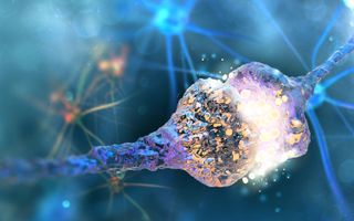
Cessation of blood flow leads to a complex cascade of pathophysiological events at the blood-vascular-parenchymal interface which evolves over time and space, and results in damage to neural cells and edema formation. Cerebral ischemic injury evokes a profound and deleterious upregulation in inflammation and triggers multiple cell death pathways, but it also induces a series of the events associated with regenerative responses, including vascular remodeling, angiogenesis, and neurogenesis. Emerging evidence suggests that epigenetic reprograming could play a pivotal role in ongoing post-stroke neurovascular unit (NVU) changes and recovery. This review summarizes current knowledge about post-stroke recovery processes at the NVU, as well as epigenetic mechanisms and modifiers (e.g., DNA methylation, histone modifying enzymes and microRNAs) associated with stroke injury, and NVU repair. It also discusses novel drug targets and therapeutic strategies for enhancing post-stroke recovery.

Introduction

Stroke is defined as an abrupt onset of focal or global neurological symptoms caused by a blockage of cerebral vessels (ischemic stroke), rupture of blood vessels (hemorrhage), or transient occlusion of small blood vessels (transient ischemic attack). It is particularly prevalent in the aging population, with people over 65 years old accounting for ∼75% of all registered cases (Hollander et al., 2003). Ischemic stroke is further subdivided based on the caliber of occluded vessels into macro- and microvascular (i.e., lacuna stroke), and based on the origin of clot-causing blockage into (a) thrombotic stroke where clot form inside brain blood vessels, and (b) thromboembolic/embolic stroke where clots form elsewhere in the body and travel toward and lodge in brain blood vessels (Reed et al., 2014; Topcuoglu et al., 2018; Tsai et al., 2018).
Pathophysiologically, cessation of blood flow leads to a complex cascade of events at the blood-vascular-parenchymal interface which evolves over time and space, and results in damage to neural cells and edema formation (Dirnagl et al., 1999; Allen et al., 2012). The central events in the hyperacute phase (within minutes and up to 6 h) include compromised mitochondrial function, anaerobic glycolysis, decreased pH (acid condition), impaired ATP production and reduced ion pump activity. As a consequence, cells swell and die, predominantly due to accumulation of lactic acid, ions (Ca2+ and Na+) and increased water influx (Dirnagl et al., 1999; Ginsberg, 2008; Nagy and Nardai, 2017).
(The following sounds an awful lot like the neuronal cascade of death. Why doesn't anyone solve this death cascade?)
There follows a cascade of events in the acute and subacute phase (hours to 7 days), including blood-brain barrier (BBB), and neurovascular unit (NVU) damage characterized by mitochondrial failure, robust production of reactive oxygen species (ROS) (superoxide O2, hydrogen peroxide, and peroxynitrite), excitotoxicity (release of glutamate from dying neurons), activation of matrix metalloproteinases (MMP2, astrocytes; MMP3, MMP9 endothelial cells and neutrophils), BBB damage triggering inflammation and blood cell infiltration (neutrophils, monocytes) that can lead to further cell death, and cellular and vasogenic edema (Enzmann et al., 2013; Posada-Duque et al., 2014; Sifat et al., 2017). The secondary damage mostly takes place in the penumbra surrounding the core infarct and its progression can extend into the chronic phase after stroke.
Paralleling these injury processes, there is activation of endogenous protective and repair mechanisms that include vascular remodeling, angiogenesis, and neurogenesis (Chopp et al., 2007; Venna et al., 2014). The degree of these ongoing repair processes on one side and persistent inflammation/damaging processes on the other determines stroke recovery and the potential risk of another stroke.
There is mounting evidence on the importance of epigenetic factors in stroke. The purpose of this review is to examine how such factors impact the cerebrovasculature in stroke injury and recovery, focusing on effects at the BBB, and NVU. There is debate over whether non-coding RNAs should be included as an epigenetic mechanism and this review will cover their effects as well as other epigenetic mechanisms.

The Blood-Brain Barrier and Neurovascular Unit in Stroke

The blood-brain barrier is a highly complex and dynamic barrier, formed by an interdependent network of brain capillary endothelial cells, endowed with barrier properties. The BBB strictly regulates paracellular permeability due to the presence of tight junctions (TJs) between endothelial cells. Those TJs are built by intricate interactions between transmembrane proteins (claudins -5, -3, -1, -12, occludin, and JAM-A), important for paracellular space occlusion, scaffolding proteins (ZO-1, -2), and the actin cytoskeleton vital for physical support and TJ function (Daneman et al., 2010; Stamatovic et al., 2016). The transcellular interactions of claudin-5 play the major role in occluding the paracellular space (Nitta et al., 2003; Ohtsuki et al., 2007). Any loosening of its adhesive interactions directly affects BBB integrity and increases paracellular permeability. The BBB role of other claudins with lower expression is still uncertain and under investigation (Tietz and Engelhardt, 2015; Sladojevic et al., 2019). BBB function is also dependent on the perivascular microenvironment, which contains cells (e.g., pericytes, astrocytes, and perivascular macrophages), neuronal endings and tissue unique extracellular matrix (Mae et al., 2011; Muoio et al., 2014). Because of this functional integration, nearly two decades ago, the concept of a BBB was broadened to a new structure, the NVU.
The neurovascular unit is composed of BBB-endowed endothelial cells and a perivascular milieu composed from cells including pericytes, smooth muscle cells, astrocytes, perivascular macrophages/microglia, neurons/neuronal endings, and extracellular matrix. The NVU mediates neurovascular coupling, modulating vessel tone (Mae et al., 2011; Muoio et al., 2014). These intimately and reciprocally linked cells and matrix generate a complex structure that regulates exchange between blood and brain, oxygen and nutrient delivery, and regional cerebral blood flow. It is essential for maintaining circulatory and brain homeostasis.
In stroke, blood-brain barrier, and neurovascular unit dysfunction actively contributes to injury pathogenesis, being a “solid substrate” for ongoing injuring processes (oxidative stress, inflammation, and cytotoxicity), contributing to ischemic core (infarct) formation in the acute phase of stroke, and facilitating the progression of injury in the subacute and chronic phases. For example, in the early (acute) phase of stroke, NVU dysfunction is characterized by disruption of BBB integrity/BBB breakdown (disassembly of TJ complex, decreases in the TJ proteins claudin-5, occludin, and ZO-1) that leads to vasogenic brain edema, a life-threatening acute stroke complication (Bauer et al., 2010; Zehendner et al., 2011; Sladojevic et al., 2014). The cell components of the NVU undergo a series of transformations during injury. Brain endothelial cells, for example, are affected very early by cytotoxic effects with dysfunction of ion channels and transporters (e.g., Na+-K+-Cl cotransporter, and Na+/H+ exchanger), release of extracellular vesicles and conversion of brain endothelial cells toward a proinflammatory and prothrombotic phenotype due to upregulation of protease-activated receptor 1 (PAR-1), tissue factor, and matrix metalloproteinases (MMPs) (Zhu et al., 2008; Bauer et al., 2010; Yamashita and Abe, 2011; Chen et al., 2015). The proinflammatory phenotype of brain endothelial cells involves an upregulation of endothelial adhesive molecules (ICAM-1, VCAM-1, P-, and E- selectins) that guide leukocyte infiltration in the acute inflammatory phase response and T and B cells infiltration in the late phase (Kleinschnitz et al., 2013; Zhou et al., 2013; Sladojevic et al., 2014). Overall, inflammation is thought to worsen acute ischemic injury, contributing to chronic focal inflammation and restricting functional recovery. However, inflammation is also involved in tissue repair.
In the hyperacute phase after stroke, pericytes may be involved in vasoconstriction, causing capillary occlusion (no-reflow phenomenon), while later, by switching to pro-inflammatory phenotype, they may enhance BBB permeability, and brain edema formation (Hall et al., 2014; Underly et al., 2017). Ischemia triggers a series of damaging reactions in astrocytes including mitochondrial dysfunction, energy depletion, ion disequilibrium, increased glutamate and Ca2+, aquaporin 4 (AQP4) channel activation, increased water permeability, and cell swelling (Friedman et al., 2009; Ito et al., 2009; Hertz et al., 2014; Mogoanta et al., 2014). It results in the release of oxidative stress products and inflammatory cytokines/chemokines (IL1, IL6, IL15, CCL2, CXCL1, CXCL10, CXCL12, and IP-10), proinflammatory associated small molecules [S100 Ca2+-binding protein B (S100B) and nitric oxide (NO)] that enhance the inflammatory post-stroke response (Yamashita et al., 2000; Hill et al., 2004; Mori et al., 2008; Shin et al., 2014; Liu H. et al., 2015; Chen et al., 2018).
Perivascular macrophages and microglia play an important role in the stroke-induced inflammatory response via production of proinflammatory cytokines (IL1|upbeta TNFα IL6, IL12) and ROS (Drake et al., 2011; Liu H. et al., 2015; Wu et al., 2016; He et al., 2019). They trigger the first line of inflammation at the NVU in the acute phase of stroke. Notable changes also occur in the extracellular matrix. At early time points (within hours), there is MMP-related basement membrane degradation with reductions in agrin, SPARC, perlecan, laminin, and fibronectin (Sole et al., 2004; Castellanos et al., 2007; Lee et al., 2011; Ji and Tsirka, 2012; Lloyd-Burton et al., 2013). This ultimately leads to increased BBB disruption, accumulation of new extracellular matrix proteins (i.e., chondroitin sulfate proteoglycan neurocan and osteopontin) and leakage of plasma proteins, such as fibrinogen, into the CNS. This mediates inflammation, edema, and potentially hemorrhagic transformation (Figure 1).

More at link. 

Amtrak stairs

These are a challenge both up and down. Down the railing is on the left side. Up I have a cup of coffee in hand, so I bounce my shoulders against the wall. Only 3 cups so far and 1 was delivered by a neighbor.  Quite steep.  The walk back to my seat with the train swaying so far hasn't resulted in me spilling my coffee or landing in someone's lap.


Amtrak travel

I'm traveling from Michigan to Salt Lake City via Amtrak. My carryon weighs about 50 lbs since it contains 6 bottles of wine. Pretty sure the selection of wine in Utah is pretty bad. Had to have a youngster hoist it into the overhead. I managed to get it down by bouncing it on the seat. 

I started out in shorts and a short sleeve shirt. With the continuous air conditioning I knew sleeping in the coach seat would be impossible. Regular travelers bring blankets.  In my manbag I had a pair of pants and jacket. Went to the bathroom to change, none were labeled handicapped, so I had a 2 by 2 foot floor space to change in. I have to sit and cross my legs to get pants and shoes on. With size 12 feet taking up most of the floor there was much swearing to get into warmer clothes. I had an aisle seat, so easy access out to the bathroom and observation car. It was on the left side so I had to forcefully grab my left arm all the time to keep it from wandering into my neighbors space.  Doing that while sleeping is a challenge.

But the leg room is terrific, maybe 1.5 feet in front of my knees as compared to flying where my knees are buried in the seat back in front of me, which is why I can no longer sleep on planes.

Life is great.

What Is the Evidence for Physical Therapy Poststroke? A Systematic Review and Meta-Analysis

My conclusion from this is that there are NO PROTOCOLS. And thus you are stuck with wild-assed guesses and guidelines. Hope you like ambiguity.  With no plan to get you 100% recovered you will be disabled for the rest of your life.  I use a lot of compensation and risk-taking to live my pretty normal life. But then I am just physically disabled a bit.

What Is the Evidence for Physical Therapy Poststroke? A Systematic Review and Meta-Analysis

Abstract

Background:
Physical therapy (PT) is one of the key disciplines in interdisciplinary stroke rehabilitation. The aim of this systematic review was to provide an update of the evidence for stroke rehabilitation interventions in the domain of PT.
Methods and Findings:
Randomized controlled trials (RCTs) regarding PT in stroke rehabilitation were retrieved through a systematic search. Outcomes were classified according to the ICF. RCTs with a low risk of bias were quantitatively analyzed. Differences between phases poststroke were explored in subgroup analyses. A best evidence synthesis was performed for neurological treatment approaches. The search yielded 467 RCTs (N=25373; median PEDro score 6 [IQR 5–7]), identifying 53 interventions. No adverse events were reported. Strong evidence was found for significant positive effects of 13 interventions related to gait, 11 interventions related to arm-hand activities, 1 intervention for ADL, and 3 interventions for physical fitness. Summary Effect Sizes (SESs) ranged from 0.17 (95%CI 0.03–0.70; I2 =0%) for therapeutic positioning of the paretic arm to 2.47 (95%CI 0.84–4.11; I2 =77%) for training of sitting balance. There is strong evidence that a higher dose of practice is better, with SESs ranging from 0.21 (95%CI 0.02–0.39; I2 =6%) for motor function of the paretic arm to 0.61 (95%CI 0.41–0.82; I2 =41%) for muscle strength of the paretic leg. Subgroup analyses yielded significant differences with respect to timing poststroke for 10 interventions. Neurological treatment approaches to training of body functions and activities showed equal or unfavorable effects when compared to other training interventions. Main limitations of the present review are not using individual patient data for meta-analyses and absence of correction for multiple testing.
Conclusions:
There is strong evidence for PT interventions favoring intensive high repetitive task-oriented and task-specific training in all phases poststroke. Effects are mostly restricted to the actually trained functions and activities. Suggestions for prioritizing PT stroke research are given.
 

Further Evidence That Acting Like An Extravert Can Boost Wellbeing

How many decades before your doctor informs you of this and has a protocol for you to accomplish this? I used to consider myself a complete introvert, moving to Michigan knowing absolutely no one required me to put myself out there and be open to any overture. Now I'm considered the life of the party and usually close it down. 

Further Evidence That Acting Like An Extravert Can Boost Wellbeing 

By Matthew Warren
Researchers have long known that people who are more extraverted tend to be happier, leading some to suggest that encouraging extraverted behaviour could improve wellbeing. Last year we reported on the first trial of such an intervention, which found that acting like an extravert for a week led to an increase in positive emotions in certain people. Now a second study appears to have replicated that result — and shown that behaving like an introvert may also reduce wellbeing.

In the new study, published in the Journal of Experimental Psychology: General, Seth Margolis and Sonja Lyubomirsky at the University of California, Riverside, asked 131 participants to alter their behaviour over a two week period to be more extraverted or introverted. For one week, participants were encouraged to act as “talkative”, “assertive” and “spontaneous” as possible; for the other, they were told to act “deliberate”, “quiet” and “reserved” (all participants completed both weeks, but half began with the extraverted week while the others began with with the introverted week). 
To encourage the participants to actually alter their behaviour, the researchers asked them to list five specific changes they planned on making, and then sent them periodic reminders of their task throughout the study.  At various points across the two weeks, the participants completed scales measuring their experience of positive and negative emotions and others aspects of wellbeing, as well as their personality traits.
Compared to baseline levels at the start of the study, participants experienced more positive emotions during the extraverted week — and also showed reduced positive emotions during the introverted week. Some other measures of wellbeing, such as feelings of connectedness and flow (the experience of being immersed in — and enjoying — an activity) were also boosted by acting extraverted and reduced by acting introverted.
However, these results didn’t hold for all measures of wellbeing. For instance, participants seemed to have reduced negative emotions compared to baseline during both interventions (although the exact pattern of results differed depending on whether participants began with the extraverted or introverted week).
The results add to the small, albeit growing, body of evidence that acting like an extravert can improve certain aspects of wellbeing — particularly measures of positive emotion. But the authors suggest that their biggest contribution is to show that acting like an introvert can also have an effect. “Given that introversion is generally not regarded as desirable or advantageous in U.S. culture … we believe our most compelling results are those showing that well-being decreases can be substantial when people act more introverted than usual,” they write.
Still, it seems too soon to suggest that we should we all begin behaving like extraverts. The study the Digest reported on last year found that people who had high trait levels of introversion didn’t report the same benefits of acting like an extravert as the rest of the participants, and actually became more fatigued and experienced more negative emotions. On the other hand, the new paper found that baseline levels of extraversion and introversion didn’t affect the results – but it’s still clear that researchers need a better understanding of how individual differences could influence the effectiveness of the intervention.
And it will also be important to figure out which behaviours are actually causing the increases or decreases in wellbeing reported in these studies. It’s not yet clear whether it was being more “talkative”, “assertive”, or “spontaneous” that resulted in an increase in wellbeing in the extraverted week, for instance, and the researchers suggest examining changes in a more specific sub-set of extraverted behaviours in the future. “We hope that research from our and others’ laboratories encourages future investigators to test the potential of behavioral interventions to spur both personality change and well-being gains,” they conclude.
Experimental manipulation of extraverted and introverted behavior and its effects on well-being
Matthew Warren (@MattbWarren) is Editor of BPS Research Digest

Research shows surprising link between weightlifting and cognition

I suppose you could ask your doctor to analyze all these and come up with an EXACT REHAB EXERCISE PROTOCOL.  But you know damn well your doctor is incapable of that, so start guessing what you should do. Aren't you glad you are paying your doctor for expertise?   

Are Aerobic Programs Similar in Design to Cardiac Rehabilitation Beneficial for Survivors of Stroke? A Systematic Review and Meta‐Analysis August 2019  

Forced, Not Voluntary, Aerobic Exercise Enhances Motor Recovery in Persons With Chronic Stroke August 2019 

Sorry Cardio Queens, Science Says Anaerobic Exercises Are Way More Efficient 

August 2019


Research shows surprising link between weightlifting and cognition


John Murphy, MDLinx | August 15, 2019
In a new study in the Journal of Applied Physiology, researchers demonstrated that weight training can overcome cognitive impairment and even jumpstart the creation of new neurons. Just three resistance-training workouts a week were enough to improve cognition and boost memory performance in “gym rats.”
Advertisement
Older ‘gym rats’ might very well be staving off age-related cognitive impairment, researchers reported.
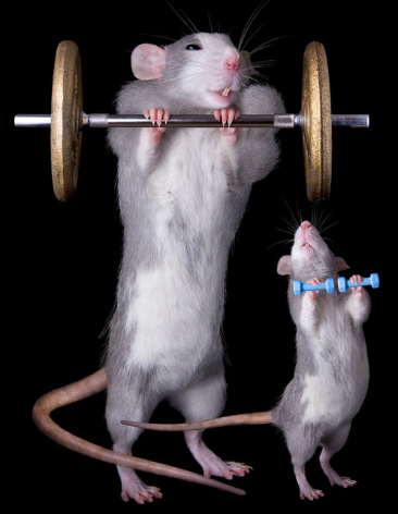
But in this study, the “gym rats” were actual rats. Lab rats, to be precise.
Of course, this doesn’t mean that humans who weight-train will get the same brain gains. Nevertheless, the researchers suspect that strengthening exercises may very well counteract age-related cognitive impairment and memory loss in humans.

Strong muscles, stronger mind

A good deal of previous research led up to this study. Other investigators have found indications that resistance training in humans heightens IGF-1 signaling—an indicator of neurogenesis—and increases neuroplasticity. But no previous researchers had ever really mapped out the molecular signaling process that connects resistance training to improved cognitive function and memory.
To discover these molecular changes, the current study researchers first needed rats with cognitive impairment. Because neuroinflammation precedes age-related cognitive impairment, the researchers created inflammation in the rats’ brains by injecting a type of fat that induced neuroinflammation in the rodents.
Then it was time for weight training. Since it’s very likely impossible to coach rats to lift little dumbbells—especially rats who are cognitively impaired—the researchers instead attached small weights to the rats’ hindquarters and trained the rodents to climb a 3-foot high ladder. Doing several sets of this activity a few times per day, 3 days per week, had the effect of strength training. Soon, the rats were building muscles. As their strength increased, the rats were given progressively heavier weights.
Another group of rats (the sedentary group) also had induced neuroinflammation, but did not have to do resistance training. A third group of rats (the control group) had sham training but weren’t given the fat injection to the brain, so they weren’t cognitively impaired.
At 5 weeks, the researchers gave the three groups of rats a memory test in a maze. As expected, the cognitively impaired rats had more trouble than the unimpaired control rats. But after a few days, the cognitively impaired rats who had done strength training matched—and in some cases exceeded—the ability of the control rats. The sedentary rats lagged far behind.

To the researchers, this indicated that weight training was able to reverse the impairment caused by neuroinflammation, even though the inflammation in their brains was still present.
Examination of the rats’ brains showed increased activity in downstream IGF-1 signaling, indicating new neurons were generated. The researchers also found molecular signs of increased neuroplasticity, suggesting that the weight-trained rats had recovered some brain function despite their cognitive impairment.
“This model offers a potential therapy that may prevent or delay the onset of mild cognitive impairment in neurodegenerative diseases that warrants further investigation,” the authors concluded.
That’s great news for rats. But will it work in humans?

Go ahead and lift weights

As the study authors wrote, more research needs to be done. In the meantime, they added, weight training to stave off age-related cognitive impairment and memory loss is certainly worth trying.
“This is an option for the elderly,” Frank W. Booth, PhD, professor, Department of Biomedical Sciences, University of Missouri, Columbia, MO, said to the Columbia Daily Tribune. Dr. Booth was one of the authors and he funded the study out of his own pocket. “You see old individuals just sitting around. If they start using their muscles, it will be helpful to society.”
In an interview with The New York Times, lead author and doctoral candidate Taylor Kelty said: “I think it’s safe to say that people should look into doing some resistance training. It’s good for you for all kinds of other reasons, and it appears to be neuroprotective. And who doesn’t want a healthy brain?”

Wednesday, August 28, 2019

Paired Associative Stimulation as a Tool to Assess Plasticity Enhancers in Chronic Stroke

Stroke survivors don't need lazy 'assessments' of MEP(Motor Evoked Potentials). We need protocols. WHEN THE HELL WILL YOU GET THERE?

Paired Associative Stimulation as a Tool to Assess Plasticity Enhancers in Chronic Stroke

Joshua Silverstein1, Mar Cortes2, Katherine Zoe Tsagaris1, Alejandra Climent3, Linda M. Gerber4, Clara Oromendia4, Pasquale Fonzetti5,6, Rajiv R. Ratan5,7,8, Tomoko Kitago1,5*, Marco Iacoboni9,10, Allan Wu10,11, Bruce Dobkin12 and Dylan J. Edwards13,14
  • 1Human Motor Recovery Laboratory, Burke Neurological Institute, White Plains, NY, United States
  • 2Department of Rehabilitation and Human Performance, Icahn School of Medicine at Mount Sinai, New York, NY, United States
  • 3Sant Joan de Deu Hospital, Department of Neurology, University of Barcelona, Barcelona, Spain
  • 4Department of Healthcare Policy and Research, Weill Cornell Medical College, New York, NY, United States
  • 5Department of Neurology, Weill Cornell Medical College, New York, NY, United States
  • 6Memory Evaluation and Treatment Service, Burke Rehabilitation Hospital, White Plains, NY, United States
  • 7Burke Neurological Institute, White Plains, NY, United States
  • 8Feil Family Brain and Mind Research Institute, Weill Cornell Medical College, New York, NY, United States
  • 9Department of Psychiatry and Biobehavioral Sciences, UCLA Semel Institute for Neuroscience and Human Behavior, Los Angeles, CA, United States
  • 10Ahmanson-Lovelace Brain Mapping Center, University of California, Los Angeles, Los Angeles, CA, United States
  • 11Department of Neurology, University of California, Los Angeles, Los Angeles, CA, United States
  • 12Department of Neurology, Geffen School of Medicine, Reed Neurologic Research Center, University of California, Los Angeles, Los Angeles, CA, United States
  • 13Moss Rehabilitation Research Institute, Elkins Park, PA, United States
  • 14School of Medical and Health Sciences, Edith Cowan University, Perth, WA, Australia
Background and Purpose: The potential for adaptive plasticity in the post-stroke brain is difficult to estimate, as is the demonstration of central nervous system (CNS) target engagement of drugs that show promise in facilitating stroke recovery. We set out to determine if paired associative stimulation (PAS) can be used (a) as an assay of CNS plasticity in patients with chronic stroke, and (b) to demonstrate CNS engagement by memantine, a drug which has potential plasticity-modulating effects for use in motor recovery following stroke.
Methods: We examined the effect of PAS in fourteen participants with chronic hemiparetic stroke at five time-points in a within-subjects repeated measures design study: baseline off-drug, and following a week of orally administered memantine at doses of 5, 10, 15, and 20 mg, comprising a total of seventy sessions. Each week, MEP amplitude pre and post-PAS was assessed in the contralesional hemisphere as a marker of enhanced or diminished plasticity. Strength and dexterity were recorded each week to monitor motor-specific clinical status across the study period.
Results: We found that MEP amplitude was significantly larger after PAS in baseline sessions off-drug, and responsiveness to PAS in these sessions was associated with increased clinical severity. There was no observed increase in MEP amplitude after PAS with memantine at any dose. Motor threshold (MT), strength, and dexterity remained unchanged during the study.
Conclusion: Paired associative stimulation successfully induced corticospinal excitability enhancement in chronic stroke subjects at the group level. However, this response did not occur in all participants, and was associated with increased clinical severity. This could be an important way to stratify patients for future PAS-drug studies. PAS was suppressed by memantine at all doses, regardless of responsiveness to PAS off-drug, indicating CNS engagement.

Introduction

The capacity of the brain to make structural, physiological, and genetic adaptations following stroke, otherwise known as plasticity, is likely to be critical for improving sensorimotor impairments and functional activities. Promotion of adaptive plasticity in the central nervous system (CNS) leading to sustained functional improvement is of paramount importance, given the personal suffering and cost associated with post-stroke disability (Ma et al., 2014). In addition to rehabilitation therapies to retrain degraded motor skills, animal and human studies have tried to augment recovery with neuropharmacologic interventions. Unfortunately, few if any have had a notable effect in patients or have come into routine use (Martinsson et al., 2007; Chollet et al., 2011; Cramer, 2015; Simpson et al., 2015). Methods to screen drugs based on their presumed mechanism of action on plasticity in human motor systems could speed translation to patients. However, there is currently no accepted method in stroke patients for evaluating the potential effectiveness or individual responsiveness to putative “plasticity enhancing” drugs in an efficient, low-cost, cross-sectional manner, in order to establish target engagement in humans and to avoid the extensive time and cost of protracted clinical trials.
Paired associative stimulation (PAS) is a safe, painless, and non-invasive technique known to result in short-term modulation of corticospinal excitability in the adult human motor system, lasting ∼90 min (Stefan et al., 2000; Wolters et al., 2003). Post-PAS excitability enhancement has been considered an LTP-like response thought to relate to transient changes in synaptic efficacy in the glutamatergic system at the N-methyl-D-aspartate (NMDA) receptor, since both human NMDA receptor deficiency (Volz et al., 2016) and pharmacological manipulation with dextromethorphan (Stefan et al., 2002) can block the effect. While PAS has been explored as a potential therapeutic intervention in patients with residual motor deficits after stroke (Jayaram and Stinear, 2008; Castel-Lacanal et al., 2009), it has not previously been investigated for its potential use as an assay of motor system plasticity in this context. Prior studies have suggested that motor practice and PAS share the same neuronal substrates, modulating LTP and LTD-like plasticity in the human motor system (Ziemann et al., 2004; Jung and Ziemann, 2009); therefore, as an established non-invasive human neuromodulation method (Suppa et al., 2017), we reasoned that PAS would be a suitable assay in the present study to examine the effect of a drug on motor system plasticity.
Here, we examine the effect of memantine, a drug used for treatment of Alzheimer’s disease, on the PAS response in patients with chronic stroke. Memantine is described pharmacologically as a low affinity, voltage dependent, non-competitive, NMDA antagonist (Rogawski and Wenk, 2003). At high concentrations, like other NMDA-R antagonists, it can inhibit synaptic plasticity. At lower, clinically relevant concentrations, memantine can, under some circumstances, promote synaptic plasticity by selectively inhibiting extra-synaptic glutamate receptor activity while sparing normal synaptic transmission, and hence may have clinical utility for rehabilitation (Xia et al., 2010). Interest in specifically using the drug for its interaction with stroke pathophysiology stems from animal models of both prevention (Trotman et al., 2015), in which pre-conditioning reduced infarct size, as well as for functional recovery, in which chronic oral administration starting >2 h post-stroke resulted in improved function through a non-neuroprotective mechanism (López-Valdés et al., 2014). In humans, memantine taken over multiple days has been used to demonstrate that the NMDA receptor is implicated in specific transcranial magnetic paired-pulse measures (Schwenkreis et al., 1999), and short-term training-induced motor map reorganization (Schwenkreis et al., 2005). In studies of neuromodulation, memantine blocked the facilitatory effect of intermittent theta-burst stimulation (iTBS) (Huang et al., 2007). Similarly, LTP-like plasticity induced by associative pairing of painful laser stimuli and TMS over primary motor cortex (M1) can also be blocked by memantine (Suppa et al., 2013). The effects of memantine on the PAS response have not yet been demonstrated, including examination of potential dose-response effects, which would be important for the potential clinical application of memantine for stroke recovery.
In our study, we set out to determine whether PAS might be a useful tool to probe the potential for plasticity after stroke in persons with chronic hemiparesis and apply PAS as an assay to look at drug effects on motor system plasticity using memantine. We hypothesized that (a) PAS would enhance corticospinal excitability in the contralesional hemisphere of stroke patients, and that (b) since PAS-induced plasticity is thought to involve a short-term change in glutamatergic synaptic efficacy, memantine would have a dose-dependent effect on PAS response. We predicted that at low doses, memantine would enhance PAS-induced plasticity through selective blockade of extrasynaptic NMDA receptors, whereas higher doses would inhibit PAS-induced plasticity.


More at link.  

Blueberries May Help Fight Alzheimer’s

So at least you have an amount, but good luck finding  freeze-dried blueberry powder. Living in Michigan I buy extra pints of blueberries in summer and freeze for the rest of the year, have about 12 pints in the freezer right now. Hope real blueberries are just as good

Blueberries May Help Fight Alzheimer’s

Blueberries, a popular fruit, already classified as a “superfruit” for its health benefits, could now also help fight dementia, new research suggests.


Blueberries may help fight Alzheimer’s disease, according to a study conducted by University of Cincinnati Academic Health Center in Ohio.

The study was conducted by the team from the Universityof Cincinnati Academic Health Center in Ohio. Lead author Robert Krikorian said:

"Our new findings corroborate those of previous animal studies and preliminary human studies, adding further support to the notion that blueberries can have a real benefit in improving memory and cognitive function in some older adults.”

Read more Does Memory Loss Always Mean Dementia?

A type of flavonoid known as anthocyanins, found in blueberries are not only responsible for its purple and blue color but, they also contribute to its numerous health benefits. Besides anthocyanins, an assorted variety of phenolic compounds can be found in blueberries. The compounds – quercetin, myricetin, kaempferol and chlorogenic acid contribute to the antioxidant capacity thereby increasing the health benefits of the superfood.


First Study Reveals Blueberries May Help Fight Alzheimer’s


One study involved 47 adults aged 68 and older, who had mild cognitive impairment, a risk condition for Alzheimer’s disease. They were given freeze-dried blueberry powder, which is equivalent to a cup of berries, or a placebo powder once a day for 16 weeks.

Dr. Krikorian reported:  

"There was improvement in cognitive performance and brainfunction in those who had the blueberry powder compared with those who took the placebo.

"The blueberry group demonstrated improved memory and improved access to words and concepts."

MRI scans conducted on the participants showed increased brain activity in those who ingested the blueberry powder, suggesting the superfoods may help fight Alzheimer’s disease.

Read more Blueberriesmay prevent diabetes, says research

Dr Krikorian said blueberries' beneficial effects could be due to flavonoids called anthocyanins, which have been shown to improve cognition in animals.

plaques and tangles in Alzheimer's


A Bigger Study Reveals Same Results


A second study involved 94 people aged 62 to 80 who did not have measurable cognitive decline but reported experiencing memory loss. They were given either blueberry powder, fish oil (containing omega-3 fatty acids believed to prevent Alzheimer's), a combination of fish oil and blueberry powder, or a placebo.

"The results were not as robust as with the first study," said Dr. Krikorian.

"Cognition was somewhat better for those with powder or fish oil separately, but there was little improvement with memory."

fMRI results for participants given blueberry powder were found to be less significant than those observed in the first study.

Read more Drinkingbeet juice boosts muscle strength in heart failure patients

Dr Krikorian says that the difference may be because the participants had less severe issues when they entered the research.

He said the two studies provide a basis for future research from which they hope to ascertain whether or not blueberries could shield against the onset of Alzheimer's symptoms. While promising, the less robust results of the second study indicate that more research will be necessary.

At present, the researchers plan to conduct a study involving participants aged 50-65, including a number of people considered to be at risk of developing Alzheimer's - people who are obese, suffer from hypertension (high blood pressure) or hypercholesterolemia (high levels of cholesterol).

Movement therapy induced neural reorganization and motor recovery in stroke: A review

Useless review. No protocols came out of it. 

Movement therapy induced neural reorganization and motor recovery in stroke: A review

Here's Why Drugs That Work So Well in Mouse Brains Often Fail Miserably in Humans

Well, years ago Dr. Michael Tymianski of the Toronto Western Hospital Research Institute in Canada referenced 1000+ failed neuroprotective clinical trials. Of course I don't know what they are, but your doctor should know every one of those failed trials. 

Everyone of those failed trials should be looked at based on this new data and rerun. But that assumes we have a great stroke association and leader we can talk to. Well instead we have fucking failures of stroke associations doing nothing to get to 100% recovery.  

Here's Why Drugs That Work So Well in Mouse Brains Often Fail Miserably in Humans

A synapse where a signal travels from one neuron to the next.
(Image: © Shutterstock)
Neuroscientists face a major obstacle in developing drugs to treat brain disorders — if the drugs work really well on mice, they often fall short when humans are treated. Now, a new study suggests a potential reason why:  Brain cells in mice turn on genes that are very different from the ones in human brain cells.
Mice and humans have evolutionarily conserved brains, meaning they have very similar brain architectures made up of similar types of brain cells. In theory, that makes mice ideal test subjects for neuroscientists, who don't typically have the ability to peer into living human brains.
Yet for mysterious reasons, treatments that worked beautifully in the mouse brain often don't pan out when tested in humans.
Related: 7 Ways to Trick Your Brain
CLOSE
To figure out why that may be, a group of scientists from the Allen Institute for Brain Science in Seattle analyzed brains donated from deceased people and brain tissue donated by epilepsy patients after brain surgery. They specifically looked at a part of the brain called the medial temporal gyrus, which is involved in language processing and deductive reasoning.
Researchers sorted through nearly 16,000 cells from this brain region and identified 75 different cell types. When they compared the human cells with a data set of mouse cells, they found that mice had counterparts that were similar to almost all of those human brain cells.
But when they looked at which genes were switched on or off inside those cells, they found stark differences between the mouse and human cells.
For example, serotonin is a neurotransmitter — or brain chemical — that regulates appetite, mood, memory and sleep. It does so by binding to brain cells via a receptor on the cell surface, which acts like a glove that is made to catch a baseball.
But a mouse's serotonin receptors are not found on the same cells that they're found in humans, the researchers discovered. So a drug that increases serotonin levels in the brain, such as those used to treat depression, might deliver it to vastly different cells in mice than in humans.
They also found differences in the expression of genes that help build connections between neurons. In essence, the cellular roadmap in our brains may look very different from what it looks like in a mouse.
"The bottom line is there are great similarities and differences between our brain and that of the mouse," co-senior author Christof Koch, the chief scientist and president of the Allen Institute for Brain Science, said in a statement. "One of these tells us that there is great evolutionary continuity, and the other tells us that we are unique."
"If you want to cure human brain diseases, you have to understand the uniqueness of the human brain," he added. The findings were published yesterday (Aug. 21) in the journal Nature.
Originally published on Live Science.