Changing stroke rehab and research worldwide now.Time is Brain! trillions and trillions of neurons that DIE each day because there are NO effective hyperacute therapies besides tPA(only 12% effective). I have 523 posts on hyperacute therapy, enough for researchers to spend decades proving them out. These are my personal ideas and blog on stroke rehabilitation and stroke research. Do not attempt any of these without checking with your medical provider. Unless you join me in agitating, when you need these therapies they won't be there.

What this blog is for:

My blog is not to help survivors recover, it is to have the 10 million yearly stroke survivors light fires underneath their doctors, stroke hospitals and stroke researchers to get stroke solved. 100% recovery. The stroke medical world is completely failing at that goal, they don't even have it as a goal. Shortly after getting out of the hospital and getting NO information on the process or protocols of stroke rehabilitation and recovery I started searching on the internet and found that no other survivor received useful information. This is an attempt to cover all stroke rehabilitation information that should be readily available to survivors so they can talk with informed knowledge to their medical staff. It lays out what needs to be done to get stroke survivors closer to 100% recovery. It's quite disgusting that this information is not available from every stroke association and doctors group.

Showing posts with label 33% chance of getting dementia/Alzheimers. Show all posts

Wednesday, August 19, 2026

Building strength in this 1 area of the body can reduce dementia risk, research shows

 Ask your competent? doctor if this is enough to counteract the dementia risk from your stroke. NO ANSWER IS INCOMPETENCE!

Your risk of dementia, has your doctor told you of this?  Your doctor is responsible for preventing this! Is s/he willing to prevent this?

1. A documented 33% dementia chance post-stroke from an Australian study?   May 2012.

2. Then this study came out and seems to have a range from 17-66%. December 2013.`    

3. A 20% chance in this research.   July 2013.

4. Dementia Risk Doubled in Patients Following Stroke September 2018 

My left hand grip strength is not good, right hand is great. Since my doctor failed at curing my left hand opening spasticity getting it stronger is difficult.

The latest here: 

Building strength in this 1 area of the body can reduce dementia risk, research shows

A wealth of research links dementia risk to lifestyle choices and chronic illnesses. Among that data are studies that consistently show physical activity, specifically building and maintaining strength, can reduce one's likelihood of cognitive decline.

out as an especially reliable indicator for cognitive health, one 2022 study found: your hands and forearms.

Improving and maintaining grip strength during middle age can lead to better neurological health later in life, Jennifer A. Schrack, Ph.D., director at the Johns Hopkins Bloomberg School of Public Health’s Center on Aging and Health, tells TODAY.com.

However, she adds, building grip strength involves much more than upper-body workouts at the gym; it’s an undertaking that calls for a full-body commitment meant to stimulate your mind for the long run.

How Grip Strength and Dementia Are Related

Your grip strength is a reflection of your total body strength, which research has repeatedly linked to overall cognitive function, says Schrack. "We use grip strength in studies because it’s easy to measure, whereas total body strength is much more difficult to measure," she explains.

Grip strength in particular can track how well the brain is communicating with the rest of the body to control motor function and sensation, she adds. It is measured with a hand dynamometer, available at physical therapists’ offices, some primary care offices and at hand specialist practices.

A 2019 study found the weaker an older adult's grip strength, the greater the likelihood of cognitive impairment. The study authors said that measuring grip strength can help identify people who may have symptoms beyond normal age-related cognitive decline and need early intervention.

A 2022 study looking at grip strength and dementia risk similarly concluded that increasing muscle strength in middle age may help maintain brain health.

Because muscle mass typically peaks between ages 30 and 35, TODAY.com previously reported, strength and coordination will progressively decline after that. It also doesn’t help that “we tend to do less as we age,” Schrack says. That lack of movement further drives this loss of muscle mass, she explains, and degeneration in muscle function often precedes mental degeneration.

When to Start Building Grip Strength

While there are benefits to being fit at all stages of life, Shrack says the most critical time to prioritize building strength to reduce dementia risk is between ages 45 and 65. Doing so combats the decline in muscle mass taking place.

In one study, the most physically active adults (in mid-life and late-life) were 41-45% less likely to develop dementia than the least active.

What is considered good grip strength? Shrack points to a 2014 study that found the average grip strength for men was between 26-32 kilograms (meaning they squeezed the equivalent of this weight with their grip), and anything less than that was considered “weak.” For women, grip strength between 16-20 kilograms was considered “intermediate,” and anything below 16 was “weak.”

How to Improve Grip Strength

Since grip strength is a reference for overall strength, improving it calls for strengthening your entire body, says Schrack. But you can also integrate grip strength into your routine with specific exercises in the same way you'd treat any other muscle group.

Your approach should depend "on a person’s age and fitness level, of course, but there’s lots of things people can do,” Schrack says.

For older populations, she recommends chair stands, wall pushups and light dumbbells. They can also squeeze stress balls or use resistance bands between their arms if they want to dedicate part of their workouts to grip strength.

Occupations, hobbies and sports, she says, can make a difference, too. Schrack’s mother is an 85-year-old organist whose hands get a challenge every day. And those who enjoy knitting, tennis or golf will definitely have their grip strength put to the test.

Younger people, on the other hand, will need something more challenging, says Schrack. Their muscles will require regular strength training and progressive overload. For grip-strength-focused exercises, Alexander Rothstein, Ed.D., assistant professor of exercise science at New York Institute of Technology’s School of Health Professions, tells TODAY.com he recommends these workouts:

  • Farmer’s carry
  • Suitcase carry
  • Bottoms-up kettlebell carry
  • Dead Hang

This article was originally published on TODAY.com

Thursday, August 13, 2026

Drug taken for common condition may be slowing Alzheimer’s decline

 With your risk of dementia post stroke, is your competent? doctor preparing testing for stroke patients to see if this would prevent Alzheimers'? NO? NOTHING DOING? PURE INCOMPETENCE THEN!

Your risk of dementia, has your doctor told you of this?  Your doctor is responsible for preventing this! Is s/he willing to prevent this?

1. A documented 33% dementia chance post-stroke from an Australian study?   May 2012.

2. Then this study came out and seems to have a range from 17-66%. December 2013.`    

3. A 20% chance in this research.   July 2013.

4. Dementia Risk Doubled in Patients Following Stroke September 2018 

The latest here: 

Drug taken for common condition may be slowing Alzheimer’s decline

A blood-thinning medication already taken by millions of people for an irregular heartbeat may also be helping to slow cognitive decline in patients with Alzheimer’s disease, according to new research from Karolinska Institutet in Sweden.

The study, published in the European Heart Journal, found that people with both atrial fibrillation and Alzheimer’s disease who were treated with newer blood thinners known as NOACs unintentionally experienced a significantly slower rate of cognitive decline than those given the older drug named warfarin or no blood thinner at all.

“There are reasons to believe that the treatment could have a positive effect on cognition, for example by improving blood flow and reducing small-scale damage in the brain,” Maria Eriksdotter, a professor at Karolinska Institutet who led the study, said in a statement.

Newsweek reached out to Karolinska Institutet for more information.

Why the Two Conditions Are Often Linked

Atrial fibrillation, a common heart rhythm disorder among older adults, frequently occurs alongside Alzheimer’s disease.

Doctors typically prescribe blood-thinning medication to atrial fibrillation patients to lower their risk of blood clots. Earlier research had suggested that anticoagulant treatment might reduce the risk of developing dementia in the first place, but scientists knew far less about how these drugs affect people who already have Alzheimer’s.

Researchers drew on data from SveDem, Sweden’s national quality register for cognitive disorders and dementia, examining 7,308 people diagnosed with both atrial fibrillation and Alzheimer’s disease.

Participants were sorted into three matched groups: those taking NOACs, those taking warfarin, and those taking no anticoagulant medication at all. Cognitive function was tracked over time using the Mini-Mental State Examination (MMSE), a standard test used to measure memory and thinking skills.

What the Results Showed

Patients on NOACs saw their cognitive decline slow by just over 0.2 MMSE points per year compared with those on warfarin or no treatment at all.

The benefits extended beyond cognition. Patients treated with NOACs also had a lower risk of death, stroke, blood clots and fractures compared with those who received no anticoagulant treatment. Warfarin was also linked to lower risks of death, stroke and blood clots, but it came with a higher risk of major bleeding than NOACs.

England-based general practitioner, Dr Mohammad Bakhtiar, spoke with Newsweek about the findings.

“Over 7,000 people is large enough that the signal is hard to dismiss as noise,” he said. “That makes the data more reliable than the usual small clinic series.

“What I take from the study is encouraging evidence for the newer anticoagulants over warfarin in this group. The cognitive difference is modest, but it sits alongside fewer strokes, clots and deaths, and less major bleeding than warfarin. For GPs deciding between a NOAC and warfarin in someone with Alzheimer’s and atrial fibrillation, that is useful.”

The researchers cautioned that, because the study was observational, it cannot prove that the drugs directly caused the slower decline. Some factors that may have influenced both which drug a patient was prescribed and their eventual health outcomes could not be fully accounted for. Some patients also may have switched between medications during the course of the study, which followed participants over time. All in all, while they remain optimistic, they acknowledge the promising results require further studies.

“We should read observational studies cautiously,” Bakhtiar added. “People who get a NOAC are often different from people who get nothing. They may be fitter, better supported, or under more active specialist care. Some patients also switched drugs during follow-up.

“So, the honest line is association, not proof. NOACs were linked with slower decline. We do not know they caused it.”

He advises that patients excited by the news do not start, stop or switch their current medications because of this paper.

The study was funded by several organizations, including the Swedish Research Council, the Swedish Brain Foundation, CIMED, ALF project funding and Karolinska Institutet.

Reference

Eriksdotter, M., et al. (2026). Oral anticoagulants, cognition, and clinical outcomes in atrial fibrillation and Alzheimer’s disease: a Swedish nationwide study. European Heart Journal. https://doi.org/10.1093/eurheartj/ehag584.

Contact Newsweek editors on this story: Marc Vargas and Gray R. Thomas


An Alzheimer’s drug cut expected cognitive decline roughly in half. It didn’t target amyloid

 You may need this; do you have a competent? doctor ready?

Your risk of dementia, has your doctor told you of this?  Your doctor is responsible for preventing this! Is s/he willing to prevent this?

1. A documented 33% dementia chance post-stroke from an Australian study?   May 2012.

2. Then this study came out and seems to have a range from 17-66%. December 2013.`    

3. A 20% chance in this research.   July 2013.

4. Dementia Risk Doubled in Patients Following Stroke September 2018 

The latest here: 

An Alzheimer’s drug cut expected cognitive decline roughly in half. It didn’t target amyloid


Researchers say the oral drug LM11A-31 slowed the decline of brain-network function in a small trial, pointing to a potential broader strategy for treating Alzheimer’s.

Could the key to treating Alzheimer’s be protecting the brain’s wiring? Potentially.

Last month, at the Alzheimer’s Association International Conference in London, Indiana University and the biopharmaceutical company PharmatrophiX presented findings that an oral drug called LM11A-31 slowed the decline of brain-network function in people with mild-to-moderate Alzheimer’s. The results, which were drawn from brain scans of 159 trial participants, point to a treatment strategy that looks well beyond the field’s longtime focus on amyloid, according to a press release.

For years, most Alzheimer’s drug development has centered on clearing amyloid plaques, and more recently tau tangles. But researchers say it’s more complicated than that.

“The main reason [to look further] is that pure amyloid beta plaque and tau tangle Alzheimer’s disease is rare,” Maria C. Carrillo, chief science officer at the Alzheimer’s Association, told Inc. The association awarded PharmatrophiX a $600,000 grant in 2013 to run an early safety trial of the oral drug LM11A-31. 

In most biologically confirmed cases, she told Inc., it’s actually “mixed dementia,” with other proteins and vascular damage present alongside the two hallmark lesions. That complexity is pushing scientists toward treatments that protect the brain itself. 

“Alzheimer’s is a complex disease driven by multiple interacting mechanisms,” Aaron Burstein, head of search and evaluation at the Alzheimer’s Drug Discovery Foundation, told Inc. The foundation has backed the drug’s lead inventor, Frank Longo, since 2000 and was an early investor in PharmatrophiX. 

“Amyloid is a validated target, but it is unlikely to be the only one,” he said.

The oral drug LM11A-31 focuses on the synapses, the junctions where brain cells communicate. It targets a receptor called p75, and according to the release, earlier studies have suggested it can make synapses more resilient to the damage caused by both amyloid and tau.

The new part is the ability to measure whether that’s actually working. The Indiana University team, led by Paul Territo, processed metabolic PET scans to build “network maps” of how efficiently different brain regions communicate.  Related video: Groundbreaking Alzheimer's treatment offers patients new hope (FOX 35 Orlando) 

“Alzheimer’s disease is not simply a disease of individual brain regions. It is a disease of disrupted brain networks,” Territo said in the release.

The analysis found dose-dependent effects, with the strongest benefits in patients on the higher 400-milligram dose, along with distinct patterns between men and women. It builds on earlier results, in which the drug cut the expected progression of cognitive decline roughly in half.

“Protecting synapses has become a central therapeutic goal,” Burstein told Inc. “This type of approach could support a precision medicine framework in which treatments target different aspects of the disease and are ultimately combined based on each patient’s biology.”

Implications for the future of Alzheimer’s treatments

Everyone involved has been careful to call the signal a starting point.

“LM11A-31’s potential will need to be confirmed in a larger clinical trial,” Burstein said. 

Investors, clinicians, and patients, he added, will want consistent evidence that the drug slows cognitive and functional decline in ways that are meaningful in daily life and sustained over time.

Carrillo agreed that the next step runs through regulators. She said that confirming the signal means a trial showing that over one to two years the treatment “slows or perhaps even stops the memory decline in comparison to a placebo group,” on terms agreed upon with the FDA.

In the release, PharmatrophiX said more planning is underway for that larger Phase 2b/3 trial. If it succeeds, LM11A-31 could become one more tool used alongside amyloid drugs.

“We know that the current approved treatments for early Alzheimer’s disease are just the beginning,” Carrillo told Inc. “More treatments are coming, each improving on the earlier treatments.”

PharmatrophiX did not respond to Inc.’s request for comment. 

This post originally appeared at inc.com.

“Click here to subscribe to the Inc. newsletter: inc.com/newsletters"

Study of Brain Immune Cells Reveals New Clues to Alzheimer’s Disease Progression

 Have your competent? doctor and hospital create protocols from this. Can't do that: PURE INCOMPETENCE!

 You'll need them because of this;

Your risk of dementia, has your doctor told you of this?  Your doctor is responsible for preventing this! Is s/he willing to prevent this?

1. A documented 33% dementia chance post-stroke from an Australian study?   May 2012.

2. Then this study came out and seems to have a range from 17-66%. December 2013.`    

3. A 20% chance in this research.   July 2013.

4. Dementia Risk Doubled in Patients Following Stroke September 2018 

The latest here: 

Study of Brain Immune Cells Reveals New Clues to Alzheimer’s Disease Progression

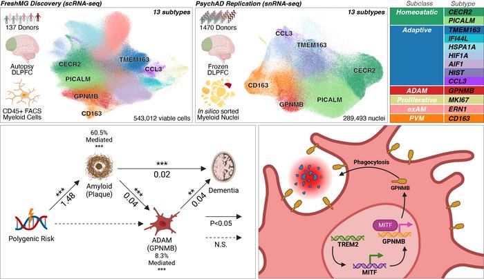
Summary: Researchers have mapped over 830,000 brain immune cells, discovering a protective subtype of microglia that expands as Alzheimer’s disease progresses. Driven by the TREM2 molecular pathway, these cells actively help clear harmful material from the brain. The findings provide a new roadmap for developing therapies focused on strengthening the brain’s natural immune defenses rather than targeting amyloid plaques alone.

Key Facts:

  • Unprecedented Scale: The study analyzed over 830,000 myeloid-origin immune cells from 1,607 donors, providing the most detailed reference to date of immune cell changes across aging and disease.
  • Protective Microglia Subtype: A specific, disease-associated subtype of microglia becomes more abundant as Alzheimer’s advances, acting to protect the brain by engulfing and clearing harmful material.
  • Crucial Molecular Pathway: The beneficial, protective effects of these immune cells rely entirely on a molecular signaling pathway involving the proteins TREM2, MITF, and GPNMB.

Source: Mount Sinai Hospital / Mount Sinai School of Medicine

The brain’s immune cells are increasingly recognized as key players in Alzheimer’s disease, but exactly how they change as the disease develops has remained unclear. Now, a groundbreaking study published in Nature Genetics provides the most comprehensive map to date of these cells.

Researchers from the Icahn School of Medicine at Mount Sinai have identified a protective subtype of brain immune cell that expands as Alzheimer’s disease progresses, uncovering the molecular pathway that enables these cells to help defend the brain. The findings offer new insights that could inform future Alzheimer’s therapies.

Led by Donghoon Lee, PhD, and Panos Roussos, MD, PhD, the team analyzed more than 830,000 myeloid-origin immune cells of the brain. This included microglia—the brain’s resident immune cells—and perivascular macrophages, which are crucial for modulating immune responses. The cells were sourced from the prefrontal cortex of 1,607 donors spanning a wide range of ages and Alzheimer’s disease pathology stages.Donghoon Lee graphical abstract.

Graphical abstract of the work. Credit Mount Sinai Health System.

By profiling brain tissue at this unprecedented scale, the team identified six subclasses comprising 13 distinct subtypes of myeloid cells, characterizing how these populations adapt during aging and disease progression.

Crucially, the researchers identified a disease-associated subtype of microglia that becomes increasingly abundant as Alzheimer’s disease advances. Rather than contributing to neurodegeneration, these cells appear to play a protective role by increasing their ability to engulf and clear harmful material from the brain.

The study further identified a molecular pathway involving the proteins TREM2, MITF, and GPNMB that is required to maintain this protective microglial state. Experiments in both human tissue and mouse models demonstrated that the beneficial effects of these cells depend strictly on TREM2 signaling.

“Our study provides the clearest picture yet of how the brain’s immune cells adapt during aging and Alzheimer’s disease,” said Donghoon Lee, PhD, Assistant Professor of Genetics and Genomic Sciences and Psychiatry at Mount Sinai, and first and corresponding author of the paper.

“By identifying the specific immune cells that appear to protect the brain—and the molecular signals they rely on—we have uncovered potential new targets for therapies aimed at slowing Alzheimer’s disease progression.”

Beyond identifying this protective microglial population, the study helps explain why genetic variants in immune-related genes such as TREM2 and APOE increase Alzheimer’s risk. Ultimately, the findings provide a roadmap for developing therapies that strengthen the brain’s natural immune defenses.

Editorial Notes:

  • This article was edited by a Neuroscience News editor.
  • Journal paper reviewed in full.
  • Additional context added by our staff.

About this Alzheimer’s disease research news

Author: Elizabeth Dowling (Media Contact)
Source: The Mount Sinai Hospital / Mount Sinai School of Medicine
Contact: Elizabeth Dowling – The Mount Sinai Hospital / Mount Sinai School of Medicine
Image: Graphical abstract credit goes to Mount Sinai Health System

Original Research: Peer-Reviewed Publication “Plasticity of human microglia and brain perivascular macrophages in aging and Alzheimer’s disease” by Donghoon Lee, Panos Roussos, et al. Nature Genetics

DOI: 10.1038/s41588-026-02716-6

Tuesday, August 11, 2026

Study of Brain Immune Cells Reveals New Clues to Alzheimer’s Disease Progression

 How will your competent? doctor use this to prevent your likely dementia? NOTHING LIKE USUAL!

Your risk of dementia, has your doctor told you of this?  Your doctor is responsible for preventing this! Is s/he willing to prevent this?

1. A documented 33% dementia chance post-stroke from an Australian study?   May 2012.

2. Then this study came out and seems to have a range from 17-66%. December 2013.`    

3. A 20% chance in this research.   July 2013.

4. Dementia Risk Doubled in Patients Following Stroke September 2018 

You do know your doctor is TOTALLY FUCKING INCOMPETENT  in everything stroke related, right?

 Your incompetent doctor knew nothing of TREM2, right?

  • NMOSD (2 posts to March 2018)

Study of Brain Immune Cells Reveals New Clues to Alzheimer’s Disease Progression

Summary: Researchers have mapped over 830,000 brain immune cells, discovering a protective subtype of microglia that expands as Alzheimer’s disease progresses. Driven by the TREM2 molecular pathway, these cells actively help clear harmful material from the brain. The findings provide a new roadmap for developing therapies focused on strengthening the brain’s natural immune defenses rather than targeting amyloid plaques alone.

Key Facts:

  • Unprecedented Scale: The study analyzed over 830,000 myeloid-origin immune cells from 1,607 donors, providing the most detailed reference to date of immune cell changes across aging and disease.
  • Protective Microglia Subtype: A specific, disease-associated subtype of microglia becomes more abundant as Alzheimer’s advances, acting to protect the brain by engulfing and clearing harmful material.
  • Crucial Molecular Pathway: The beneficial, protective effects of these immune cells rely entirely on a molecular signaling pathway involving the proteins TREM2, MITF, and GPNMB.

Source: Mount Sinai Hospital / Mount Sinai School of Medicine

The brain’s immune cells are increasingly recognized as key players in Alzheimer’s disease, but exactly how they change as the disease develops has remained unclear. Now, a groundbreaking study published in Nature Genetics provides the most comprehensive map to date of these cells.

Researchers from the Icahn School of Medicine at Mount Sinai have identified a protective subtype of brain immune cell that expands as Alzheimer’s disease progresses, uncovering the molecular pathway that enables these cells to help defend the brain. The findings offer new insights that could inform future Alzheimer’s therapies.

Led by Donghoon Lee, PhD, and Panos Roussos, MD, PhD, the team analyzed more than 830,000 myeloid-origin immune cells of the brain. This included microglia—the brain’s resident immune cells—and perivascular macrophages, which are crucial for modulating immune responses. The cells were sourced from the prefrontal cortex of 1,607 donors spanning a wide range of ages and Alzheimer’s disease pathology stages.

Donghoon Lee graphical abstract.
Graphical abstract of the work. Credit Mount Sinai Health System.

By profiling brain tissue at this unprecedented scale, the team identified six subclasses comprising 13 distinct subtypes of myeloid cells, characterizing how these populations adapt during aging and disease progression.

Crucially, the researchers identified a disease-associated subtype of microglia that becomes increasingly abundant as Alzheimer’s disease advances. Rather than contributing to neurodegeneration, these cells appear to play a protective role by increasing their ability to engulf and clear harmful material from the brain.

The study further identified a molecular pathway involving the proteins TREM2, MITF, and GPNMB that is required to maintain this protective microglial state. Experiments in both human tissue and mouse models demonstrated that the beneficial effects of these cells depend strictly on TREM2 signaling.

“Our study provides the clearest picture yet of how the brain’s immune cells adapt during aging and Alzheimer’s disease,” said Donghoon Lee, PhD, Assistant Professor of Genetics and Genomic Sciences and Psychiatry at Mount Sinai, and first and corresponding author of the paper.

“By identifying the specific immune cells that appear to protect the brain—and the molecular signals they rely on—we have uncovered potential new targets for therapies aimed at slowing Alzheimer’s disease progression.”

Beyond identifying this protective microglial population, the study helps explain why genetic variants in immune-related genes such as TREM2 and APOE increase Alzheimer’s risk. Ultimately, the findings provide a roadmap for developing therapies that strengthen the brain’s natural immune defenses.

Editorial Notes:

  • This article was edited by a Neuroscience News editor.
  • Journal paper reviewed in full.
  • Additional context added by our staff.

About this Alzheimer’s disease research news

Author: Elizabeth Dowling (Media Contact)
Source: The Mount Sinai Hospital / Mount Sinai School of Medicine
Contact: Elizabeth Dowling – The Mount Sinai Hospital / Mount Sinai School of Medicine
Image: Graphical abstract credit goes to Mount Sinai Health System

Original Research: Peer-Reviewed Publication “Plasticity of human microglia and brain perivascular macrophages in aging and Alzheimer’s disease” by Donghoon Lee, Panos Roussos, et al. Nature Genetics
DOI: 10.1038/s41588-026-02716-6

Thursday, August 6, 2026

Gut microbiome shifts may begin before Alzheimer’s symptoms appear

 Then have your competent? doctor test for this so THOSE EXACT PREVENTION PROTOCOLS CAN BE INITIATED! Don't have any, do they? PURE INCOMPETENCE! 

Why you need them;

1. A documented 33% dementia chance post-stroke from an Australian study?   May 2012.

2. Then this study came out and seems to have a range from 17-66%. December 2013.`    

3. A 20% chance in this research.   July 2013.

4. Dementia Risk Doubled in Patients Following Stroke September 2018 

Gut microbiome shifts may begin before Alzheimer’s symptoms appear

A new review traces how diet, gut bacteria, and their metabolites may intersect with the silent biological changes that precede cognitive decline.

In a recent narrative review published in the journal Nutrients, researchers across Australia summarized evidence on the gut microbiota, microbial metabolites, and dietary patterns in relation to early Alzheimer’s disease (AD). The review did not generate or analyze new data.

AD has a long preclinical stage when metabolic alterations, amyloid accumulation, and neuroinflammation develop while people remain cognitively unimpaired (CU). Understanding these biological changes can help elucidate mechanisms pertinent to early prevention. Recent findings suggest that early AD pathology may be accompanied by alterations in the gut microbiota and other systemic changes. Diet plays an important role in gut microbiota composition and metabolic output.

Diet-induced changes in the microbiota may influence gut–brain communication relevant to metabolic and neuroinflammatory regulation. Research also links long-term dietary patterns to the risk of cognitive impairment and AD. Nevertheless, the relevance of microbiome–diet interactions to microbial functional pathways in preclinical AD remains poorly defined. In this narrative review, researchers summarized evidence linking the gut microbiota, microbial metabolites, and diet to early AD.

AD: Diagnosis and Therapeutic Context

Research advances have enabled AD detection based on biological markers without relying solely on clinical symptoms. Cerebrospinal fluid (CSF) biomarkers and amyloid positron emission tomography (PET) imaging enable the detection of pathology in CU individuals. Standardized amyloid PET quantification, such as the Centiloid scale, can help stratify people along the preclinical continuum and facilitate comparisons across studies.

Current AD treatments offer limited benefit once cognitive decline is established. Common symptomatic treatments, including memantine, donepezil, galantamine, and rivastigmine, provide modest cognitive and functional improvements. Further, while disease-modifying therapies, for example, lecanemab, have been developed for early-stage AD, they are restricted to select populations and have limited clinical benefit.

As such, most available treatments are initiated only after substantial damage has occurred, thereby reducing their capacity to meaningfully modify disease progression. This therapeutic limitation constitutes a substantial challenge in AD treatment, underscoring the need for further research and attention toward earlier AD stages.

Gut–Brain Communication

The gut–brain axis (GBA) is the bidirectional communication network between the central nervous system (CNS) and the gastrointestinal system. It plays an important role in regulating physiological processes, including brain function, metabolism, and immune activity. Alterations in the microbiota composition can modify metabolic and inflammatory conditions in ways that may increase vulnerability to neurodegenerative processes.

Microbial metabolites are an important component of gut–brain communication. Short-chain fatty acids (SCFAs), the most studied metabolites, regulate immune cell differentiation, metabolic pathways, epithelial barrier integrity, and cytokine production. SCFAs can modulate neuronal function and neuroinflammatory processes through these actions. Emerging evidence suggests that GBA disruptions may contribute to the metabolic and inflammatory disturbances seen in early AD. However, direct human evidence in preclinical AD remains limited, and SCFA effects may depend on their concentration and biological context.

Gut Microbiome Dysregulation in AD

A growing body of research suggests that AD is associated with changes in gut microbiota function and composition. In particular, reduced abundance of SCFA-producing genera, such as Eubacterium, Roseburia, and Faecalibacterium, has been commonly reported in AD and cognitive impairment. Meanwhile, some studies report an increased relative abundance of Escherichia/Shigella and other Proteobacteria, which are associated with inflammatory signaling, metabolic imbalance, and oxidative stress.

Microbiota functional alterations in AD include disruptions in fermentation pathways, changes in lipid and amino acid metabolism, and decreases in SCFA biosynthesis. Early changes in microbial metabolic pathways and SCFA-producing taxa have been reported in a small number of studies involving CU individuals with biomarker evidence of amyloid pathology. Although studies have reported inconsistent findings, they suggest that the earliest stages of AD may be characterized by gut microbial dysregulation.

Dietary Patterns Associated With AD

Dietary Approaches to Stop Hypertension (DASH) and the Mediterranean diet (MD) are associated with improved cognitive, cardiovascular, and metabolic outcomes in aging populations. Both emphasize increased intake of plant-based foods, while limiting processed and red meats, added sugars, and saturated fats. Higher adherence to these diets is linked to reduced cerebral amyloid burden, slower cognitive decline, and improved vascular function in older individuals.

The Mediterranean-DASH Intervention for Neurodegenerative Delay (MIND) diet, which combines elements from DASH and MD, has similarly been associated with slower accumulation of AD-related pathology. Further, the prudent dietary pattern has been associated with improved inflammatory and metabolic profiles in aging populations. It is characterized by high intake of fruits, whole grains, vegetables, fish, low-fat dairy, and legumes. The prudent dietary pattern has been associated with higher microbial diversity and enrichment of SCFA-producing taxa.

Concluding Remarks

Collectively, available research suggests that alterations in the composition and metabolic activity of the gut microbiota may be associated with early AD pathology. Reductions in SCFA-producing taxa and alterations in microbial metabolic pathways have been observed in a limited number of studies involving CU populations with amyloid pathology.

However, the current evidence base has important limitations. Most studies have involved populations with AD or mild cognitive impairment rather than biomarker-confirmed preclinical AD, and much of the available evidence is cross-sectional, preventing conclusions about the direction of the relationship. Sequencing has generally been limited to the genus level, while few studies have jointly assessed diet, gut microbiota, microbial functional pathways, SCFAs, and amyloid pathology. As a narrative review, this work also focused primarily on SCFAs rather than other potentially relevant microbial metabolites.

Future studies should prioritize longitudinal, species-level metagenomic analyses in CU individuals grouped by amyloid status. These studies should assess microbial functional pathways, SCFAs, and diet within the same cohort. Researchers should also investigate fecal microbiota transplantation as a potential intervention in experimental AD models.

Journal reference:
  • Dissanayaka DMS, Rainey-Smith SR, Sohrabi HR, et al. (2026). Gut Microbiome Changes in Preclinical Alzheimer’s Disease. Nutrients, 18(15):2469. DOI: 10.3390/nu18152469. https://www.mdpi.com/2072-6643/18/15/2469

Wednesday, August 5, 2026

Midlife Vascular Health Adds Nearly 13 Dementia-Free Years

 My blood pressure didn't spike until age 60, now controlled, the rest I'm good at. But ask your competent? doctor if your stroke negates these factors! NO answer; PURE FUCKING INCOMPETENCE!

1. A documented 33% dementia chance post-stroke from an Australian study?   May 2012.

2. Then this study came out and seems to have a range from 17-66%. December 2013.`    

3. A 20% chance in this research.   July 2013.

4. Dementia Risk Doubled in Patients Following Stroke September 2018 

Midlife Vascular Health Adds Nearly 13 Dementia-Free Years

Summary: Maintaining healthy blood pressure, avoiding diabetes, and not smoking between ages 48 and 68 is associated with nearly 13 additional years of dementia-free life.

While the presence of all three vascular risk factors significantly accelerated dementia onset and reduced dementia-free survival across all groups, the researchers noted key demographic variations: women lived longer without dementia than men regardless of risk profile, and white participants experienced longer dementia-free lifespans than Black participants under similar risk conditions.

Key Facts

  • 13-Year Healthspan Gain: Individuals free of hypertension, diabetes, and smoking from ages 48 to 68 lived an average of nearly 13 additional years without developing dementia compared to those with all three risk factors (30 years vs. 17 years post-baseline).
  • Broad Cohort Scope: Analyzed 12,409 participants (average baseline age of 56) followed over a 26-year tracking period, during which 3,008 developed dementia and 5,238 died dementia-free.
  • Sex-Based Differences: Women consistently maintained longer dementia-free survival than men across equivalent risk categories (18.1 years for women with all three risk factors vs. 16.6 years for men).
  • Racial Disparities: White participants with all three risk factors lived an average of 19.6 dementia-free years compared to 16.0 years for Black participants, emphasizing the need for targeted midlife interventions in vulnerable populations.
  • Critical Prevention Window: Emphasizes age 48 to 68 as a crucial developmental window where aggressive vascular risk modification directly safeguards cognitive reserve in later life.

Source: NYU

Having normal blood pressure, no diabetes, and not smoking from age 48 to 68, are associated with nearly 13 additional years on average of dementia-free life, a new study shows.

Led by NYU Langone Health researchers, the new work also found that, no matter how many risk factors they had, women lived longer without dementia than men, and that white study participants had more dementia-free years than Black participants.

This shows a man and a brain.
Avoiding midlife vascular risk factors adds nearly 13 additional years of dementia-free life. Credit: Neuroscience News

Published online Aug. 5 in Neurology, the study results underscore the dual impact of the identified midlife risk factors, the presence of which was linked to both accelerated dementia onset and shortened overall dementia-free lifespans.

“Our findings argue that people need to actively avert these factors in midlife as a strategy for preserving brain health for more than a decade,” said study senior investigator Josef Coresh, MD, PhD, founding director, Optimal Aging Institute, NYU Langone.

“Discovering new ways to delay dementia is crucial with 42 percent of Americans at risk for developing the condition at any time after age 55.” Dr. Coresh is also the Terry and Mel Karmazin Professor, Department of Population Health, NYU Langone.

For the new study, the researchers analyzed data in the community-based Atherosclerosis Risk in Communities (ARIC) Study, which started in 1986 and followed participants over decades into late life, measuring midlife vascular risk factors and dementia.

The current study looked at 12,409 people with an average age of 56 who were free of dementia. Participants were assessed for three risk factors: high blood pressure, diabetes, and smoking. They were then followed for an average of 26 years. During that time, 3,008 developed dementia and 5,238 people died without developing dementia. Those who had no risk factors lived without developing dementia nearly 13 years longer than people with the three risk factors (30 years total after the study’s start versus 17 years).

The team also sought to determine how cardiovascular risk factors in midlife influenced dementia-free survival across demographics. Women with all three risk factors lived an average of 18.1 years from the start of tracking without developing dementia, compared to 16.6 years for male study participants. White participants with all three risk factors lived an average of 19.6 years without developing dementia, compared to 16 years for Black participants.

“These results suggest that vascular risk reduction may benefit all groups, but that certain populations, such as Black adults, may especially benefit from targeted prevention efforts,” Dr. Coresh added. “Hopefully, these results will encourage people to stop smoking and watch their vascular health closely from age 48 on.”

Importantly, the study was not designed to prove that avoiding these vascular risk factors caused the observed delay in dementia — only that they were associated with each other. Another limitation of the study was that the risk factor measurement was performed just once, so changes over time were not captured.

Funding: This study was supported by National Institutes of Health (NIH) grants K24HL152440 and K01DK138273, and the ARIC study was funded by grants NIH U01HL096812, U01HL096814, U01HL096899, U01HL096902 and U01HL096917.

Along with Dr. Coresh, study authors are Jiaqi Hu, Institute for Hospital Management, Tsinghua University in Beijing (lead author); Jason Smith, University of North Carolina School of Medicine; A. Richey Sharrett, Elizabeth Selvin and Michael Fang, Department of Epidemiology, Johns Hopkins Bloomberg School of Public Health; Rebecca F. Gottesman, Stroke Branch, National Institute of Neurological Disorders and Stroke; Pamela Lutsey, Division of Epidemiology and Community Health, School of Public Health, University of Minnesota; Thomas H. Mosley Jr., MIND Center, University of Mississippi Medical Center School of Medicine, Jackson; and Jordan Weiss, Department of Medicine (Division of Precision Medicine) and Optimal Aging Institute, NYU Grossman School of Medicine.

Key Questions Answered:

Q: Why are midlife vascular factors so influential on dementia risk decades later?

A: Midlife hypertension, diabetes, and smoking cause cumulative structural damage to the brain’s microvasculature. Chronic hypoperfusion, small vessel disease, inflammation, and blood-brain barrier breakdown reduce cognitive resilience and accelerate pathological protein accumulation, laying the groundwork for clinical dementia late in life.

Q: What did the study reveal regarding health disparities between demographic groups?

A: Even when carrying the same three vascular risk factors, Black participants experienced shorter dementia-free lifespans (16.0 years) than white participants (19.6 years). This highlights that systemic healthcare disparities, social determinants of health, and differences in baseline disease severity likely amplify the cognitive burden of vascular risk in Black communities.

Q: Did the study establish that treating these risk factors directly prevents dementia?

A: The study demonstrated a strong prospective association rather than direct causation. Because risk factors were measured at baseline, the observational design does not capture how midlife treatment changes over time impact dementia trajectories, though the robust 26-year tracking strongly supports midlife risk reduction as a primary preventative strategy.

Editorial Notes:

  • This article was edited by a Neuroscience News editor.
  • Journal paper reviewed in full.
  • Additional context added by our staff.

About this dementia and aging research news

Author: David March
Source: NYU
Contact: David March – NYU
Image: The image is credited to Neuroscience News

Original Research: The findings will appear in Neurology