Changing stroke rehab and research worldwide now.Time is Brain! trillions and trillions of neurons that DIE each day because there are NO effective hyperacute therapies besides tPA(only 12% effective). I have 523 posts on hyperacute therapy, enough for researchers to spend decades proving them out. These are my personal ideas and blog on stroke rehabilitation and stroke research. Do not attempt any of these without checking with your medical provider. Unless you join me in agitating, when you need these therapies they won't be there.

What this blog is for:

My blog is not to help survivors recover, it is to have the 10 million yearly stroke survivors light fires underneath their doctors, stroke hospitals and stroke researchers to get stroke solved. 100% recovery. The stroke medical world is completely failing at that goal, they don't even have it as a goal. Shortly after getting out of the hospital and getting NO information on the process or protocols of stroke rehabilitation and recovery I started searching on the internet and found that no other survivor received useful information. This is an attempt to cover all stroke rehabilitation information that should be readily available to survivors so they can talk with informed knowledge to their medical staff. It lays out what needs to be done to get stroke survivors closer to 100% recovery. It's quite disgusting that this information is not available from every stroke association and doctors group.

Showing posts with label alzheimers cause. Show all posts

Saturday, September 19, 2026

Potential root cause of Alzheimer’s may have just been discovered

 With your risk of dementia post stroke will your incompetent? doctor do nothing with this?

Your doctor is responsible for preventing this! Is s/he willing to prevent this?

1. A documented 33% dementia chance post-stroke from an Australian study?   May 2012.

2. Then this study came out and seems to have a range from 17-66%. December 2013.`    

3. A 20% chance in this research.   July 2013.

4. Dementia Risk Doubled in Patients Following Stroke September 2018 

The latest here:

Potential root cause of Alzheimer’s may have just been discovered

A small shift in a brain molecule may explain how Alzheimer’s disease turns from an abnormal nerve connection to a condition affecting memories, a new study suggests.

According to previous research, abnormal protein clumps known as amyloid plaques and tau tangles are the key drivers of Alzheimer’s.

The clumps are a hallmark of a wider breakdown in which nerve cells become overactive, lose their connections and see supporting cells turn reactive. But how these changes link to cognitive decline remains one of the disease’s central mysteries.

The new study, from the Institute of Basic Science in South Korea, says a receptor protein molecule, ERBB4, may be the missing link.

An abnormal activation of this molecule in nerve cells called excitatory neurons can initiate several Alzheimer’s-related problems at once.

Researchers found that a distinct population of excitatory neurons emerged early in the Alzheimer’s process and they carried the ERBB4 receptor. The receptor allows nerve cells to respond to external signals.

In the study, researchers first probed two types of brain cells, called astrocytes and microglia, which support neurons and help remove unwanted connections. Researchers have long suspected that the normal clean-up functions of astrocytes and microglia are compromised in Alzheimer’s disease.

Researchers found that in mice with Alzheimer’s symptoms, astrocytes and microglia removed increasing numbers of healthy excitatory neuron connections.

They also found that non-neuronal cells in the brain, known as glial cells, were unbalancing the signals meant for accelerating nerve activity and those for stopping it.

This kind of imbalance sometimes appears before major cognitive symptoms become obvious in Alzheimer’s patients.
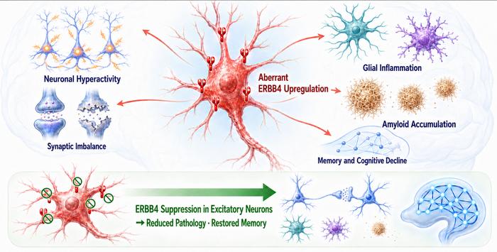
Schematic illustration of the proposed disease mechanism
Schematic illustration of the proposed disease mechanism (Institute for Basic Science)

To understand the root cause of this imbalance, researchers looked at which genes were active within individual cell nuclei.

In healthy brains, the ERBB4 receptor is found mainly in neurons that inhibit signals and help apply brakes to brain activity. But in Alzheimer’s, ERBB4 was found in excitatory neurons responsible for driving brain activity.

The findings indicate that Alzheimer’s may not only damage neurons, but also push some of them into an abnormal state.

Researchers discovered that a single intervention targeting the ERBB4 receptors could address many of the Alzheimer’s symptoms.

Distinct population of early responsive excitatory neurons that was selectively enriched in the Alzheimer's disease model
Distinct population of early responsive excitatory neurons that was selectively enriched in the Alzheimer's disease model (Institute for Basic Science)

They used gene-editing technology to eliminate ERBB4 receptors from excitatory neurons in the hippocampus, the brain region key to forming memories.

The intervention quieted hyperactive nerve cells and corrected several abnormalities.

The mice's astrocytes and microglia became less reactive, their brain plaque build-up declined and the mice performed better on tests of memory and spatial cognition.

Researchers noticed that when ERBB4 was activated in a small number of excitatory neurons in otherwise healthy mice, the rodents developed excessive brain activity and cognitive impairment.


They examined post-mortem tissue from nearly 450 people and found that ERBB4 expression was elevated in excitatory neurons affected by Alzheimer’s disease.

“These findings identify aberrant ERBB4 expression in excitatory neurons as an early driver of AD pathophysiology and a potential therapeutic target across neurodegenerative diseases,” scientists concluded.

Thursday, August 13, 2026

Study of Brain Immune Cells Reveals New Clues to Alzheimer’s Disease Progression

 Have your competent? doctor and hospital create protocols from this. Can't do that: PURE INCOMPETENCE!

 You'll need them because of this;

Your risk of dementia, has your doctor told you of this?  Your doctor is responsible for preventing this! Is s/he willing to prevent this?

1. A documented 33% dementia chance post-stroke from an Australian study?   May 2012.

2. Then this study came out and seems to have a range from 17-66%. December 2013.`    

3. A 20% chance in this research.   July 2013.

4. Dementia Risk Doubled in Patients Following Stroke September 2018 

The latest here: 

Study of Brain Immune Cells Reveals New Clues to Alzheimer’s Disease Progression

Summary: Researchers have mapped over 830,000 brain immune cells, discovering a protective subtype of microglia that expands as Alzheimer’s disease progresses. Driven by the TREM2 molecular pathway, these cells actively help clear harmful material from the brain. The findings provide a new roadmap for developing therapies focused on strengthening the brain’s natural immune defenses rather than targeting amyloid plaques alone.

Key Facts:

  • Unprecedented Scale: The study analyzed over 830,000 myeloid-origin immune cells from 1,607 donors, providing the most detailed reference to date of immune cell changes across aging and disease.
  • Protective Microglia Subtype: A specific, disease-associated subtype of microglia becomes more abundant as Alzheimer’s advances, acting to protect the brain by engulfing and clearing harmful material.
  • Crucial Molecular Pathway: The beneficial, protective effects of these immune cells rely entirely on a molecular signaling pathway involving the proteins TREM2, MITF, and GPNMB.

Source: Mount Sinai Hospital / Mount Sinai School of Medicine

The brain’s immune cells are increasingly recognized as key players in Alzheimer’s disease, but exactly how they change as the disease develops has remained unclear. Now, a groundbreaking study published in Nature Genetics provides the most comprehensive map to date of these cells.

Researchers from the Icahn School of Medicine at Mount Sinai have identified a protective subtype of brain immune cell that expands as Alzheimer’s disease progresses, uncovering the molecular pathway that enables these cells to help defend the brain. The findings offer new insights that could inform future Alzheimer’s therapies.

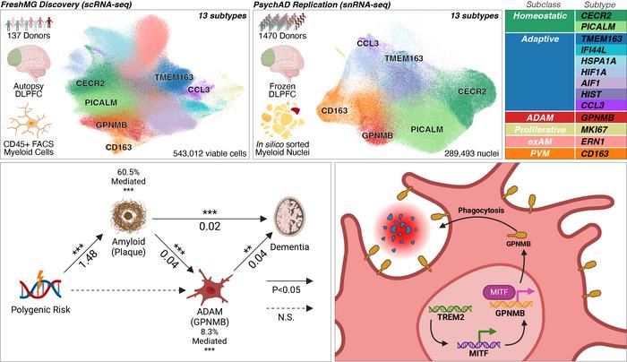
Led by Donghoon Lee, PhD, and Panos Roussos, MD, PhD, the team analyzed more than 830,000 myeloid-origin immune cells of the brain. This included microglia—the brain’s resident immune cells—and perivascular macrophages, which are crucial for modulating immune responses. The cells were sourced from the prefrontal cortex of 1,607 donors spanning a wide range of ages and Alzheimer’s disease pathology stages.Donghoon Lee graphical abstract.

Graphical abstract of the work. Credit Mount Sinai Health System.

By profiling brain tissue at this unprecedented scale, the team identified six subclasses comprising 13 distinct subtypes of myeloid cells, characterizing how these populations adapt during aging and disease progression.

Crucially, the researchers identified a disease-associated subtype of microglia that becomes increasingly abundant as Alzheimer’s disease advances. Rather than contributing to neurodegeneration, these cells appear to play a protective role by increasing their ability to engulf and clear harmful material from the brain.

The study further identified a molecular pathway involving the proteins TREM2, MITF, and GPNMB that is required to maintain this protective microglial state. Experiments in both human tissue and mouse models demonstrated that the beneficial effects of these cells depend strictly on TREM2 signaling.

“Our study provides the clearest picture yet of how the brain’s immune cells adapt during aging and Alzheimer’s disease,” said Donghoon Lee, PhD, Assistant Professor of Genetics and Genomic Sciences and Psychiatry at Mount Sinai, and first and corresponding author of the paper.

“By identifying the specific immune cells that appear to protect the brain—and the molecular signals they rely on—we have uncovered potential new targets for therapies aimed at slowing Alzheimer’s disease progression.”

Beyond identifying this protective microglial population, the study helps explain why genetic variants in immune-related genes such as TREM2 and APOE increase Alzheimer’s risk. Ultimately, the findings provide a roadmap for developing therapies that strengthen the brain’s natural immune defenses.

Editorial Notes:

  • This article was edited by a Neuroscience News editor.
  • Journal paper reviewed in full.
  • Additional context added by our staff.

About this Alzheimer’s disease research news

Author: Elizabeth Dowling (Media Contact)
Source: The Mount Sinai Hospital / Mount Sinai School of Medicine
Contact: Elizabeth Dowling – The Mount Sinai Hospital / Mount Sinai School of Medicine
Image: Graphical abstract credit goes to Mount Sinai Health System

Original Research: Peer-Reviewed Publication “Plasticity of human microglia and brain perivascular macrophages in aging and Alzheimer’s disease” by Donghoon Lee, Panos Roussos, et al. Nature Genetics

DOI: 10.1038/s41588-026-02716-6

Tuesday, August 11, 2026

Study of Brain Immune Cells Reveals New Clues to Alzheimer’s Disease Progression

 How will your competent? doctor use this to prevent your likely dementia? NOTHING LIKE USUAL!

Your risk of dementia, has your doctor told you of this?  Your doctor is responsible for preventing this! Is s/he willing to prevent this?

1. A documented 33% dementia chance post-stroke from an Australian study?   May 2012.

2. Then this study came out and seems to have a range from 17-66%. December 2013.`    

3. A 20% chance in this research.   July 2013.

4. Dementia Risk Doubled in Patients Following Stroke September 2018 

You do know your doctor is TOTALLY FUCKING INCOMPETENT  in everything stroke related, right?

 Your incompetent doctor knew nothing of TREM2, right?

  • NMOSD (2 posts to March 2018)

Study of Brain Immune Cells Reveals New Clues to Alzheimer’s Disease Progression

Summary: Researchers have mapped over 830,000 brain immune cells, discovering a protective subtype of microglia that expands as Alzheimer’s disease progresses. Driven by the TREM2 molecular pathway, these cells actively help clear harmful material from the brain. The findings provide a new roadmap for developing therapies focused on strengthening the brain’s natural immune defenses rather than targeting amyloid plaques alone.

Key Facts:

  • Unprecedented Scale: The study analyzed over 830,000 myeloid-origin immune cells from 1,607 donors, providing the most detailed reference to date of immune cell changes across aging and disease.
  • Protective Microglia Subtype: A specific, disease-associated subtype of microglia becomes more abundant as Alzheimer’s advances, acting to protect the brain by engulfing and clearing harmful material.
  • Crucial Molecular Pathway: The beneficial, protective effects of these immune cells rely entirely on a molecular signaling pathway involving the proteins TREM2, MITF, and GPNMB.

Source: Mount Sinai Hospital / Mount Sinai School of Medicine

The brain’s immune cells are increasingly recognized as key players in Alzheimer’s disease, but exactly how they change as the disease develops has remained unclear. Now, a groundbreaking study published in Nature Genetics provides the most comprehensive map to date of these cells.

Researchers from the Icahn School of Medicine at Mount Sinai have identified a protective subtype of brain immune cell that expands as Alzheimer’s disease progresses, uncovering the molecular pathway that enables these cells to help defend the brain. The findings offer new insights that could inform future Alzheimer’s therapies.

Led by Donghoon Lee, PhD, and Panos Roussos, MD, PhD, the team analyzed more than 830,000 myeloid-origin immune cells of the brain. This included microglia—the brain’s resident immune cells—and perivascular macrophages, which are crucial for modulating immune responses. The cells were sourced from the prefrontal cortex of 1,607 donors spanning a wide range of ages and Alzheimer’s disease pathology stages.

Donghoon Lee graphical abstract.
Graphical abstract of the work. Credit Mount Sinai Health System.

By profiling brain tissue at this unprecedented scale, the team identified six subclasses comprising 13 distinct subtypes of myeloid cells, characterizing how these populations adapt during aging and disease progression.

Crucially, the researchers identified a disease-associated subtype of microglia that becomes increasingly abundant as Alzheimer’s disease advances. Rather than contributing to neurodegeneration, these cells appear to play a protective role by increasing their ability to engulf and clear harmful material from the brain.

The study further identified a molecular pathway involving the proteins TREM2, MITF, and GPNMB that is required to maintain this protective microglial state. Experiments in both human tissue and mouse models demonstrated that the beneficial effects of these cells depend strictly on TREM2 signaling.

“Our study provides the clearest picture yet of how the brain’s immune cells adapt during aging and Alzheimer’s disease,” said Donghoon Lee, PhD, Assistant Professor of Genetics and Genomic Sciences and Psychiatry at Mount Sinai, and first and corresponding author of the paper.

“By identifying the specific immune cells that appear to protect the brain—and the molecular signals they rely on—we have uncovered potential new targets for therapies aimed at slowing Alzheimer’s disease progression.”

Beyond identifying this protective microglial population, the study helps explain why genetic variants in immune-related genes such as TREM2 and APOE increase Alzheimer’s risk. Ultimately, the findings provide a roadmap for developing therapies that strengthen the brain’s natural immune defenses.

Editorial Notes:

  • This article was edited by a Neuroscience News editor.
  • Journal paper reviewed in full.
  • Additional context added by our staff.

About this Alzheimer’s disease research news

Author: Elizabeth Dowling (Media Contact)
Source: The Mount Sinai Hospital / Mount Sinai School of Medicine
Contact: Elizabeth Dowling – The Mount Sinai Hospital / Mount Sinai School of Medicine
Image: Graphical abstract credit goes to Mount Sinai Health System

Original Research: Peer-Reviewed Publication “Plasticity of human microglia and brain perivascular macrophages in aging and Alzheimer’s disease” by Donghoon Lee, Panos Roussos, et al. Nature Genetics
DOI: 10.1038/s41588-026-02716-6

Monday, July 27, 2026

Scientists found three gene variants that may change when Alzheimer's begins

 FYI.

Scientists found three gene variants that may change when Alzheimer's begins

Researchers identified three gene variants linked to the timing and progression of inherited Alzheimer’s. 

The study, published in The Lancet Neurology, focused on autosomal dominant Alzheimer’s disease (ADAD), a rare inherited form of Alzheimer’s caused by mutations in one of three genes: APP, PSEN1 or PSEN2.  

Although the condition accounts for only about 1 percent of Alzheimer’s cases, people who inherit these mutations are highly likely to develop the disease, often at a relatively young age. 

One long-standing mystery has been why Alzheimer’s symptoms can begin at different ages, even among people carrying the same disease-causing mutation.  

To investigate, researchers analyzed whole-genome sequencing data from 101 people with symptomatic ADAD and compared their genetic profiles with more than 5,000 individuals without a known ADAD mutation. The team discovered significant associations involving three genetic regions linked to the genes CNIH4, CCNG1, and RHOJ.  

Each appeared to modify Alzheimer’s risk or progression independent of the underlying inherited mutation. 

One of the strongest findings involved a variant in CCNG1, which was associated with an earlier age of dementia onset. Carriers of the risk allele developed dementia roughly a decade earlier than those without it.  

The same variant was also linked to higher levels of TDP-43, a protein implicated in several neurodegenerative diseases, and signs of accelerated brain aging on MRI scans. 

The researchers also found that a variant in RHOJ was associated with biological markers of more severe Alzheimer’s disease. Individuals carrying the risk allele had higher levels of total tau and phosphorylated tau 181 in cerebrospinal fluid and a lower Aβ42/Aβ40 ratio—changes commonly associated with Alzheimer’s pathology.  

Meanwhile, a variant within CNIH4 showed one of the strongest associations with Alzheimer’s risk among mutation carriers, suggesting it may play an important role in how the disease develops. 

Senior author Dr. Cyril P. Pottier of the Washington University School of Medicine said identifying genetic modifiers could help explain why disease trajectories differ so widely among people with inherited Alzheimer’s.  

Researchers hope that understanding these pathways may eventually improve genetic counseling, refine clinical trial design, and uncover new therapeutic targets.  

Although the findings were made in people with a rare inherited form of Alzheimer’s disease, the researchers also examined whether the variants were associated with age at onset in thousands of people with sporadic Alzheimer’s disease, the far more common form of the condition. 

The results add to a growing body of evidence that genes beyond the well-known Alzheimer’s risk factors can influence how the disease unfolds.  

By revealing biological pathways that can either accelerate or potentially delay decline, scientists hope to identify new strategies for preventing or slowing Alzheimer’s—not only in families with inherited disease, but in the broader population as well.  

Reference

Patel M, Feng W, Mckay N et al. Identification of genetic modifiers of autosomal dominant Alzheimer’s disease: a genome-wide association study. The Lancet Neurology, 25, 581-590.

Contact Newsweek editors on this story: Kara Dolman and Sam Wilson.

Related Articles

Start your unlimited Newsweek trial

Wednesday, July 1, 2026

Scientists discover how Alzheimer’s may spread in brain—and way to slow it

 You might need this, is your doctor competent in following research in the field of  stroke and dementia?

Your risk of dementia, has your doctor told you of this?  Your doctor is responsible for preventing this! Is s/he willing to prevent this?

1. A documented 33% dementia chance post-stroke from an Australian study?   May 2012.

2. Then this study came out and seems to have a range from 17-66%. December 2013.`    

3. A 20% chance in this research.   July 2013.

4. Dementia Risk Doubled in Patients Following Stroke September 2018 

The latest here: 

Scientists discover how Alzheimer’s may spread in brain—and way to slow it

Scientists believe they have identified a key mechanism behind the spread of Alzheimer’s disease through the brain, a discovery that could open the door to new treatments aimed at slowing the condition rather than simply clearing away toxic proteins after damage is done.

Researchers at the University of Utah Health found that a brain protein called Arc, which normally helps neurons communicate with one another, may be inadvertently helping the disease spread. The protein appears to carry toxic Tau—a hallmark of Alzheimer’s—out of damaged brain cells and into healthy ones.

Dr. Christopher U. Missling, president of Anavex Life Sciences, a biotechnology company focused on central nervous system disorders including Alzheimer’s, told Newsweek the breakthrough findings reframe how scientists think about the disease.

“For decades, research has focused on the toxic buildup of Tau inside neurons; this study reframes the problem by showing how Tau may exploit the brain’s own communication machinery, specifically the Arc protein and its extracellular vesicle system, to spread between cells,” Missling said.

He added that the discovery “underscores how normal synaptic signaling proteins can become hijacked in disease, blurring the line between physiological and pathological communication,” and that targeting this transport system might one day help slow or contain Alzheimer’s spread rather than trying to eliminate Tau entirely.

How Tau Hitches a Ride

Alzheimer’s disease is driven by the buildup of Tau, a protein that clumps into sticky tangles inside neurons, disrupting their internal machinery and eventually killing them. As Tau spreads to new regions of the brain, memory loss and cognitive decline worsen.

To understand how this spread happens, researchers compared mice with Alzheimer’s-like disease to mice that also lacked the Arc protein. They found Arc plays an essential role in moving Tau between cells.

Normally, Arc packages itself into tiny structures called extracellular vesicles, which shuttle between neurons carrying important cellular signals. But the study found that toxic Tau can hitch a ride inside these same vesicles, using Arc to travel from a diseased neuron into a healthy one, where it can trigger new tangles to form.

When Arc was removed from the mice, the vesicles carried far less Tau, and the disease no longer spread effectively between neurons.

A Protective Role Complicates the Picture

The results were not entirely straightforward. Arc also appears to do the positive job of helping neurons survive longer in the early stages of disease by allowing them to expel excess toxic Tau.

That finding suggests simply blocking Arc altogether may not be the answer. Instead, researchers believe future treatments should focus on preventing the Tau-carrying vesicles from entering healthy neurons, while still allowing damaged cells to expel their toxic waste.

A Potential New Target for Alzheimer’s Therapies

The team also detected extracellular vesicles containing both Arc and Tau in human brain tissue, suggesting the same process could be at work in people. Researchers caution that significant further study, especially on humans, is needed before the findings could lead to any treatment, but they say the discovery offers a promising new target for future therapies aimed at slowing, rather than eliminating, the disease’s spread through the brain by intercepting Tau containing extracellular vesicles after they leave diseased neurons, before they reach healthy ones. 

The study was published in Cell on June 29.

Contact Newsweek editors on this story: Kara Dolman and Emma Lee-Sang.

Related Articles

Start your unlimited Newsweek trial

Monday, June 22, 2026

New trigger for Alzheimer's disease may have been found

 Have your competent? doctor analyze these other possibilities AND DELIVER EXACT PREVENTION PROTOCOLS! Can't do that, you don't have a functioning stroke doctor!

 January 2024

New trigger for Alzheimer's disease may have been found

A new study is raising questions about one of the most widely held ideas in Alzheimer’s research—suggesting the disease may not start with plaques in the brain after all. 

Researchers at the University of California, Riverside (UCR) say the earliest changes could instead happen inside nerve cells, where two key proteins appear to interfere with each other. 

For years, much of the focus has been on amyloid beta, or a-beta, because it forms clumps in the brains of people with Alzheimer’s. That link appeared well supported, especially since genetic mutations that increase a-beta levels are known to cause early-onset forms of the disease. 

But attempts to treat Alzheimer’s by removing these clumps have been largely unsuccessful, with thousands of trials failing to stop or reverse its progression. 

Michael Kane, chief medical officer at Indiana Center for Recovery, told Newsweek that the findings of this study should not be seen as dismissing the amyloid theory entirely, but rather as refining it.

“I see these findings less as a rejection of the amyloid theory and more as a possible link between amyloid beta and tau,” he said. 

Connection Between Amyloid Beta and Tau 

Scientists have long known that another protein, tau, is also involved. Both a-beta and tau build up in the brains of people diagnosed with Alzheimer’s, yet how they are connected has remained unclear. 

“In addition to having dementia, Alzheimer’s diagnosis requires both a-beta and tau buildup in the brain,” said study lead author Ryan Julian, a chemistry professor at UCR. “But many labs focus on the role of one and ignore the other.” 

The new study, published in Proceedings of the National Academy of Sciences, Nexus, looks at what happens when the two proteins are present inside the same cell. 

Kane said this connection is one of the most significant aspects of the research.

“Amyloid beta and tau have both been central to Alzheimer’s research for decades, but the field has struggled to explain exactly how they interact. This study gives scientists a more specific place to look,” he said.   

What Happens Inside Nerve Cell 

Tau normally supports structures called microtubules, which act as internal pathways, helping nerve cells move essential materials to where they are needed. 

The researchers found that the part of tau that attaches to these structures is very similar to amyloid beta. That similarity led them to examine whether a-beta could attach to microtubules in the same way. 

Using a fluorescent marker to track the protein, the team observed that a-beta can bind to microtubules with similar strength to tau. 

“Our work shows amyloid beta and tau compete for the same binding sites on microtubules, and that a-beta can prevent tau from functioning correctly,” Julian said. 

Kane said this mechanism could represent an earlier stage of disease development than previously recognised.

“The damage may start earlier, with the cell’s machinery becoming less stable,” he said.   

Disruption That May Come First 

The researchers suggest that this competition could be an important early step. 

If a-beta builds up inside a neuron, it may push tau away from the microtubules. Without tau in place, the cell’s transport system may begin to break down. 

At the same time, tau may start to change behavior—clumping together and moving into areas of the cell where it is not normally found. 

This points to a different way of thinking about the disease. Instead of protein buildup being the starting cause on its own, the two processes may be part of a wider problem inside cells. 

Kane cautioned that while the explanation is biologically plausible, it remains a working model. “A plausible mechanism is not the same as proof that this is what drives Alzheimer’s in patients,” he said.   

Ageing May Play Role 

The study also highlights a process called autophagy, which normally clears unwanted proteins from cells, including a-beta. 

As this process becomes less efficient with age, autophagy may begin to accumulate inside neurons. This could increase the chances of it competing with tau and interfering with normal cell function. 

What It Could Mean Going Forward 

The findings may help explain why some earlier approaches to treatment have struggled to make a difference. 

They also suggest that future research may look more closely at how these proteins interact inside cells, rather than focusing only on removing them once they have formed clumps. 

Julian said the idea helps bring together different strands of research. 

“This idea helps make sense of many results that previously seemed unrelated,” he said. “It gives us a clearer picture of what may be going wrong inside neurons and where new treatments might start.” 

Kane said the study could point scientists toward new types of therapeutic strategies, but warned against overstating its immediacy.

“It could point researchers toward targets inside the neuron, such as protecting microtubules or preventing amyloid beta from interfering with tau—but I would not describe it as an immediate treatment breakthrough,” he said.  

He added that the most important next step is confirming whether the process occurs in people.

“Researchers need to know when it happens, who it happens in, and whether it tracks with memory loss or functional decline over time,” he said.  

Kane also urged caution in interpreting the findings.

“The most useful part of this study is that it moves the conversation from what we see after neurons are already damaged to what may be going wrong inside the neuron earlier,” he said. “That is where better treatments may eventually come from.” 

Related Articles

Start your unlimited Newsweek trial

Tuesday, October 14, 2025

Obesity-linked fat vesicles accelerate amyloid aggregation in Alzheimer’s disease

It is YOUR DOCTOR'S TOTAL RESPONSIBILITY TO GET YOU 100% RECOVERED SO YOU DON'T GAIN WEIGHT!

My doctor obviously knew nothing about weight gain post stroke. He didn't reference body metabolism slowing down after age 50 and my limited exercise ability which I used to do to excess allowing me to eat as I wanted. This incompetence led me to a 35 lb. weight gain which I'm still working to conquer.  I'm still overweight but not obese.

Obesity-linked fat vesicles accelerate amyloid aggregation in Alzheimer’s disease

Scientists find fat-derived particles could accelerate Alzheimer’s, connecting obesity to toxic brain changes.

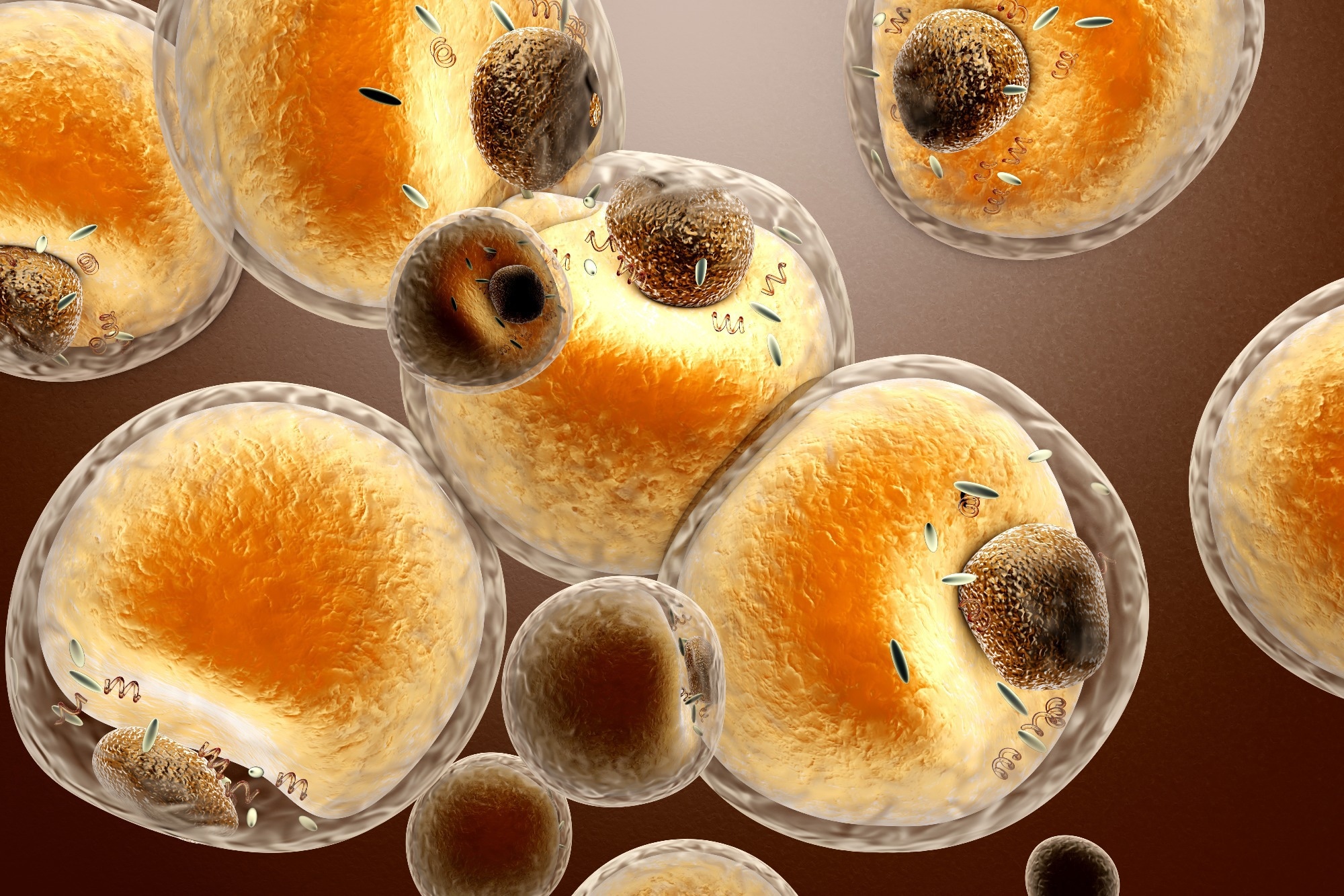
obesity-liked fat vesicles 

Study: Decoding adipose-brain crosstalk: Distinct lipid cargo in human adipose-derived extracellular vesicles modulates amyloid aggregation in Alzheimer's disease. Image Credit: Spectral-Design / Shutterstock.com

Alzheimer’s disease (AD) could affect 82 million people by 2050. The brain is rich in fats, which comprise a significant portion of the myelin sheath and neuronal membranes. Disruptions in lipid metabolism—whether due to genetic factors or environmental influences—can increase the risk of AD, particularly when linked to obesity.

In a recent study published in Alzheimer’s & Dementiaresearchers discuss the role of extracellular vesicles as a potential link between obesity and Alzheimer’s disease (AD).

How fat dysregulation leads to amyloid pathology

During obesity, lipotoxicity, which reflects abnormally high lipid levels, can damage brain tissue by causing inflammation. This condition is worsened by the presence of adipokines, which are chemicals released from fat tissue that activate immune-inflammatory pathways.

According to the amyloid cascade hypothesis, neurodegeneration in AD begins with the formation of the amyloid-β (Aβ) 40 and 42 peptides. Following their secretion into the extracellular space, these peptides aggregate under certain conditions to form small oligomers or fibrils.

Aβ fibrillization, the process by which Aβ fibrils are generated, precedes amyloid plaque formation, a characteristic feature of AD brains. Plaques are lipid-enriched and can be produced during fat-dependent Aβ peptide condensation, which leads to brain inflammation, neuronal injury, inadequate energy supply, and oxidative stress.

Extracellular vesicles (EVs) from fat cells are membrane-bound molecules that contain fat released from different regions throughout the body. Often originating from peripheral fat tissue, these EVs may cross the blood-brain barrier to change the balance of fats in the brain, which increases the risk of neurodegenerative disease.

Aβ fibrilization is affected by genetic and environmental factors, including lipid-laden EVs from peripheral fatty tissue.

The current study's researchers removed and purified EVs, mostly exosomes, from subcutaneous and visceral fat samples obtained from lean and obese people. In addition to quantifying the complete array of fat molecules present in these EVs, in vitro Aβ aggregation was quantified using purified Aβ40 and Aβ42 peptides in fat-rich environments that resemble the brain milieu in health and disease.

Study findings

EVs obtained from obese individuals were characterized by a distinctive lipid profile that reflects their role as fat carriers originating from subcutaneous and visceral fat tissues. Purified EVs isolated from obese people had higher concentrations of lysophosphatidylcholine (LPC) and sphingomyelin (SM), which suggests abnormal fat metabolism.