Changing stroke rehab and research worldwide now.Time is Brain! trillions and trillions of neurons that DIE each day because there are NO effective hyperacute therapies besides tPA(only 12% effective). I have 523 posts on hyperacute therapy, enough for researchers to spend decades proving them out. These are my personal ideas and blog on stroke rehabilitation and stroke research. Do not attempt any of these without checking with your medical provider. Unless you join me in agitating, when you need these therapies they won't be there.

What this blog is for:

My blog is not to help survivors recover, it is to have the 10 million yearly stroke survivors light fires underneath their doctors, stroke hospitals and stroke researchers to get stroke solved. 100% recovery. The stroke medical world is completely failing at that goal, they don't even have it as a goal. Shortly after getting out of the hospital and getting NO information on the process or protocols of stroke rehabilitation and recovery I started searching on the internet and found that no other survivor received useful information. This is an attempt to cover all stroke rehabilitation information that should be readily available to survivors so they can talk with informed knowledge to their medical staff. It lays out what needs to be done to get stroke survivors closer to 100% recovery. It's quite disgusting that this information is not available from every stroke association and doctors group.

Showing posts with label brain iron. Show all posts

Wednesday, September 10, 2025

Brain Iron Levels Predict Cognitive Decline Risk

 Your competent? doctor already knew about this and had the dietician CREATE EXACT DIET PROTOCOLS, RIGHT? NO? So, you having a fucking incompetent doctor then? RUN AWAY!


Diets That Reduce Brain Iron May Delay Cognitive Decline December 2024 

The latest here:

Brain Iron Levels Predict Cognitive Decline Risk

Summary: A new study demonstrates that brain iron levels, measured with a special MRI technique, can predict cognitive decline years before symptoms of Alzheimer’s disease appear. Researchers followed 158 cognitively healthy older adults and found that higher iron levels in memory-related regions of the brain were associated with a greater risk of mild cognitive impairment.

This risk was even higher when participants also showed amyloid buildup, highlighting the interaction of multiple factors in dementia. The findings suggest QSM MRI could serve as a valuable tool for early detection and a pathway to new treatments targeting brain iron.

Key Facts

  • Iron Detection: Quantitative susceptibility mapping (QSM) MRI precisely measures brain iron noninvasively.
  • Risk Link: Higher iron levels in key brain regions predicted mild cognitive impairment and faster decline.
  • Dual Target: Iron may act as both a biomarker for early diagnosis and a potential treatment target.

Source: RSNA

A special MRI technique that detects iron levels in different regions of the brain can predict the onset of mild cognitive impairment and cognitive decline in cognitively unimpaired older adults, potentially creating a pathway to earlier interventions, according to a study published today in Radiology.  

Wednesday, June 18, 2025

Platelet Membrane-Based Nanoparticles for Targeted Delivery of Deferoxamine to Alleviate Brain Injury Induced by Ischemic Stroke

 There is much earlier research on nano stuff for recovery. HAS YOUR INCOMPETENT? DOCTOR DONE NOTHING WITH THIS? So, you DON'T have a functioning stroke doctor, do you? And complete incompetence(for over a decade!) in the stroke medical world which seems to have NO idea on how to solve stroke! They are all blithering idiots straight from the pages of Monty Python. Like this:

Monty Python's Flying Circus - Upper Class Twit of the Year (1971)
  • nanoparticles (83 posts to June 2012)
  • nanomissiles (1 post to June 2022)
  • nanoneedles (2 post to March 2015)
  • nanopatch (1 post to August 2024)
  • nanopeptide (1 post to October 2017)
  • nanophytomedicine (1 post to May 2021)
  • nanopropellers (1 post to May 2020)
  • nanoRobots (14 posts to March 2012)
  • nanorockets (1 post to December 2016)
  • nanorods (1 post to January 2016)
  • nanosensors (3 posts to November 2017)
  • nanospear (1 post to June 2018)
  • nanospheres (1 post to June 2025)
  • nanostructures (1 post to November 2019)
  • nanosubmarine (1 post to November 2015)
  • nanotechnology (9 posts to December 2014)
  • nanotherapeutic (5 posts to April 2015)
  • nanotubes(5 posts to February 2012)
  • Nanovesicles (2 posts to November 2021)
  • nanowires (23 posts to January 2012)
  • Nanozymes (2 posts to June 2024)
  • Platelet Membrane-Based Nanoparticles for Targeted Delivery of Deferoxamine to Alleviate Brain Injury Induced by Ischemic Stroke

    Authors Wang PLv XTian SYang W Feng MChang SYou LChang YZ

    Received 8 January 2025

    Accepted for publication 8 June 2025

    Published 16 June 2025 Volume 2025:20 Pages 7533—7548

    DOI https://doi.org/10.2147/IJN.S516316

    Checked for plagiarism Yes

    Review by Single anonymous peer review

    Peer reviewer comments 2

    Editor who approved publication: Professor Jie Huang


    Peina Wang,1,2,* Xin Lv,1,* Siyu Tian,1 Wen Yang,2 Mudi Feng,1 Shiyang Chang,2 Linhao You,1 Yan-Zhong Chang1

    1Laboratory of Molecular Iron Metabolism, Key Laboratory of Animal Physiology, Biochemistry and Molecular Biology of Hebei Province, Ministry of Education Key Laboratory of Molecular and Cellular Biology, Department of Physiology, College of Life Science, Hebei Normal University, Shijiazhuang, Hebei Province, 050024, People’s Republic of China; 2Department of Histology and Embryology, College of Basic Medical Sciences, Hebei Medical University, Shijiazhuang, Hebei Province, 050017, People’s Republic of China

    *These authors contributed equally to this work

    Correspondence: Yan-Zhong Chang; Peina Wang, Email chang7676@163.com; 19301641@hebmu.edu.cn

    Background: Timely thrombolysis serves as the primary therapeutic approach for ischemic stroke, one of the most serious global public health problems, although reperfusion can cause severe ischemia reperfusion (I/R) injury. Oxidative stress and activation of cell death pathways are the main mechanisms of I/R injury. Our previous studies have demonstrated that iron overload stimulates the generation of reactive oxygen species and facilitates the activation of iron-dependent ferroptosis in the pathogenesis of I/R injury. Removal of excess free iron by deferoxamine (DFO), an iron chelator, may inhibit iron toxicity and reverse I/R-induced neurological deficits. Despite its therapeutic potential, DFO’s clinical translation for I/R injury is hampered by rapid systemic clearance, suboptimal bioavailability, and a lack of ischemic lesion-targeting ability. Nanoscale delivery platforms enabling targeted DFO release in stroke lesions may overcome these pharmacokinetic barriers and enhance clinical outcomes.
    Methods: On the basis of the properties of liposomes in carrying hydrophilic substances and crossing the leaky blood–brain barrier in cerebral I/R, we first encapsulated DFO within traditional liposomes to improve its biocompatibility. Subsequently, inspired by the natural homing properties of platelets to damaged blood vessels during I/R injury, the isolated platelet membranes were coated onto the DFO-liposomes, thus endowing the nanodrug with the ability to target stroke lesion.
    Results: Our results demonstrate that Platesome-DFO exhibits accurate lesion-targeting ability and significantly decreases lesion iron content, thereby preventing neuronal ferroptosis and ultimately reversing neurological deficits in I/R mice.
    Conclusion: Platesome-DFO provides a novel therapeutic approach for cerebral I/R injury by regulating brain iron status and iron-dependent pathways, highlighting its promising application in the clinical treatment of cerebral I/R injury.

    Keywords: ischemic stroke, iron, ferroptosis, deferoxamine, platelet membrane, nanomedicine

    Introduction

    Stroke is an acute and severe cerebrovascular disease with high morbidity, mortality, and medical cost. It has been reported that ischemic stroke, which results from a lack of blood supply to the brain, accounts for approximately 84% of all stroke cases.1 Irreversible neurological injury can be avoided only if the blocked blood vessels are reperfused within the therapeutic time window. However, ischemia/reperfusion (I/R) can immediately cause dysregulation of oxidation and antioxidation, disrupt the balance between the generation and scavenging of reactive oxygen species (ROS), eventually leading to the accumulation of ROS in the ischemic brain, which exacerbates the activation of inflammatory responses and results in secondary neurological damage.2 Therefore, I/R injury is considered the main culprit and an inevitable obstacle in the therapy of ischemic stroke. Despite the development of various drugs and functional nanoparticles with ROS scavenging ability, effective treatment options for I/R injury remain disappointingly limited.

    Many studies have indicated that a higher iron status is a critical risk factor associated with neuronal damage following cerebral I/R. Standard clinical analysis has repeatedly shown that elevated plasma ferritin levels are associated with poor outcomes in patients with ischemic stroke.3,4 Iron overload exacerbates brain edema and hemorrhagic transformation in ischemic stroke patients receiving thrombolytic therapy with tissue plasminogen activator.5,6 Mechanistically, our previous research has demonstrated that excessive iron exacerbates neuronal damage by catalyzing the Fenton reaction to convert superoxide and hydroxyl radicals into highly reactive toxic radicals or catalyzing lipid peroxidation as a cofactor of lipid oxidation enzymes.7–9 Of note, we and others have recently described the induction of neuronal ferroptosis, a newly identified form of regulated cell death resulting from the catastrophic accumulation of iron-dependent lipid reactive oxygen species, in I/R brains.8,10–12 Therefore, iron is a key factor that stimulates ROS generation and facilitates the subsequent activation of cell death pathways, particularly the ferroptosis pathway, in I/R injury. Iron depletion via chelator may protect neuronal cells against ferroptosis and is expected to emerge as a promising strategy for the treatment of cerebral I/R.

    Among the clinically available iron chelators, deferoxamine (DFO) has long been used to remove excess iron in iron overload diseases, such as the secondary iron overload that afflicts thalassemia patients, and has been shown to be the most effective option with the most favorable toxicity profile.13,14 Moreover, DFO has demonstrated its protective effects in animal stroke models of I/R as well as in clinical studies in which ischemic stroke patients administered DFO exhibited improved outcomes.5,15–17 However, the poor bioavailability and the extremely short plasma half-life (20 min in humans) of DFO limits its use in stroke treatment.18,19 More important, at the high doses needed to achieve effective concentrations, DFO can cause serious side effects, including renal and liver complications.13 Another limitation of DFO for use in stroke treatment is that systemic administration does not specifically target to the injured region of the I/R brain. New delivery approaches that can target deliver DFO to the injured brain region and improve the local DFO concentration are expected to provide significant improvements in stroke outcomes.

    Despite advancements in stroke-targeted nanoplatforms, such as polymeric carriers, metallic nanoparticles, and carbon-based systems, current materials face persistent translational barriers, including compromised biosafety profiles, suboptimal therapeutic efficacy, and insufficient blood–brain barrier penetrability.20,21 Recently, cell membrane-based biomimetic cloaking has emerged as a new strategy for enhancing synthetic nanoparticles delivery, leveraging retained membrane functionalities to overcome systemic biological barriers while preserving immunoevasive properties. Platelets (PLT) play a critical role in the development and progression of thrombosis. Previous studies have shown that the platelet membrane exhibits properties in binding to injured vasculature and has advantages for targeting ischemic brain regions.22–26 Therefore, we propose a new type of PLT membrane-coated biomimetic nanodevice to deliver DFO specifically to the ischemic brain. On the basis of the properties of liposomes in carrying hydrophilic substances and crossing the leaky blood–brain barrier in cerebral I/R,27 we first encapsulated DFO within traditional liposomes through hydration to improve its biocompatibility. Subsequently, the isolated PLT membranes were coated onto the DFO-liposomes by coextrusion, endowing this nanodrug with stroke lesion targeting ability (Figure 1a). This PLT membrane-cloaked, DFO-loaded nanoliposome, named Platesome-DFO, can deliver DFO to the targeted lesion, increasing intracellular drug concentrations to markedly higher levels than those achievable through systemic administration. Particularly, we confirmed the chelation of excess iron by Plateletsome-DFO, and the consequent inhibition of iron-dependent lipid peroxidation and ferroptosis in the cerebral I/R brain (Figure 1b). 2,3,5-triphenyltetrazolium chloride (TTC) staining measurement from mouse models demonstrated that the Platesome-DFO treatment effectively alleviated I/R-induced brain damage. Our Platesome-DFO formulation offers a viable strategy for specifically targeting lesions to inhibit neuronal death, demonstrating potential to improve outcomes in cerebral I/R injury.

    Figure 1 Schematic illustration of Platesome-DFO structure and its proposed protective mechanism in ischemic stroke. (a) The Platesome-DFO was fabricated by coextrusion of platelet membranes and Liposome-DFO to form nanoparticles that inherit the natural characteristics of the platelet membrane, including targeting damaged blood vessels and immune escape capabilities, by the presence of specific membrane proteins, such as CD36, CD41, and CD47. (b) After intravenous injection, Platesome-DFO is delivered to the injured brain region after stroke, followed by the release of DFO, which chelates excessive iron and inhibits ferroptosis of neuronal cells.


    More at link.

    Friday, December 13, 2024

    Diets That Reduce Brain Iron May Delay Cognitive Decline


    Will this be enough to get your competent? doctor to instruct the dietician to incorporate this into hospital meals?  My opinion of doctor competence is having EXACT 100% RECOVERY PROTOCOLS! What is your doctor's opinion of their competence?

    Diets That Reduce Brain Iron May Delay Cognitive Decline

    Summary: New research suggests that certain nutrients may lower iron buildup in the brain, a factor linked to cognitive decline in aging. Excess non-heme iron, which accumulates over time, contributes to oxidative stress and can impair memory and executive function.

    Over three years, participants with higher intake of antioxidants, vitamins, and iron-chelating nutrients showed less brain iron accumulation and better cognitive performance. These findings highlight the potential of diets like the Mediterranean or DASH to support brain health and combat age-related cognitive decline.

    Key Facts:

    • Iron’s Role: Excess brain iron, especially non-heme iron, is linked to poor memory and executive function in aging.
    • Nutritional Impact: Higher intake of antioxidants, vitamins, and iron-chelating nutrients reduces brain iron buildup.
    • Dietary Potential: Diets rich in these nutrients, such as the Mediterranean or DASH diets, may protect against cognitive decline.

    Source: University of Kentucky

    Researchers at the University of Kentucky have found that incorporating specific nutrients into a regular diet may reduce iron buildup in the brain — a factor associated with cognitive decline in normal aging.

    The study, titled “Exploring the links among brain iron accumulation, cognitive performance, and dietary intake in older adults: A longitudinal MRI study,” was published in Neurobiology of Aging.

    The work was supported by multiple grants from the National Institutes of Health’s National Institute on Aging and National Institute of Neurological Disorders and Stroke.

    Video credit: Neuroscience News

    “It’s crucial to understand how diet and other lifestyle factors impact the risk of Alzheimer’s and related dementias as we age,” said Brian Gold, Ph.D., professor in the Department of Neuroscience in the College of Medicine, faculty in the Sanders-Brown Center on Aging and principal investigator of the study.

    “This study is an example of how we can encourage healthier lifestyle choices to help combat some risk factors that can affect brain health,” said Gold.

    In this project, researchers specifically looked at non-heme iron, which is critical for brain health. This type of iron does not bind with storage proteins and, with age and in excess, can contribute to oxidative stress, potentially affecting neuronal integrity and cognition.

    Excessive brain iron has been linked to poor cognitive performance, even in normal aging.

    “Despite mounting evidence connecting iron overload to negative cognitive outcomes, there are currently no established methods for reducing brain iron accumulation in older adults,” said Valentinos Zachariou, Ph.D., an assistant professor in the Department of Behavioral Science in the College of Medicine and first author of the paper.

    This study builds on the research team’s previous work that found that higher intake of antioxidants, vitamins, iron-chelating nutrients and polyunsaturated fatty acids correlated with lower brain iron levels and better working memory performance.(So where is the protocol located so survivors can train their stroke medical 'professionals' in their use?)

    “We still had important questions that remained unanswered in that initial investigation, particularly regarding the long-term effectiveness of these nutrients and their potential to reduce age-related brain iron accumulation,” said Zachariou.

    For a follow-up study, Gold and Zachariou worked with the same research team, including the Department of Neuroscience’s Colleen Papas, Ph.D., Christopher Bauer, Ph.D., and Elayna Seago.

    The team reassessed brain iron concentrations approximately three years later in the same cohort of older adults. They measured brain iron levels with a specific MRI technique called quantitative susceptibility mapping.

    Researchers also analyzed a month’s worth of dietary information and cognitive performance, which was evaluated using neuropsychological tests of episodic memory (memories of specific events) and executive function.

    “Our results revealed a broad network of cortical and subcortical brain regions where iron accumulation occurred over the three-year period,” said Zachariou.

    “These regional increases in iron levels were associated with poorer episodic memory and executive function at the follow-up time-point.”

    “However, participants who had higher baseline intake of antioxidants, vitamins, iron-chelating nutrients, and polyunsaturated fatty acids showed significantly less iron accumulation over the three-year period,” said Gold.

    The research team said the findings offer valuable insights for future clinical trials aimed at evaluating the impact of similar nutritional intake on brain iron accumulation and cognitive function.

    Further study of iron accumulation and cognition effects of healthy diets rich in the nutrients examined in this study, such as the Mediterranean or DASH diets, would be highly beneficial.

    Funding: Research reported in this publication was supported by the National Institute on Aging of the National Institutes of Health under Award Numbers R01AG055449, R01AG068055, P30AG072946 and P30AG028383; by the National Institute of Neurological Disorders and Stroke of the National Institutes of Health under Award Number RF1NS122028; and the Office of Research Infrastructure Programs of the National Institutes of Health under Award Number S10OD023573.

    The content is solely the responsibility of the authors and does not necessarily represent the official views of the National Institutes of Health.

    About this diet and brain health research news

    Author: Lindsay Travis
    Source: University of Kentucky
    Contact: Lindsay Travis – University of Kentucky
    Image: The image is credited to Neuroscience News

    Original Research: Open access.
    Exploring the links among brain iron accumulation, cognitive performance, and dietary intake in older adults: A longitudinal MRI study” by Brian Gold et al. Neurobiology of Aging

    Wednesday, April 19, 2023

    Newfound Link Between Alzheimer’s and Iron Could Lead to New Medical Interventions

     Nothing on how to prevent this iron in the brain or even how it got there. So for right now useless information. I take an iron pill daily right before donating blood so I don't fall below that hemoglobin test.

    Maybe this?

    Diet affects brain iron levels differently in men and women, UB pilot study shows

    The latest here:

    Newfound Link Between Alzheimer’s and Iron Could Lead to New Medical Interventions

    Summary: A new imaging probe revealed an increase in iron redox in brain areas where amyloid plaques occur in the brain in Alzheimer’s disease.

    Source: UT Austin

    There is a growing body of evidence that iron in the brain may play a role in Alzheimer’s disease.

    Lending weight to that idea, a new imaging probe has for the first time shown that in the same regions of the brain where the amyloid beta plaques associated with Alzheimer’s occur, there is also an increase in iron redox, meaning the iron in these regions is more reactive in the presence of oxygen.

    Their imaging probe could yield even more details about the causes of Alzheimer’s and help in the search for new drugs to treat it.

    A team from The University of Texas at Austin and the University of Illinois at Urbana-Champaign published a study today on the new imaging technique and findings in Science Advances.

    “The link between iron redox and Alzheimer’s disease has been a black box,” said Yi Lu, corresponding author and professor of chemistry at UT Austin. “The most exciting part to me is that we now have a way to shine light into this black box so that we can begin to understand this whole process in much more detail.”

    About a decade ago, scientists discovered ferroptosis, a process in the body that is dependent on elevated iron levels, leads to cell death and plays a key role in neurodegenerative diseases, such as Alzheimer’s.

    Using magnetic resonance imaging on living Alzheimer’s patients, scientists have observed that these patients tend to have elevated iron levels in the brain, although that method doesn’t differentiate between different forms of iron.

    Together, these findings suggested that iron might play a role in destroying brain cells in Alzheimer’s patients.

    For the new study, the researchers developed DNA-based fluorescent sensors that can detect two different forms of iron (Fe2+ and Fe3+) at the same time in cell cultures and in brain slices from mice genetically modified to mimic Alzheimer’s. One sensor glows green for Fe2+ and the other glows red for Fe3+. This is the first imaging technique that can simultaneously detect both forms of iron in cells and tissue while also indicating their quantity and spatial distribution.

    “The best part about our sensor is that we can now visualize the changes of Fe2+ and Fe3+ and their ratios in each location,” said Yuting Wu, a co-first author of the study and a postdoctoral researcher in Lu’s lab at UT Austin. “We can change one parameter at a time to see if it changes the plaques or the oxidative states of iron.”

    That ability could help them better understand why there is an increased ratio of Fe3+ to Fe2+ in the location of amyloid beta plaques and whether increased iron redox is involved in forming the plaques.

    Another key question is whether the iron redox is directly involved in cell death in Alzheimer’s, or simply a byproduct. The researchers plan to explore this question in Alzheimer’s mice. If further research determines that iron and its redox changes indeed cause cell death in Alzheimer’s patients, that information could provide a potential new strategy for drug development.

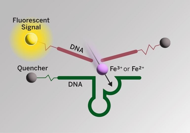
    This is a diagram from the study
    Schematic of a novel iron sensor. When a short strand of DNA called a DNAzyme (green) binds to a specific form of iron (e.g., Fe3+ or Fe2+), the DNAzyme cuts a second strand of DNA (red) and releases a fluorescent signal (yellow) that indicates visually the presence of the specific form of iron. Credit: David Steadman/University of Texas at Austin

    In other words, perhaps a drug that change the ratio Fe3+ to Fe2+ could help protect brain cells. The new imaging probe could be used to test how well drug candidates work at changing the ratio.

    To develop the sensors, the scientists first hired a commercial lab to produce a library of 100 trillion short DNA strands, through a chemical process called oligonucleotide synthesis.

    They then conducted a screening process to find those strands that recognize — or in chemistry parlance “bind tightly to and conduct a catalytic reaction with” — a specific form of iron and not any other forms.

    To complete the sensors, other components were added including molecules called fluorophores that glow in a specific color when the probe recognizes the specific form of iron.

    Lu, who moved his lab to UT Austin from the University of Illinois at Urbana-Champaign in the summer of 2021, collaborated with researchers there including professor of chemistry Liviu Mirica.

    Funding: This work was supported by the National Institutes of Health, the Alzheimer’s Association and the Robert A. Welch Foundation. Lu holds the Richard J.V. Johnson – Welch Regents Chair in Chemistry.

    About this Alzheimer’s disease research news

    Author: Marc Airhart
    Source: UT Austin
    Contact: Marc Airhart – UT Austin
    Image: The image is credited to David Steadman/University of Texas at Austin

    Original Research: Open access.
    Simultaneous Fe2+/Fe3+ imaging shows Fe3+ over Fe2+ enrichment in Alzheimer’s disease mouse brain” by Yi Lu et al. Science Advances


    Abstract

    Simultaneous Fe2+/Fe3+ imaging shows Fe3+ over Fe2+ enrichment in Alzheimer’s disease mouse brain

    Visualizing redox-active metal ions, such as Fe2+ and Fe3+ ions, are essential for understanding their roles in biological processes and human diseases. Despite the development of imaging probes and techniques, imaging both Fe2+ and Fe3+ simultaneously in living cells with high selectivity and sensitivity has not been reported.

    Here, we selected and developed DNAzyme-based fluorescent turn-on sensors that are selective for either Fe2+ or Fe3+, revealing a decreased Fe3+/Fe2+ ratio during ferroptosis and an increased Fe3+/Fe2+ ratio in Alzheimer’s disease mouse brain.

    The elevated Fe3+/Fe2+ ratio was mainly observed in amyloid plaque regions, suggesting a correlation between amyloid plaques and the accumulation of Fe3+ and/or conversion of Fe2+ to Fe3+.

    Our sensors can provide deep insights into the biological roles of labile iron redox cycling.

    Sunday, December 19, 2021

    Brain iron deposition is linked with cognitive severity in Parkinson’s disease

     With your risk of Parkinsons you'll want your doctor to test for this and have removal protocols in place.

    Your risk of Parkinsons here:

    Parkinson’s Disease May Have Link to Stroke March 2017

    How brain iron accumulates here:

    Brain Iron Accumulation in Atypical Parkinsonian Syndromes: in vivo MRI Evidences for Distinctive Patterns

     Possible brain iron removal here:

    The Efficacy of Iron Chelators for Removing Iron from Specific Brain Regions and the Pituitary—Ironing out the Brain

    Don't listen to me, I'm not medically trained, your doctors should be intimately knowledgeable about all of this, ask them. 

    The latest here:

     
     George Edward Calver Thomas1,
    1. Louise Ann Leyland1,
    2. Anette-Eleonore Schrag2,3,
    3. Andrew John Lees4,
    4. Julio Acosta-Cabronero5,
    5. Rimona Sharon Weil1,6
    1. Correspondence to Dr Rimona Sharon Weil, Dementia Research Centre, London WC1N 3BG, UK; r.weil@ucl.ac.uk

    Abstract

    Background 

    Dementia is common in Parkinson’s disease (PD) but measures that track cognitive change in PD are lacking. Brain tissue iron accumulates with age and co-localises with pathological proteins linked to PD dementia such as amyloid. We used quantitative susceptibility mapping (QSM) to detect changes related to cognitive change in PD.

    Methods 

    We assessed 100 patients with early-stage to mid-stage PD, and 37 age-matched controls using the Montreal Cognitive Assessment (MoCA), a validated clinical algorithm for risk of cognitive decline in PD, measures of visuoperceptual function and the Movement Disorders Society Unified Parkinson’s Disease Rating Scale part 3 (UPDRS-III). We investigated the association between these measures and QSM, an MRI technique sensitive to brain tissue iron content.

    Results 

    We found QSM increases (consistent with higher brain tissue iron content) in PD compared with controls in prefrontal cortex and putamen (p<0.05 corrected for multiple comparisons). Whole brain regression analyses within the PD group identified QSM increases covarying: (1) with lower MoCA scores in the hippocampus and thalamus, (2) with poorer visual function and with higher dementia risk scores in parietal, frontal and medial occipital cortices, (3) with higher UPDRS-III scores in the putamen (all p<0.05 corrected for multiple comparisons). In contrast, atrophy, measured using voxel-based morphometry, showed no differences between groups, or in association with clinical measures.

    Conclusions 

    Brain tissue iron, measured using QSM, can track cognitive involvement in PD. This may be useful to detect signs of early cognitive change to stratify groups for clinical trials and monitor disease progression.

    https://creativecommons.org/licenses/by/4.0/

    This is an open access article distributed in accordance with the Creative Commons Attribution 4.0 Unported (CC BY 4.0) license, which permits others to copy, redistribute, remix, transform and build upon this work for any purpose, provided the original work is properly cited, a link to the licence is given, and indication of whether changes were made. See: https://creativecommons.org/licenses/by/4.0/.

    Introduction

    Dementia affects up to 50% of patients with Parkinson’s disease (PD)1 but patients vary in the timing and severity of cognitive involvement and useful quantitative tools to track cognitive change in PD are required. PD dementia is thought to be caused by the combination of amyloid, tau and α-synuclein, but the reasons for selective vulnerability of particular brain regions in PD dementia remain unclear.2

    Neuroimaging measures sensitive to PD cognition are important to track change in clinical trials and detect early neuroanatomical correlates of cognitive involvement. Conventional neuroimaging, which uses MRI to assess volume loss caused by neuronal cell death, is poorly sensitive in PD as cell death at a large scale occurs only at later disease stages.3 Techniques sensitive to brain tissue microstructure are better suited to detect brain changes linked to cognitive involvement in PD.

    A potential mechanism for selective vulnerability in PD dementia is excess brain iron accumulation.4 Iron is ubiquitous in numerous biological processes in normal ageing as well as in neurodegeneration.5 Brain iron accumulation is seen with age, in part due to increased blood-brain-barrier permeability,6 especially affecting the basal ganglia.7–9 The toxic potential of excess iron lies in its ability to generate reactive oxygen species,10 which damage DNA,11 irreversibly modify proteins via highly reactive aldehydes12 and stimulate release of iron from storage proteins leading to generation of further reactive oxygen species.5 This can ultimately end in iron-mediated cell death.13 Excess brain iron is also important in key pathophysiological pathways specific to PD.9 Notably, free radical species generated through iron overload interact with α-synuclein to promote Lewy-related pathology14 and produce neurotoxic by-products via catalysation of dopamine oxidation reactions.15 Increased iron is seen in the substantia nigra at post mortem in PD16 and in vivo using transcranial sonography.17

    Of key significance, brain iron co-localises with Alzheimer’s pathology, particularly amyloid and tau,18 which are key predictors of PD dementia.19 Therefore, detecting levels of brain iron could be a sensitive way to identify brain tissue already affected by the earliest processes that ultimately lead to PD dementia.20

    Quantitative susceptibility mapping (QSM) is an emerging MRI technique which detects local variations in iron content.21 22 QSM is sensitive to magnetic susceptibility differences between chemical species, which are captured by the signal phase of MRI gradient echo sequences. QSM recovers local susceptibility sources giving rise to magnetic field perturbations which are increased in basal ganglia regions in PD,20 but has never been used across the whole brain to track cognitive changes in PD.

    Outcomes relating to progression of cognitive impairment are of particular interest. Recently, risk algorithms combined clinical information to predict cognitive change over time.23 Visual changes are also emerging as early markers of cognitive change in PD.24 Whether structural brain changes are more strongly linked with clinical risk scores or visual deficits before onset of dementia is not yet known.

    Here, we used QSM to measure cognitive-related changes in 100 patients with PD without dementia. We hypothesised that magnetic susceptibility values reflecting brain tissue iron would be higher (1) in mesial temporal structures in relation to poorer cognitive ability; (2) in posterior and prefrontal cortical regions in relation to higher risk of dementia, measured using algorithmic scores and finally, (3)in basal ganglia regions in relation to motor change.


    Sunday, March 15, 2020

    Iron in brain shows cognitive decline in people with Parkinson's

    Useless, NOTHING on how to reduce such iron accumulation.  You do need to worry about this. 

    Parkinson’s Disease May Have Link to Stroke March 2017

    Iron in brain shows cognitive decline in people with Parkinson's

    MedicalXpress Breaking News-and-Events | February 21, 2020
    A cutting-edge MRI technique to detect iron deposits in different brain regions can track declines in thinking, memory and movement in people with Parkinson's disease, finds a new UCL-led study.
    The findings, published in the Journal of Neurology, Neurosurgery, and Psychiatry, suggest that measures of iron might eventually help predict which people with Parkinson will develop .

    "Iron in the brain is of growing interest to people researching such as Parkinson and dementias. As you get older, iron accumulates in the brain, but it's also linked to the build-up of harmful brain proteins, so we're starting to find evidence that it could be useful in monitoring , and potentially even in diagnostics," said the study's lead author, Dr. Rimona Weil (UCL Queen Square Institute of Neurology).
    The study involved 97 people with Parkinson disease, who had been diagnosed within the last 10 years, along with 37 people without the condition, as a control (comparison) group. They were tested for their thinking and memory as well as for their motor function.
    Parkinson disease is a progressive condition of brain degeneration resulting in tremors, stiffness and slowness of movement. Close to 50% of people with the condition end up developing dementia, but the timing and severity vary substantially.
    Currently there are no reliable measures to track Parkinson progression in the brain, so clinicians rely on monitoring symptoms. Conventional brain imaging fails to track progression until quite a late stage, when large-scale brain volume loss can be detected.
    Iron accumulates in people's brains as part of the normal aging process, partly due to increased permeability in the blood-brain barrier. Excess iron can have toxic effects leading to proteins being irreversibly modified. Recent studies have found that when proteins linked to Alzheimer disease (amyloid and tau, which are also linked to Parkinson dementia) build up, iron also accumulates in the affected brain areas.
    In the current study, researchers used a new technique, called quantitative susceptibility mapping, to map iron levels in the brain based on MRI (magnetic resonance imaging) scans. They found that iron accumulation in the hippocampus and thalamus brain regions was associated with poor memory and thinking scores. Iron in the putamen brain region was associated with poor movement scores, suggesting a more advanced stage of the disease.
    In Parkinson disease, the hippocampus and thalamus are known to be associated with thinking and memory, and the putamen with movement scores, so the researchers say it's very promising that iron deposition was specifically detected in those areas.
    The findings suggest that iron deposition could be valuable to track if a treatment is working in a clinical trial, and might eventually be helpful for early diagnosis of Parkinson or other neurodegenerative diseases.
    Dr. Weil has previously found in a 2019 study that a suite of vision tests may be helpful to predict cognitive decline in Parkinson. She and her colleagues hope that further research will determine if the vision tests and iron measures could be helpful to predict which people with dementia are likely to develop dementia.
    First author, PhD student George Thomas (UCL Queen Square Institute of Neurology), said: "It's really promising to see measures like this which can potentially track the varying progression of Parkinson disease, as it could help clinicians devise better treatment plans for people based on how their condition manifests."
    Co-author Dr. Julio Acosta-Cabronero (Tenoke Ltd. and Wellcome Centre for Human Neuroimaging, UCL) added: "We were surprised at how well the iron levels measured in different regions of the brain with MRI were correlated with cognitive and motor skills. We hope that brain iron measurement could be useful for a wide range of conditions, such as to gauge dementia severity or to see which brain regions are affected by other movement, neuromuscular and neuroinflammatory disorders, stroke, and drug abuse."
    The researchers are now following up the same study participants to see how their disease is progressing, whether they develop dementia, and how such measures correlate with changes in levels over time.
    To read more, click here.

    Saturday, September 8, 2018

    Iron Overload Exacerbates the Risk of Hemorrhagic Transformation After tPA (Tissue-Type Plasminogen Activator) Administration in Thromboembolic Stroke Mice

    A simple question. Has your stroke hospital implemented ANYTHING FROM STROKE RESEARCH in the past 30 years, other than the 88% failure rate to fully recover using tPA? I bet your stroke hospital doesn't publish all their failure points in stroke. Yes this is in mice but if they don't followup and engage with researchers to followup this with human testing then they just prove their incompetency once again.

    Iron Overload Exacerbates the Risk of Hemorrhagic Transformation After tPA (Tissue-Type Plasminogen Activator) Administration in Thromboembolic Stroke Mice

    Originally publishedStroke. 2018;49:2163–2172

    Abstract

    Background and Purpose—

    Recanalization with tPA (tissue-type plasminogen activator) is the only pharmacological therapy available for patients with ischemic stroke. However, the percentage of patients who may receive this therapy is limited by the risk of hemorrhagic transformation (HT)—the main complication of ischemic stroke. Our aim is to establish whether iron overload affects HT risk, to identify mechanisms that could help to select patients and to prevent this devastating complication.

    Methods—

    Mice fed with control or high-iron diet were subjected to thromboembolic stroke, with or without tPA therapy at different times after occlusion. Blood samples were collected for determination of malondialdehyde, matrix metalloproteinases, and fibronectin. Brain samples were collected 24 hours after occlusion to determine brain infarct and edema size, hemorrhage extension, IgG extravasation, and inflammatory and oxidative markers (neutrophil infiltration, 4-hydroxynonenal, and matrix metalloproteinase-9 staining).

    Results—

    Despite an increased rate of recanalization, iron-overload mice showed less neuroprotection after tPA administration. Importantly, iron overload exacerbated the risk of HT after early tPA administration, accelerated ischemia-induced serum matrix metalloproteinase-9 increase, and enhanced basal serum lipid peroxidation. High iron increased brain lipid peroxidation at most times and neutrophil infiltration at the latest time studied.

    Conclusions—

    Our data showing that iron overload increases the death of the compromised tissues, accelerates the time of tPA-induced reperfusion, and exacerbates the risk of HT may have relevant clinical implications for a safer thrombolysis. Patients with stroke with iron overload might be at high risk of HT after fibrinolysis, and, therefore, clinical studies must be performed to confirm our results.(Whom in stroke leadership have you contacted to get this followup research initiated? Or did you do nothing because it is not your responsibility?)

    Introduction

    Stroke is a leading cause not only of death but also of long-term disability and dementia in developed countries. Recanalization with tPA (tissue-type plasminogen activator) is the only pharmacological therapy available for patients with ischemic stroke.1,2 However, to limit hemorrhagic transformation (HT)—the main complication of intravenous thrombolysis, this drug is used only under restrictive conditions.3 The percentage of patients with stroke who receive intravenous tPA remains between 5% and 10%, and successful recanalization is achieved approximately in <40% of the treated patients—another factor that limits the benefits of the therapy. Endovascular thrombectomy with new-generation devices improves clinical outcome in patients with large anterior cerebral arterial occlusions who have contraindications or have failed intravenous tPA revascularization.4 Because of the mechanic retrieval of the clot by thrombectomy, effective recanalization is achieved in >80% of the patients, extending the beneficial effect of the thrombolysis. The meta-analysis of clinical trials has shown similar proportion of symptomatic intracranial hemorrhage in the thrombectomy and control groups, most of them treated with intravenous tPA,4 so intracerebral bleeding remains the main complication of revascularization therapies.
    During an ischemic episode, the blood-brain barrier (BBB) is damaged and undergoes structural alterations that contribute to brain injury. Several studies have focused on elucidating the mechanisms by which the vasculature is altered during stroke. Among others, oxidative stress, activation of proteases, and infiltration of circulating white cells seem to play an important role in short-term BBB damage and HT, in particular after tPA-induced recanalization.5
    Because of its double redox nature, iron is an essential element that catalyzes processes such as mitochondrial respiration, oxygen transport in blood,6,7 and neurotransmitter synthesis in the brain. Paradoxically, this bivalence also makes iron potentially toxic because by undergoing Haber-Weiss reactions, it can generate reactive oxygen species, which damage cellular substrates. Iron toxicity is generally avoided by highly regulated homeostatic mechanisms that keep iron under its less reactive, ferric form (Fe3+), and chelated by specific binding proteins. These controlling mechanisms are especially important in brain, which maintains iron levels constant by the regulation of its uptake through the BBB. Even in disorders causing systemic iron accumulation, such as hemochromatosis, brain iron levels remain unaltered.8 However, it is widely accepted that iron homeostasis is impaired early after cerebral ischemia, being one of the first mediators of damage in the ischemic cascade.9 Importantly, iron overload in patients has been described to be associated to poor outcome after stroke.10–13 Experimental studies confirm this detrimental effect14,15 and suggest that iron overload could accelerate the damage of the compromised tissue during acute ischemia.16 BBB could be also more vulnerable in patients with iron overload—a fact supported by some clinical studies that found high levels of iron associated with a higher rate of HT after intravenous tPA therapy.10,17
    The aim of this study is to establish experimental evidence of iron overload on the HT risk, to identify the mechanisms involved in this effect and to find some related prognostic markers that could help to avoid this feared complication.