Changing stroke rehab and research worldwide now.Time is Brain! trillions and trillions of neurons that DIE each day because there are NO effective hyperacute therapies besides tPA(only 12% effective). I have 523 posts on hyperacute therapy, enough for researchers to spend decades proving them out. These are my personal ideas and blog on stroke rehabilitation and stroke research. Do not attempt any of these without checking with your medical provider. Unless you join me in agitating, when you need these therapies they won't be there.

What this blog is for:

My blog is not to help survivors recover, it is to have the 10 million yearly stroke survivors light fires underneath their doctors, stroke hospitals and stroke researchers to get stroke solved. 100% recovery. The stroke medical world is completely failing at that goal, they don't even have it as a goal. Shortly after getting out of the hospital and getting NO information on the process or protocols of stroke rehabilitation and recovery I started searching on the internet and found that no other survivor received useful information. This is an attempt to cover all stroke rehabilitation information that should be readily available to survivors so they can talk with informed knowledge to their medical staff. It lays out what needs to be done to get stroke survivors closer to 100% recovery. It's quite disgusting that this information is not available from every stroke association and doctors group.

Showing posts with label neuronal connections. Show all posts

Wednesday, January 14, 2026

Brain Conductors Find Precise Connection to Target Cells via Protein Handshake

 Hoew will your competent? doctor EXACTLY USE THIS TO GET YOU 100% RECOVERED? 

Do you prefer your doctor, hospital and board of director's incompetence NOT KNOWING? OR NOT DOING? Your choice; let them be incompetent or demand action!

Brain Conductors Find Precise Connection to Target Cells via Protein Handshake

Summary: Researchers have identified two specific proteins, gliomedin and CNTNAP4, that act as a “handshake” mechanism allowing inhibitory chandelier cells to connect precisely with excitatory pyramidal neurons. This connection is vital for maintaining electrical balance in the brain. Disruptions in this process are linked to neurological disorders such as epilepsy, schizophrenia, and autism.

Source: Ohio State University

The brain’s ability to process information relies on a delicate balance between neurons that send “go” signals and those that send “stop” signals. Now, researchers have discovered exactly how the “conductors” of this orchestra find their way to the podium.

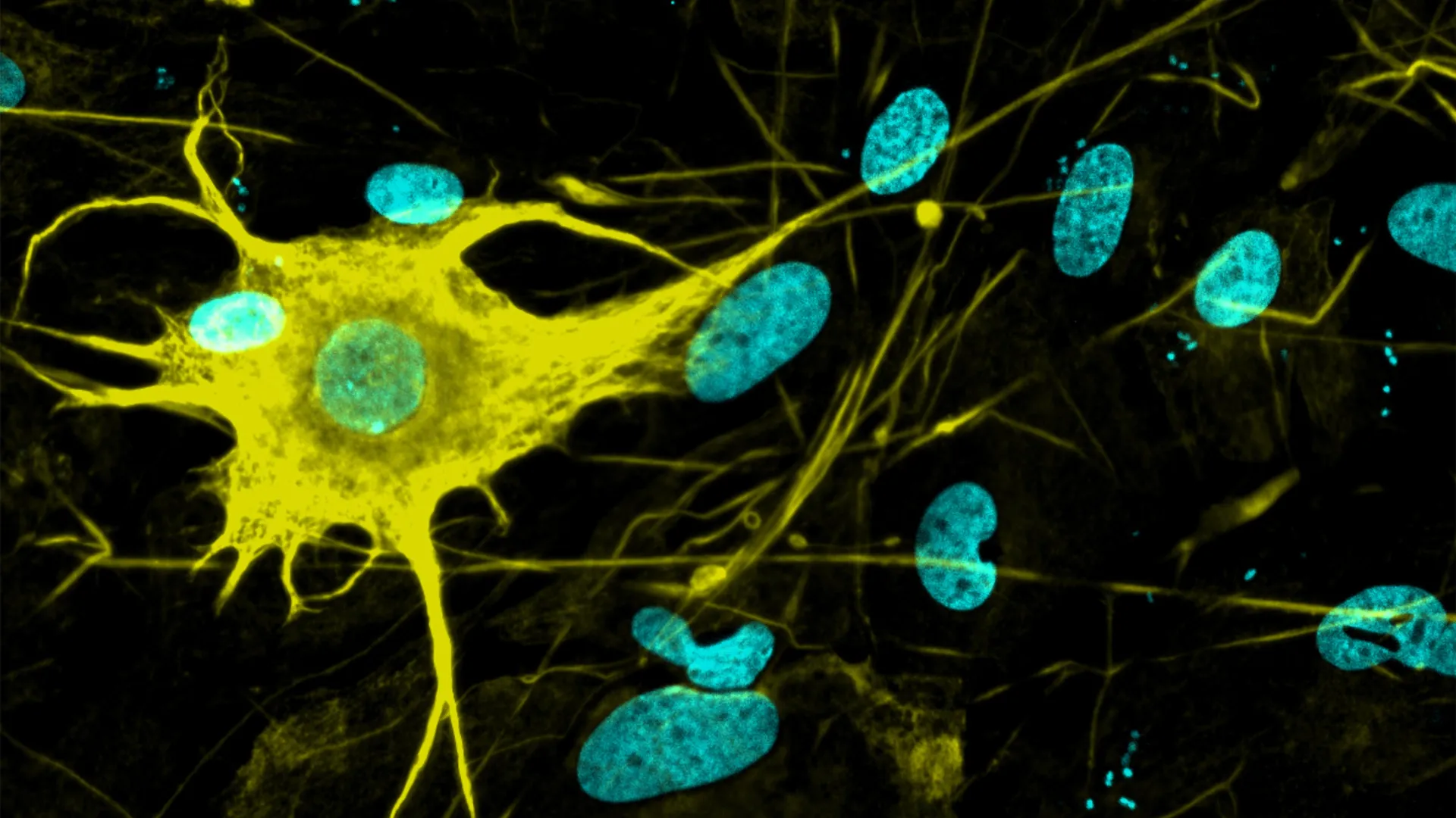
A new study from Ohio State University reveals how chandelier cells—a class of inhibitory interneurons—link up with their target excitatory cells. The team identified two specific molecules that must be present to enable a “handshake” between the cells, allowing synapses to form.

Chandelier cells are critical for brain function. They connect to a specific location on target excitatory neurons (pyramidal cells) called the axon initial segment. By grabbing this “handle,” chandelier cells can powerfully suppress the activity of the excitatory neurons, effectively preventing runaway electrical signals.

“These inhibitory interneurons shape and balance local circuit activity – they are the modulators, coordinators, the conductors of the orchestra,” said Yasufumi Hayano, lead author and postdoctoral scholar at Ohio State University. “From our results, we’ve concluded that interaction between two specific proteins regulates the specificity of their synapse formation.”

Loss of coordination between these cell types is associated with severe neurological and psychiatric disorders, including epilepsy, depression, autism, and schizophrenia.

The Molecular Handshake

The researchers discovered that the connection relies on a precise molecular interaction. The study identified two key proteins:

  • CNTNAP4: Located on the chandelier cells (the “conductors”).
  • Gliomedin: Located on the axon initial segment of the target neurons.

When these two proteins meet, they facilitate the formation of the synapse. Using visualizations in the brains of young mice, the team observed that when the genes for gliomedin were removed, the chandelier cells failed to form adequate connections with their targets. The “handshake” was broken, leaving the “conductors” unable to control the orchestra.

Implications for Neurological Disorders

Because the axon initial segment is the exact site where neurons generate action potentials (the signals used to communicate), chandelier cells have a disproportionately strong influence on brain activity. They essentially control the “faucet” of information flow.

“This is basic neuroscience, but there might be an impact for neuronal disorders,” said Hayano. “If this process is disrupted, what happens? If we lose those genes, which neuronal disorder might occur? We still don’t know, but those possibilities should be explored.”

Senior author Hiroki Taniguchi noted that understanding these developmental mechanisms is the first step toward identifying therapeutic targets for conditions where brain circuitry is imbalanced.


About this neuroscience research news

Author: Media Relations
Source: Ohio State University
Contact: Emily Caldwell – Ohio State University
Image: The image is credited to Ohio State University

Original Research: Closed access.
“The highly localized interaction between Neurofascin-186 and Gliomedin promotes subcellular innervation by the chandelier cell” by Yasufumi Hayano et al. The Journal of Neuroscience

Sunday, July 13, 2025

Researchers grow 400+ brain cell types—a leap for Alzheimer’s and Parkinson’s research

 You lost hundreds of millions of neurons, think your competent? doctor can ensure research produces the quantity needed? And the types you need? AND get them connected?

Researchers grow 400+ brain cell types—a leap for Alzheimer’s and Parkinson’s research

Date:
July 12, 2025
Source:
ETH Zurich
Summary:
Scientists at ETH Zurich have broken new ground by generating over 400 types of nerve cells from stem cells in the lab, far surpassing previous efforts that produced only a few dozen. By systematically experimenting with combinations of morphogens and gene regulators, the researchers replicated the vast diversity of neurons found in the human brain. This breakthrough holds major promise for studying neurological diseases like Alzheimer’s and Parkinson’s, creating more accurate models for drug testing, and eventually even enabling neuron replacement therapies.
Share:
FULL STORY

Nerve cells are not just nerve cells. Depending on how finely we distinguish, there are several hundred to several thousand different types of nerve cell in the human brain according to the latest calculations. These cell types vary in their function, in the number and length of their cellular appendages, and in their interconnections. They emit different neurotransmitters into our synapses and, depending on the region of the brain - for example, the cerebral cortex or the midbrain - different cell types are active.

When scientists produced nerve cells from stem cells in Petri dishes for their experiments in the past, it was not possible to take their vast diversity into account. Until now, researchers had only developed procedures for growing a few dozen different types of nerve cell in vitro. They achieved this using genetic engineering or by adding signalling molecules to activate particular cellular signalling pathways. However, they never got close to achieving the diversity of hundreds or thousands of different nerve cell types that actually exists.

"Neurons derived from stem cells are frequently used to study diseases. But up to now, researchers have often ignored which precise types of neuron they are working with," says Barbara Treutlein, Professor at the Department of Biosystems Science and Engineering at ETH Zurich in Basel. However, this is not the best approach to such work. "If we want to develop cell culture models for diseases and disorders such as Alzheimer's, Parkinson's and depression, we need to take the specific type of nerve cell involved into consideration."

Systematic screening was the key to success

Treutlein and her team have now successfully produced over 400 different types of nerve cell. In doing so, the scientists have paved the way for more precise basic neurological research with cell culture experiments.

The ETH researchers achieved this by working with a culture of human induced pluripotent stem cells that had been generated from blood cells. In these cells, they used genetic engineering to activate certain neuronal regulator genes and treated the cells with various morphogens, a special class of signalling molecules. Treutlein and her team took a systematic approach, using seven morphogens in different combinations and concentrations in their screening experiments. This resulted in almost 200 different sets of experimental conditions.

Morphogens

Morphogens are messengers that are known from research into embryonic development. They are not distributed uniformly within an embryo but occur in a variety of concentrations forming spatial patterns. In this way, they define the position of cells within the embryo, for example whether a cell is near the body axis or in the back, abdomen, head or torso. Accordingly, morphogens help to determine what grows where in the embryo.

The researchers used various analyses to prove that they had produced over 400 different types of nerve cell in their experiment. They examined the RNA (and therefore genetic activity) at the level of individual cells, as well as the external appearance of cells and their function: for example, which type of cell appendage they had in which quantities and which electric nerve impulses they emitted.

The researchers then compared their data with information from databases of neurons from the human brain. By doing this, they were able to identify the types of nerve cell that had been created, such as those found in the peripheral nervous system or brain cells and the part of the brain they come from, whether they perceive pain, cold or movement, and so on.

In-vitro neurons for active ingredient research

Treutlein clarifies that they are still a long way off producing all types of nerve cell that exist in vitro. Nonetheless, the researchers now have access to a much larger number of different cell types than they had before.

They would like to use in-vitro nerve cells to develop cell culture models for studying serious neurological conditions, including schizophrenia, Alzheimer's, Parkinson's, epilepsy, sleep disorders and multiple sclerosis. Cell culture models of this kind are also of great interest in pharmaceutical research for testing the effects of new active compounds in cell cultures without animal testing, with the ultimate aim of one day being able to cure these conditions.

In the future, the cells could also be used for cell replacement therapy, which involves replacing sick or dead nerve cells in the brain with new human cells.

But there is a challenge to overcome before this can happen: the researchers often produced a mixture of multiple different types of nerve cell in their experiments. They are now working to optimise their method so that each experimental condition only produces one specific cell type. They already have some initial ideas as to how this might be achieved.

Story Source:

Materials provided by ETH Zurich. Note: Content may be edited for style and length.


Journal Reference:

  1. Hsiu-Chuan Lin, Jasper Janssens, Benedikt Eisinger, Philipp Hornauer, Ann-Sophie Kroell, Malgorzata Santel, Maria Pascual-Garcia, Ryoko Okamoto, Kyriaki Karava, Zhisong He, Marthe Priouret, Manuel Schröter, J. Gray Camp, Barbara Treutlein. Human neuron subtype programming via single-cell transcriptome-coupled patterning screens. Science, 2025; 389 (6756) DOI: 10.1126/science.adn6121

Cite This Page:

ETH Zurich. "Researchers grow 400+ brain cell types—a leap for Alzheimer’s and Parkinson’s research." ScienceDaily. ScienceDaily, 12 July 2025. <www.sciencedaily.com/releases/2025/07/250711224316.htm>.

Explore More

from ScienceDaily

RELATED STORIES


Friday, August 27, 2021

Study Shows Psilocybin Regrowing Brain Tissue Damaged by Depression

Just maybe you want a 10% increase in the number of neuronal connections after your stroke, so use your likely depression as a reason to get this prescribed from your doctor. If they won't do it, ask for EXACT PROTOCOLS that delivers the neuronal connections and dendritic spines you need to recover. 

Post stroke depression(33% chance).

Study Shows Psilocybin Regrowing Brain Tissue Damaged by Depression

Depression impacts millions of Americans, with over 17% of adults suffering a major depressive issue. Depression can cause feelings of sadness and despair and potentially a loss of interest in things that once caused happiness. The mental and physical issues caused by this mental health condition can understandably hinder one’s work productivity and social relationships. The good news is that depression is treatable and advancements in psychedelics are potentially opening up new treatment avenues. A recent study by Yale researchers shows that psilocybin, the psychoactive ingredient in “magic mushrooms” may actually regrow brain tissue damaged by depression, as covered by Interesting Engineering:

“A psychedelic drug called psilocybin, which shows up naturally in some mushrooms, has shown signs of increasing durable connections between neurons in mouse brains, according to a new study published in the journal Neuron.

“In other words, the damage depression does to your brain might be reversible with psychedelic mushrooms, and scientists think the trip itself could play a vital role.

“‘We not only saw a 10% increase in the number of neuronal connections, but also they were on average about 10% larger, so the connections were stronger as well,’ said the study’s lead author Alex Kwan, who is also an associate professor of both psychiatry and neuroscience at Yale, in an embargoed release shared with IE. Earlier laboratory experiments hinted that psilocybin, in addition to the anesthetic ketamine, can reduce the effects of depression. But this latest research showed these compounds also increase the density of dendritic spines, creating small protrusions on nerve cells capable of enhancing the way information transmits from one neuron to the next. Depression and chronic stress are known factors in the reduction of these crucial neuronal connections.”

While legalized psilocybin therapy is likely years away for most, Oregon is leading the way following the passage of Measure 109. In less than two years, state-licensed and regulated psilocybin therapy will be available to help treat a whole host of mental health ailments, including depression. As more studies demonstrate the benefit of magic mushrooms and other psychedelics, Oregon needs to continue to be on the forefront of this growing medical revolution, and other states will certainly follow. Just as federal cannabis prohibition is about to crumble, we have an opportunity to create a similar movement for psychedelics, improving lives along the way.


Friday, May 15, 2020

Plasticity and Response to Action Observation: A Longitudinal fMRI Study of Potential Mirror Neurons in Patients With Subacute Stroke

I couldn't tell if actual movements were measured or just some nebulous neuronal activation. You are probably better off reading all these earlier posts on action observation. 

Plasticity and Response to Action Observation: A Longitudinal fMRI Study of Potential Mirror Neurons in Patients With Subacute Stroke 

First Published March 18, 2014 Research Article Find in PubMed


Background.
Action observation has been suggested as a possible gateway to retraining arm motor function post stroke. However, it is unclear if the neuronal response to action observation is affected by stroke and if it changes during the course of recovery.  
Objective.
To examine longitudinal changes in neuronal activity in a group of patients with subacute stroke when observing and executing a bimanual movement task.  
Methods.
 Eighteen patients were examined twice using 3-T functional magnetic resonance imaging; 1 to 2 weeks and 3 months post stroke symptom onset. Eighteen control participants were examined once. Image time series were analyzed (SPM8) and correlated with clinical motor function scores.  
Results.
During action observation and execution, an overlap of neuronal activation was observed in the superior and inferior parietal lobe, precentral gyrus, insula, and inferior temporal gyrus in both control participants and patients (P < .05; false discovery rate corrected). The neuronal response in the observation task increased from 1 to 2 weeks to 3 months after stroke. Most activated clusters were observed in the inferior temporal gyrus, the thalamus and movement-related areas, such as the premotor, supplementary and motor cortex (BA4, BA6). Increased activation of cerebellum and premotor area correlated with improved arm motor function. Most patients had regained full movement ability.  
Conclusions.
Plastic changes in neurons responding to action observation and action execution occurred in accordance with clinical recovery.(But did you measure clinical recovery? That is the only useful thing for survivors.) The involvement of motor areas when observing actions early and later after stroke may constitute a possible access to the motor system.

The reorganization of motor areas after stroke has been examined in numerous imaging studies, focusing primarily on possible correlations between task execution and recovery of motor function, for example, reviews by Johansen-Berg1 and Rehme et al.2 Results from behavioral and neurophysiological studies have suggested that mere observation of movement facilitates motor memory function and motor performance both in healthy individuals and patients with stroke.3-5 The potential mutual relationship between task execution and task observation is commonly referred to as mirror activity. More specifically, neurons that are activated both when a task is executed and when a similar task is observed are referred to as mirror neurons, and were first reported in macaques more than 2 decades ago.6
The existence of mirror neurons in humans, however, remains controversial; for example, the review by Turella et al.7 One point of discussion is why neurons with mirror properties are seemingly more widespread in the human brain in contrast to the spatially well-defined regions originally reported in macaques.8 It has been pointed out that large differences in choice of methodology such as the lack of inclusion of both an action observation and action execution task, combined with large differences in task selection can possibly account for many of these discrepancies.9 Neurons responding to the observation of hand actions have been reported in the premotor and supplementary motor cortex, the inferior frontal gyrus, the inferior parietal cortex and in the posterior middle temporal gyrus in healthy adults.10
There is a considerable interest in exploiting mirror neurons to improve motor function after stroke. Initial studies applying the observation of motor activities as a treatment approach have shown promising results, indicating that action observation could be a possible avenue to retraining motor function after stroke.3-5 However, little is known about reorganization processes in neurons with mirror activity after stroke. With focus on rehabilitation, it is important to investigate if similar neuronal responses exist in patients with lesions affecting the motor network. In the current study, functional magnetic resonance imaging (fMRI) based on blood oxygenation level–dependent contrast was used to investigate the neuronal activation when observing and executing a bimanual motor task in a group of patients with subacute stroke.
The main objective of this study was to identify for the first time a possible action observation/action execution matching system in patients with stroke during the course of recovery. An overlap of neurons involved in execution and observation in both patients and healthy control participants was expected, and that overlapping clusters of neurons would include brain regions that are typically associated with the mirror neuron system, such as premotor cortex, inferior frontal gyrus and inferior parietal cortex.

More at link.

Monday, January 15, 2018

Caltech researchers develop new method to see neural connections in living flies

Now if we could just accomplish this in humans. We could see which neurons are talking but no one is listening and map exactly the dead and damaged areas.  Using that knowledge to target axonal sprouting and dendrite branching to overcome those missing connections. Targeting interventions using nanotechnology to correct specific neurons could then be possible. I think grandiose ideas, but at least I think about solving stroke rather than sitting on my ass like the non-existent stroke leaders we have.
https://www.news-medical.net/news/20180112/Caltech-researchers-develop-new-method-to-see-neural-connections-in-living-flies.aspx

The human brain is composed of billions of neurons wired together in intricate webs and communicating through electrical pulses and chemical signals. Although neuroscientists have made progress in understanding the brain's many functions--such as regulating sleep, storing memories, and making decisions--visualizing the entire "wiring diagram" of neural connections throughout a brain is not possible using currently available methods. But now, using Drosophila fruit flies, Caltech researchers have developed a method to easily see neural connections and the flow of communications in real time within living flies. The work is a step forward toward creating a map of the entire fly brain's many connections, which could help scientists understand the neural circuits within human brains as well.
A paper describing the work appears online in the December 12 issue of eLife. The research was done in the laboratory of Caltech research professor Carlos Lois.
"If an electrical engineer wants to understand how a computer works, the first thing that he or she would want to figure out is how the different components are wired to each other," says Lois. "Similarly, we must know how neurons are wired together in order to understand how brains work."
When two neurons connect, they link together with a structure called a synapse, a space through which one neuron can send and receive electrical and chemical signals to or from another neuron. Even if multiple neurons are very close together, they need synapses to truly communicate.
The Lois laboratory has developed a method for tracing the flow of information across synapses, called TRACT (Transneuronal Control of Transcription). Using genetically engineered Drosophila fruit flies, TRACT allows researchers to observe which neurons are "talking" and which neurons are "listening" by prompting the connected neurons to produce glowing proteins.
With TRACT, when a neuron "talks"--or transmits a chemical or electrical signal across a synapse--it will also produce and send along a fluorescent protein that lights up both the talking neuron and its synapses with a particular color. Any neurons "listening" to the signal receive this protein, which binds to a so-called receptor molecule--genetically built-in by the researchers--on the receiving neuron's surface. The binding of the signal protein activates the receptor and triggers the neuron it's attached to in order to produce its own, differently colored fluorescent protein. In this way, communication between neurons becomes visible. Using a type of microscope that can peer through a thin window installed on the fly's head, the researchers can observe the colorful glow of neural connections in real time as the fly grows, moves, and experiences changes in its environment.
Many neurological and psychiatric conditions, such as autism and schizophrenia, are thought to be caused by altered connections between neurons. Using TRACT, scientists can monitor the neuronal connections in the brains of hundreds of flies each day, allowing them to make comparisons at different stages of development, between the sexes, and in flies that have genetic mutations. Thus, TRACT could be used to determine how different diseases perturb the connections within brain circuits. Additionally, because neural synapses change over time, TRACT allows the monitoring of synapse formation and destruction from day to day. Being able to see how and when neurons form or break synapses will be critical to understanding how the circuits in the brain assemble as the animal grows, and how they fall apart with age or disease.
TRACT can be localized to focus in on the wiring of any particular neural circuit of interest, such as those that control movement, hunger, or vision. Lois and his group tested their method by examining neurons within the well-understood olfactory circuit, the neurons responsible for the sense of smell. Their results confirmed existing data regarding this particular circuit's wiring diagram. In addition, they examined the circadian circuit, which is responsible for the waking and sleeping cycle, where they detected new possible synaptic connections.
TRACT, however, can do more than produce wiring diagrams. The transgenic flies can be genetically engineered so that the technique prompts receiving neurons to produce proteins that have a function, rather than colorful proteins that simply trace connections.
"We could use functional proteins to ask, 'What happens in the fly if I silence all the neurons that receive input from this one neuron?'" says Lois. "Or, conversely, 'What happens if I make the neurons that are connected to this neuron hyperactive?' Our technique not only allows us to create a wiring diagram of the brain, but also to genetically modify the function of neurons in a brain circuit."
Previous methods for examining neural connections were time consuming and labor intensive, involving thousands of thin slices of a brain reconstructed into a three-dimensional structure. A laboratory using these techniques could only yield a diagram for a single, small piece of fruit-fly brain per year. Additionally, these approaches could not be performed on living animals, making it impossible to see how neurons communicated in real time.
Because the TRACT method is completely genetically encoded, it is ideal for use in laboratory animals such as Drosophila and zebrafish; ultimately, Lois hopes to implement the technique in mice to enable the neural tracing of a mammalian brain. "TRACT is a new tool that will allow us to create wiring diagrams of brains and determine the function of connected neurons," he says. "This information will provide important clues towards understanding the complex workings of the human brain and its diseases."

Tuesday, August 15, 2017

Carbon nanotubes found safe for reconnecting damaged neurons

When will this be available for stroke? I don't care that this is for spinal cord injury. Pictures at link. 

Carbon nanotubes found safe for reconnecting damaged neurons


May offer future hope for patients with spinal-cord injury
July 5, 2017

(credit: Polina Shuvaeva/iStock)
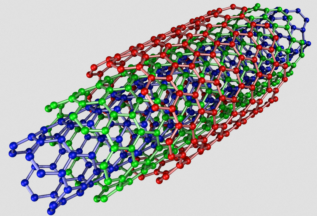
Multiwall carbon nanotubes (MWCNTs) could safely help repair damaged connections between neurons by serving as supporting scaffolds for growth or as connections between neurons.
That’s the conclusion of an in-vitro (lab) open-access study with cultured neurons (taken from the hippcampus of neonatal rats) by a multi-disciplinary team of scientists in Italy and Spain, published in the journal Nanomedicine: Nanotechnology, Biology, and Medicine.

A multi-walled carbon nanotube (credit: Eric Wieser/CC)
The study addressed whether MWCNTs that are interfaced to neurons affect synaptic transmission by modifying the lipid (fatty) cholesterol structure in artificial neural membranes.
Significantly, they found that MWCNTs:
  • Facilitate the full growth of neurons and the formation of new synapses. “This growth, however, is not indiscriminate and unlimited since, as we proved, after a few weeks, a physiological balance is attained.”
  • Do not interfere with the composition of lipids (cholesterol in particular), which make up the cellular membrane in neurons.
  • Do not interfere in the transmission of signals through synapses.
The researchers also noted that they recently reported (in an open access paper) low tissue reaction when multiwall carbon nanotubes were implanted in vivo (in live animals) to reconnect damaged spinal neurons.
The researchers say they proved that carbon nanotubes “perform excellently in terms of duration, adaptability and mechanical compatibility with tissue” and that “now we know that their interaction with biological material, too, is efficient. Based on this evidence, we are already studying an in vivo application, and preliminary results appear to be quite promising in terms of recovery of lost neurological functions.”
The research team comprised scientists from SISSA (International School for Advanced Studies), the University of Trieste, ELETTRA Sincrotrone, and two Spanish institutions, Basque Foundation for Science and CIC BiomaGUNE.

Abstract of Sculpting neurotransmission during synaptic development by 2D nanostructured interfaces

Carbon nanotube-based biomaterials critically contribute to the design of many prosthetic devices, with a particular impact in the development of bioelectronics components for novel neural interfaces. These nanomaterials combine excellent physical and chemical properties with peculiar nanostructured topography, thought to be crucial to their integration with neural tissue as long-term implants. The junction between carbon nanotubes and neural tissue can be particularly worthy of scientific attention and has been reported to significantly impact synapse construction in cultured neuronal networks. In this framework, the interaction of 2D carbon nanotube platforms with biological membranes is of paramount importance. Here we study carbon nanotube ability to interfere with lipid membrane structure and dynamics in cultured hippocampal neurons. While excluding that carbon nanotubes alter the homeostasis of neuronal membrane lipids, in particular cholesterol, we document in aged cultures an unprecedented functional integration between carbon nanotubes and the physiological maturation of the synaptic circuits.