Changing stroke rehab and research worldwide now.Time is Brain! trillions and trillions of neurons that DIE each day because there are NO effective hyperacute therapies besides tPA(only 12% effective). I have 523 posts on hyperacute therapy, enough for researchers to spend decades proving them out. These are my personal ideas and blog on stroke rehabilitation and stroke research. Do not attempt any of these without checking with your medical provider. Unless you join me in agitating, when you need these therapies they won't be there.

What this blog is for:

My blog is not to help survivors recover, it is to have the 10 million yearly stroke survivors light fires underneath their doctors, stroke hospitals and stroke researchers to get stroke solved. 100% recovery. The stroke medical world is completely failing at that goal, they don't even have it as a goal. Shortly after getting out of the hospital and getting NO information on the process or protocols of stroke rehabilitation and recovery I started searching on the internet and found that no other survivor received useful information. This is an attempt to cover all stroke rehabilitation information that should be readily available to survivors so they can talk with informed knowledge to their medical staff. It lays out what needs to be done to get stroke survivors closer to 100% recovery. It's quite disgusting that this information is not available from every stroke association and doctors group.

Showing posts with label Revascularization. Show all posts

Saturday, April 26, 2025

Effectiveness of Transcarotid vs Transfemoral Carotid Stenting for Stroke Prevention

 Why do this? Verify the Circle of Willis is complete and close the offending artery. No complications from stenting or TCAR. I'm not medically trained, so ask your doctor EXACTLY WHY THEY WANT TO DO THESE PROCEDURES! Revenue generation I bet. And I bet you don't get a guarantee of no complications; that tells you about their confidence in these procedures!

Like other types of carotid revascularization, TCAR carries a risk of:
  • Injury of your carotid artery.
  • Bleeding around your neck wound.
  • Swelling around the cut in your neck.
  • Nerve damage.
  • Stroke.
  • Heart attack.
  • Death.


Here is why your doctor needs to GUARANTEE NO complications from stenting!

Effectiveness of Transcarotid vs Transfemoral Carotid Stenting for Stroke Prevention

Key Points

Question  What is the comparative risk of stroke after transcarotid artery revascularization (TCAR) vs transfemoral carotid artery stenting (TF-CAS) for patients with carotid artery stenosis?

Findings  In this comparative effectiveness study of 5798 asymptomatic and 4721 symptomatic patients who underwent carotid stenting, TCAR was associated with a lower risk of stroke than was TF-CAS. This finding was consistent in both asymptomatic and symptomatic patients and was durable over a 3-year interval.

Meaning  With no completed or enrolling randomized clinical trial to evaluate TCAR, these comparative stroke risk results can inform future procedure choice for patients who are considering carotid artery stenting.

Abstract

Importance  The effectiveness of surgical transcarotid artery revascularization (TCAR) compared with percutaneous transfemoral carotid artery stenting (TF-CAS) for stroke prevention beyond the periprocedural period is poorly quantified.

Objective  To compare the risk of stroke after TCAR vs TF-CAS.

Design, Setting, and Participants  This retrospective cohort study used data from the Vascular Implant Surveillance and Outcomes Network (VISION), a procedural registry linked to Medicare claims data that captures clinical, procedural, and outcome data on patients who underwent carotid stenting. Patients who underwent TCAR or TF-CAS between October 1, 2016, and December 31, 2019, and were captured in the VISION database were included. Data were analyzed between January and June 2024.

Exposure  Type of carotid stenting (TCAR vs TF-CAS).

Main Outcomes and Measures  The primary outcomes were any stroke, including both periprocedural and during follow-up, defined using a validated claims code list, and death. Asymptomatic and symptomatic patients were analyzed separately. Kaplan-Meier analysis was used to calculate the cumulative incidence of the outcomes, and a multivariable Cox proportional hazards model was used to determine hazard ratios (HRs).

Results  There were 5798 asymptomatic patients (mean [SD] age, 74.6 [7.7] years; 3631 male [62.6%]; 3482 underwent TCAR; 2316 underwent TF-CAS) and 4721 symptomatic patients (mean [SD] age, 74.2 [8.3] years; 2969 male [62.9%]; 2377 underwent TCAR; 2344 underwent TF-CAS) who underwent carotid stenting. Patients who underwent TCAR were older, more likely to be female, and less likely to have had a prior ipsilateral carotid revascularization procedure. Among asymptomatic patients, the Kaplan-Meier 3-year risk of stroke was lower after TCAR (5.1%; 95% CI, 3.0%-7.1%) than TF-CAS (9.2%; 95% CI, 7.7%-10.7%) (log-rank P < .001). The composite 3-year stroke or death risk after TCAR was 22.6% (95% CI, 18.8%-26.3%), compared with 31.4% (95% CI, 28.3%-34.3%) after TF-CAS (log-rank P < .001). Compared with TCAR, the adjusted HR of stroke after TF-CAS among asymptomatic patients was 1.69 (95% CI, 1.25-2.28; P < .001). Among patients with symptomatic carotid stenosis, the 3-year stroke risk was also lower for TCAR (16.6%; 95% CI, 12.1%-20.9%) than for TF-CAS (20.9%; 95% CI, 17.5%-24.1%) (log-rank P < .001). The composite 3-year stroke or death risk after TCAR was 35.9% (95% CI, 30.1%-41.2%), compared with 41.5% (95% CI, 37.6%-45.1%) after TF-CAS (log-rank P < .001). Compared with TCAR, the adjusted HR for stroke after TF-CAS among symptomatic patients was 1.42 (95% CI, 1.17-1.73; P < .001). Sensitivity analyses yielded similar results.

Conclusions and Relevance  In this comparative effectiveness study, TCAR was associated with a lower risk of stroke than TF-CAS. This finding was consistent in both asymptomatic and symptomatic patients and durable over a 3-year interval. These findings can inform procedure choices for patients considering carotid artery stenting.

More at link.

Tuesday, April 30, 2024

Association Between MR‐Based Thrombus Imaging Characteristics and Endovascular Therapy Outcome in Acute Ischemic Stroke: A Systematic Review and Meta‐Analysis

 If your definition of successful revascularization is not 100% recovery then you'll regret your useless research when you are the 1 in 4 per WHO that has a stroke!

Association Between MR‐Based Thrombus Imaging Characteristics and Endovascular Therapy Outcome in Acute Ischemic Stroke: A Systematic Review and Meta‐Analysis

Originally publishedhttps://doi.org/10.1161/SVIN.123.001142Stroke: Vascular and Interventional Neurology. 2024;4:e001142

Abstract

Background

Prediction of successful revascularization and achieving a favorable functional outcome may help determine the optimal treatment strategy and improve the management of stroke. A growing body of literature has implicated a predictive value for thrombus imaging characteristics for stroke outcomes.(And all this is fucking useless since it does nothing to get to 100% recovery! I'D HAVE YOU ALL FIRED!)

Methods

We conducted an electronic search using PubMed, Ovid MEDLINE, and EMBASE, previously published meta‐analyses, and systematic review studies that intervened by endovascular thrombectomy or intravenous thrombolysis following large vessel occlusion stroke from 2000 to 2023 and involved magnetic resonance‐based thrombus imaging, then screened 2007 studies against our eligibility criteria. We extracted the enrollees’ characteristics and the association between clot features and radiological and functional outcome measures.

Results

Thirty‐three studies were found eligible, with a total number of 6902 enrollees. Susceptibility vessel sign was found in 3531 subjects (51.2%). Nine studies involved only the administration of intravenous thrombolysis, whereas 24 studies intervened by endovascular thrombectomy. Seventeen studies found at least an association between thrombus imaging characteristics and successful revascularization, whereas the others reported no association. only 13 studies found at least one thrombus characteristic associated with functional outcome, while the others showed no association between  the thrombus characteristics and functional outcome after stroke. Pooled meta‐analysis of studies that involved endovascular thrombectomy with or without intravenous thrombolysis showed a statistically significant association between the presence of susceptibility vessel sign and both successful reperfusion (odds ratio [OR]: 1.57 [1.09–2.27]; P = 0.02) and favorable functional outcome (OR: 1.76 [1.17–2.66]; P = 0.007).

Conclusion

The presence of susceptibility vessel sign on magnetic resonance‐based clot imaging was associated with functional outcome and successful reperfusion following thrombectomy.

Tuesday, February 23, 2021

Registry: More Options for Blocked Carotids Mean Better Outcomes

In my opinion being not medically trained they missed the most important option.

  1. Determine if the Circle of Willis is complete.

  2. If yes, then completely close up the offending artery.

  3. Your other three arteries supplying the Circle of Willis provide enough blood for you to function just fine. 

  4. This is why I would never consider a carotid endarterectomy as long as the Circle of Willis is complete.

  5. Cognitive Dysfunction and Mortality After Carotid Endarterectomy

  6. Risks of TCAR are here: Transcarotid Artery Revascularization,  you don't want them to happen and they put a stent in besides.

  7. And this problem tied to stents:Plaque Protrusion Tied to Stroke in Carotid Stenting 3% rate


 The latest here:

Registry: More Options for Blocked Carotids Mean Better Outcomes


Adoption of TCAR may be good for carotid revascularization in general

A computer rendering of the transcarotid artery revascularization procedure

Centers adding transcarotid artery revascularization (TCAR) to their treatment options had improved perioperative outcomes for patients with carotid artery stenosis, according to the Vascular Quality Initiative (VQI) registry.

Major adverse cardiovascular event (MACE) rates -- counting in-hospital stroke, MI, and death at 30 days -- were similar between patients undergoing TCAR and those getting surgical carotid endarterectomy (CEA; 2.3% vs 2.4%, P=0.91), reported a group led by Jesse Columbo, MD, MS, of Dartmouth-Hitchcock Medical Center in Lebanon, New Hampshire.

Despite such similar results, VQI centers had 10% less MACE in their overall carotid revascularization programs in the year after adopting TCAR than if they had stuck with the surgical option alone (OR 0.90, 95% CI 0.81-0.99), according to a difference-in-difference analysis by Columbo's group, published online in JAMA Open Network.

"This finding suggests that this new technology may have allowed proceduralists to select patients for whom TCAR may be superior to CEA, while still performing CEA on patients for whom that procedure was appropriate," study authors said.

"Overall, while observational in nature, these data imply that both TCAR and CEA may be reasonable treatment choices for patients undergoing carotid revascularization and providers may be able to choose the modality they feel best aligns with the patient's clinical presentation and anatomy," they concluded.

Moreover, the observed 10% MACE reduction, already "impressive," may in fact be an underestimation of the benefits of TCAR adoption, because the study included only VQI hospitals, which record only 10% of the CEAs performed in the U.S., commented Peter Groeneveld, MD, of the University of Pennsylvania in Philadelphia, in an invited commentary.

In contrast, device sales records indicate that the registry captures more than 95% of TCAR procedures.

TCAR was introduced to the U.S. market with the 2015 FDA approval of the Enroute stent system from Silk Road Medical.

The carotid procedure is designed to be a lower-risk alternative to surgery for patients at high risk due to anatomic or medical challenges. It is also designed with no need to traverse the aortic arch and carotid lesion prior to embolic protection, unlike transfemoral carotid stenting.

Whether TCAR is truly noninferior to CEA has not been proven in a randomized controlled trial. Transfemoral carotid stenting has been linked to more periprocedural events but no difference in outcomes at 10 years compared with surgery.

"An ongoing challenge for cardiovascular surgeons and interventionalists is to ascertain whether new therapeutic options for patients with a severe cardiovascular disease improve clinical outcomes across the full spectrum of patients with the disease. There are no guarantees that new therapies will produce net benefits across broad populations," Groeneveld cautioned.

He cited the examples of percutaneous coronary intervention, which may have "inappropriately shifted numerous patients away from the better treatment option" of coronary artery bypass grafting, and transcatheter aortic valve replacement, which conversely increased the accessibility of treatment while "potentially improving outcomes across the full spectrum of patients with aortic valve disease."

In general, better clinical outcomes for heart procedure recipients will require a "collective learning process" that harnesses data collection and judicious interpretation of statistical analyses, according to Groeneveld.

The study was a retrospective analysis of a national quality improvement registry maintained by the Society for Vascular Surgery. Included were people who underwent carotid procedures in 2015-2019. Those who got transfemoral carotid stenting were excluded.

A total of 86,027 patients at 469 participating VQI centers constituted the study cohort. Although only 8.9% got TCAR, TCAR procedures had jumped from 0.7% of all carotid procedures in 2015 to 17.0% in 2019. Accordingly, adoption of TCAR rose from 15 VQI centers in 2015 to 247 in 2019.

The TCAR group was significantly older on average (73.1 vs 70.6 years) and included fewer women (36.4% vs 39.5%) than the CEA group.

Unobserved confounding was a major potential limitation to the study.

"The primary threat to this study's validity is whether TCAR adoption by a hospital was associated with other concurrent quality-of-care initiatives that were the actual effectors of lower MACE rates. For example, it would not be surprising if TCAR-adopting hospitals were on a different trajectory for surgical quality than hospitals that did not adopt TCAR or did so slowly," according to Groeneveld.

A randomized trial "is likely the only way to dispel any lingering doubts regarding TCAR's comparative effectiveness" against surgery, but the "preponderance of existing observational data and evidence from single-group trials certainly supports TCAR's noninferiority to CEA," the editorialist acknowledged.

  • author['full_name']

    Nicole Lou is a reporter for MedPage Today, where she covers cardiology news and other developments in medicine. Follow

Disclosures

Columbo and Groeneveld had no disclosures.

Study coauthors reported ties to the NIH, the Patient-Centered Outcomes Research Institute, and Silk Road Medical.

Wednesday, January 29, 2020

Silent Cerebral Infarctions During Revascularisation Procedures Associated With Worse Cognitive Impairment

This is why I think you really need to have a discussion with your doctor as to why this needs to be done. If your Circle of Willis is complete it would seem to make more sense to just close up the artery completely rather than risk more brain damage. My right carotid artery was completely closed for at least 10 years before collaterals appeared and I suffered no cognitive impairment from that lack of blood source.  Push your doctor hard on that question. Will they guarantee no adverse events from the procedure they do? 

Silent Cerebral Infarctions During Revascularisation Procedures Associated With Worse Cognitive Impairment

By Eric Ramos

BARCELONA, Spain -- January 26, 2020 -- Patients with new silent cerebral infarctions during revascularisation procedures for carotid artery disease appear to have worse cognitive impairment than patients who do not develop new lesions, according to a study presented here at The International Conference of the European Society of Cardiology (ESC) Council on Stroke.

Patients who developed new ischaemic lesions scored significantly worse on the Mini Mental State Examination (MMSE) at their follow-up visit, compared with MMSE scores taken right before and after the procedure.

Patients undergoing carotid artery angioplasty and stenting have high risk of stroke or transient ischaemic attack intra-procedurally,” explained Alexandros Evangeliou, MD, Hippokration General Hospital, Thessaloniki, Greece. “Even though ischaemic brain lesions are detected on post-procedural imaging, most of these patients do not have neurological deficits.”

To clarify the association between intra-procedural ischaemic lesions and cognitive impairment, the researchers examined the neurological outcomes of 31 patients aged 56 to 78 years who successfully underwent carotid artery angioplasty and stenting for carotid artery disease.

All patients underwent a neurological clinical examination, and Mini-Mental-State-Examination (MMSE) and National Institutes of Health Stroke Scale (NIHSS) questionnaires were administered before and after surgery, and at the follow-up visit. Cerebral diffusion-weighted MRI was performed before and after revascularisation.

After a median follow-up time of 9 months, 8 (25.8%) patients showed new ischaemic lesions on MRI.

There were no differences in NIHSS scores before and after the procedure or at the follow-up visit. The authors noted that this was expected because the NIHSS score is used to quantify stroke severity in clinically evident strokes, not silent ones.

However, the MMSE score at the follow-up visit (24.7 ± 5) was significantly lower than scores recorded before the revascularisation procedure (27.2 ± 2.7; P = .049) and after (28.3 ± 1.53; P = .029).

“The MMSE score correlated with MRI imaging and can be used in this setting to assess prognosis,” said Dr. Evangeliou.

[Presentation title: Evaluation of Silent Cerebral Infarction Prognosis in Patients With Carotid Artery Disease Undergoing Carotid Artery Revascularization Procedure]

Monday, December 14, 2015

Imaging-based selection for revascularization in acute ischemic stroke

Once again our researchers are looking at only figuring out how to treat the better patients rather than all stroke patients. A great stroke association would not stand for this crappy situation. 

Imaging-based selection for revascularization in acute ischemic stroke

Puetz V, Barlinn K, Bodechtel U, Campbell B, Linn J, Gerber J; Current Opinion in Neurology (Dec 2015)

PURPOSE OF REVIEW With the positive results of recent endovascular thrombectomy (EVT) trials, intravenous thrombolysis (IVT) and EVT provide physicians with two majorly effective acute treatment options for patients with acute ischemic stroke. IVT and EVT can be used as a single treatment or as a combined IVT/EVT treatment approach. This review summarizes how imaging findings can help in selecting stroke patients who are likely to benefit from these revascularization therapies.
RECENT FINDINGS IVT applied within 4.5 h from symptom onset remains the mainstay of acute stroke therapy and was also applied to most patients in the randomized EVT trials. Recent studies have failed to demonstrate the effectiveness of IVT in later time windows. Vascular imaging is crucial to identify patients with a target intracranial occlusion prior to EVT. Patients with a small ischemic core, with good leptomeningeal collaterals or with evidence of penumbral tissue may particularly benefit from EVT. These imaging findings may also identify patients who benefit from EVT if applied more than 6 h from symptom onset.
SUMMARY Pretherapeutic imaging findings help in identifying stroke patients who are likely to benefit from endovascular stroke therapies, and may identify patients who benefit from revascularization therapies in later time windows

Monday, September 7, 2015

Newer devices reduce aspiration times, improve outcomes after stroke treatment

And once again these supposedly smart people do not discuss anything about solving the neuronal cascade of death.  Does no one have two functioning neurons to rub together?
http://www.healio.com/cardiology/intervention/news/online/%7B2a000be6-74c8-4c1d-874c-c61dae654e75%7D/newer-devices-reduce-aspiration-times-improve-outcomes-after-stroke-treatment?utm_source=maestro&utm_medium=email&utm_campaign=cardiology%20news

The use of newer, larger catheters for endovascular treatment of acute ischemic stroke was associated with shorter aspiration time and improved functional outcomes, according to data presented at the Society for NeuroInterventional Surgery Annual Meeting.
Researchers evaluated data on 932 patients (mean age, 66 years) with stroke who underwent endovascular treatment with the Penumbra Aspiration System (Penumbra Inc.). The patients were culled from six prospective and retrospective trials of the device.
“The aim … was to evaluate if there was a corresponding improvement in procedural times over the course of the evolution of these devices,” the researchers wrote in the abstract.
Outcomes of interest included clinical outcomes at 90 days, as indicated by modified Rankin scale, and aspiration time.
All patients received treatment within 8 hours of stroke onset. The mean time to presentation was 2.1 hours and the mean time to groin access was 4.4 hours. Upon admission, patients had a median NIH Stroke Scale score of 18 and a TIMI score of 0 to 1. The middle cerebral artery accounted for three-quarters of vessel locations.
After undergoing endovascular treatment, 84% of patients achieved revascularization to a TIMI score of 2 to 3.
Factors associated with shorter aspiration time included female sex, use of larger catheters, and timely presentation and treatment, according to univariable analysis. Shorter aspiration time was associated with improved outcomes (P = .0044). Younger patients, women, those with a lower baseline NIH Stroke Scale, shorter procedure times and revascularization to a TIMI score of 2 to 3 also were linked to improved outcomes.
Multivariable analysis adjusted for age, baseline NIH Stroke Scale and status after revascularization indicated an association between aspiration time and 90-day outcomes (P = .003), with a greater risk for poor outcome with increasing aspiration time.
“As each new generation of devices provides improved tools for our treatment arsenal, we are able to adjust our approach to maximize efficiencies and results for our patients,” Donald Frei, MD, president of the Society of NeuroInterventional Surgery and neurointerventionalist at Swedish Medical Center in Englewood, Colorado, said in a press release. “Thankfully, each generation … has proven to reduce procedure time, which raises the chance of a successful operation.” – by Adam Taliercio
Reference:
Frei D, et al. Implementation of new technological advances of endovascular treatment in acute ischemic stroke provides overall improvement in procedural times. Presented at: Society of NeuroInterventional Surgery Annual Meeting; July 27-30, 2015; San Francisco.

Thursday, July 2, 2015

Acute Ischemic Stroke Treatment, Part 2: Treatment “Roles of Capillary Index Score, Revascularization and Time”

More bloviating and talking about what they don't know. You're fucking screwed if you have a stroke right now. The authors need to read up on the neuronal cascade of death. They wouldn't be allowed to receive any funding from my organization with that lack of knowledge.
http://journal.frontiersin.org/article/10.3389/fneur.2015.00117/full?
  • 1Department of Neuro Interventional Surgery, Akron General Medical Center, Akron, OH, USA
  • 2Department of Research, Akron General Medical Center, Akron, OH, USA
Due to recent results from clinical intra-arterial treatment for acute ischemic stroke (IAT-AIS) trials such as the interventional management of stroke III, IAT-AIS and the merit of revascularization have been contested. Even though intra-arterial treatment (IAT) has been shown to improve revascularization rates, a corresponding increase in good outcomes has only recently been noted. Even though a significant percentage of patients achieve good revascularization in a timely manner, results do not translate into good clinical outcomes (GCOs). Based on a review of the literature, the authors suspect limited GCOs following timely and successful revascularization are due to poor patient selection(wrong, wrong, wrong) that led to futile and possibly even harmful revascularization. The capillary index score (CIS) is a simple angiography-based scale that can potentially be used to improve patient selection to prevent revascularization being performed on patients who are unlikely to benefit from treatment. The CIS characterizes presence of capillary blush related to collateral flow as a marker of residual viable tissue, with absence of blush indicating the tissue is no longer viable due to ischemia. By only selecting patients with a favorable CIS for IAT, the rate of GCOs should consistently approach 80–90%. Current methods of patient selection are primarily dependent on time from ischemia. Time from cerebral ischemia to irreversible tissue damage seems to vary from patient to patient; so focusing on viable tissue based on the CIS rather than relying on an artificial time window seems to be a more appropriate approach to patient selection.

Introduction

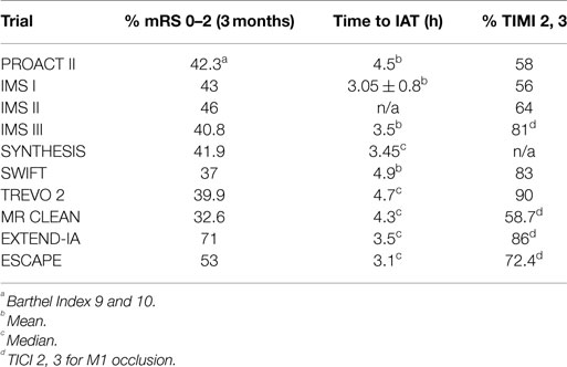
The interventional management of stroke (IMS) III trial (1) showed non-superiority of intra-arterial (IA) revascularization combined with intra venous (IV) tissue plasminogen activator (tPA) treatment over IV tPA alone, and the systemic thrombolysis for acute ischemic stroke (SYNTHESIS) trial demonstrated similar lack of favorable clinical outcomes for IA versus IV tPA therapy (2). This is despite the high revascularization rate in the IA arms in these trials. The role of intra-arterial treatment for acute ischemic stroke (IAT-AIS) has been contested. Paradoxically, however, the benefit of revascularization to clinical outcomes is convincingly attested to in prior literature. In a recent meta-analysis of 998 patients with clinical follow-up at 3 months, good clinical outcome was found in 58% of revascularized patients as compared to 24.8% in non-revascularized patients (3). When revascularization occurred within the first 6 h, good clinical outcomes (GCOs) were found in 50.9% of revascularized patients as compared to 11.1% in non-revascularized patients. Other authors reached similar conclusions. Even in the IMS III trial, better revascularization using the modified thrombolysis in cerebral infarction (mTICI) score led to better outcomes than those for patients who achieved lesser revascularization (1). This data were recently resolved with the publication of newer trials. In MR CLEAN, EXTEND-IA, and ESCAPE, good recanalization rates were achieved in 58.7, 86, and 72.4% of patients, respectively, with accompanying GCO rates at 32.6, 71, and 53%, respectively (46). While these results demonstrate IA superiority with higher recanalization rates than with IVT, there are still a significant number of patients who achieved good and timely revascularization that did not also achieve GCOs. So if better revascularization improves outcome and IA treatment has a better revascularization rate than IV treatment, how can we explain the lack of GCOs in some of these patients?

Revascularization and Outcome

Revascularization is defined as the restoration of anterograde blood flow to the ischemic area through the recently occluded artery. Currently, this is reported using the mTICI score, with mTICI of 2b or 3 being considered successful revascularization (7). The aim of revascularization is to produce clinical improvement through restoring the cerebral blood flow (CBF) level to greater than the critical threshold of 23 ml/100 g/min of viable brain tissue (8). This should translate into a permanent resolution of AIS symptoms by saving the ischemic tissue before it progresses to irreversible damage. So if perfect revascularization is achieved (mTICI = 3) in a timely manner, i.e., before ischemia becomes irreversible, clinical improvement should be achieved for almost all patients, as well as for the majority of patients with less effective revascularization (mTICI = 2b). However, review of the literature reveals that only around 50% of patients in whom we obtained timely recanalization (mTICI 2b, 3) will achieve a good clinical outcome (Table 1) (1, 2, 913). Attempting to solve the paradox regarding why all technically successful revascularizations do not translate into GCOs should help us improve our revascularization strategy.

more at link.

Acute ischemic stroke treatment, part 1: patient selection “The 50% barrier and the capillary index score”

This current strategy misses the whole problem of the neuronal cascade of death. Their assumption is wrong.  Doesn't ANYONE  in stroke read at all?
http://journal.frontiersin.org/article/10.3389/fneur.2015.00083/full?
  • 1Summit Neurovascular Specialists, Akron, OH, USA
  • 2Department of Research, Akron General Medical Center, Akron, OH, USA
  • 3Department of Cell Biology and Physiology, University of North Carolina School of Medicine, Chapel Hill, NC, USA
The current strategy for intra-arterial treatment (IAT) of acute ischemic stroke focuses on minimizing time from ictus to revascularization and maximizing revascularization. Employing this strategy has yet to lead to improved rates of successful outcomes. However, the collateral blood supply likely plays a significant role(Why?) in maintaining viable brain tissue during ischemia. Based on our prior work, we believe that only approximately 50% of patients are genetically predisposed to have sufficient collaterals for a good outcome following treatment, a concept we call the 50% barrier. The Capillary Index Score (CIS) has been developed as a tool to identify patients with a sufficient collateral blood supply to maintain tissue viability prior to treatment. Patients with a favorable CIS (f CIS) may be able to achieve a good outcome with IAT beyond an arbitrary time window. The CIS is incorporated into a proposed patient treatment algorithm. For patients suffering from a large stroke without aphasia, a non-enhanced head CT should be followed by CT angiography (CTA). For patients without signs of stroke mimics or visible signs of structural changes due to large irreversible ischemia, CTA can help confirm the vascular occlusion and location. The CIS can be obtained from a diagnostic cerebral angiogram, with IAT offered to patients categorized as f CIS.

Introduction

The current strategy for acute ischemic stroke (AIS) treatment is based on two pillars: time from ictus to revascularization (TIR) and revascularization success as measured by the modified thrombolysis in cerebral ischemia scale (mTICI). The assumption is that clinical outcome following AIS is dependent on the interaction of these two factors. The shorter the TIR and the higher the mTICI, the better the outcome. It follows that the strategy behind current intra-arterial treatment for acute ischemic stroke (IAT-AIS) is the faster and more complete the revascularization, the better the clinical outcome. However, despite the recent impressive improvement in revascularization rates and decrease in time to revascularization, until recently the clinical improvement rate remained unchanged at approximately 40–45% (Table 1) with a ratio of good clinical outcome (GCO) in treatment vs. control arms of approximately 1.7 (111). Recent trials have published GCOs above 50% in the treatment arm, but with the same ratio of GCOs between the treated and untreated arms around 1.7 (12, 13). How we can explain this consistency? A fresh look at our strategy and selection criteria is obviously warranted.
TABLE 1
www.frontiersin.org Table 1. Clinical outcomes across IAT-AIS trials.

Physiological Background and the 50% Barrier
Normal cerebral blood flow (CBF) is 50–55 ml/100 g/min (14, 15). AIS induces a rapid and sustained reduction in CBF. Clinical signs of ischemia generally become apparent when CBF drops below 23 ml/100 g/min (16). If residual CBF (rCBF) further decreases to 15–16 ml/100 g/min, the cortical-evoked potential ceases within seconds (16). The rate of depression of the evoked potential (EP) amplitude (expressed in units of percent of control/min) is highly correlated with the residual flow, following a linear relationship with the regression line intercepting the flow axis at 15.2 ml/100 g/min (17). The data strongly suggest a threshold-like relationship also exists between the amplitude of the EP and local blood flow. If flow is greater than approximately 16 ml/100 g/min the EP is not affected, but at flows less than approximately 12 ml/100 g/min the EP is abolished (17). Neither the clinical signs of ischemia nor cessation of the EP is synonymous with cell death, but cessation of the EP is one of the final stages before irreversible injury (infarction). Its physiological purpose is to conserve energy by decreasing cell metabolism to the minimal level possible; however, cell death ensues thereafter.
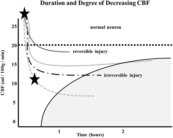
Similarly, the relationship between time to irreversible damage and rCBF is well-documented (18). In one study, rCBF in monkeys was measured in the ischemic area with time after occlusion until irreversible tissue damage occurred (16). An infarction threshold was observed relating the rCBF to time between the initial drop in CBF to irreversible ischemia (Figure 1). This work confirmed prior studies using the neuronal EP and showed that when rCBF reached a low level of around 10 ml/100 g/min, the available time to salvage the brain tissue was extremely short (<1 h) (16).
 
FIGURE 1
www.frontiersin.org
Figure 1. Depth of ischemia and time to irreversible cerebral damage: time to irreversible cerebral damage depends on the depth of ischemia, which depends on the pial collateral supply to the ischemic territory. Since different patients have different collaterals, the depth of ischemia will vary among patients, as will the time available for therapy to salvage the tissue (16).
 
 
The depth of ischemia, i.e., the level of rCBF, will vary from patient to patient depending on the available retrograde pial collaterals to the ischemic area. The major determinants of the amount of collateral perfusion are the number and diameter of these pial collaterals, plus perfusion pressure and resistance above and below the collateral network. Greater collateral numbers and diameters sustain a higher rCBF, thus more salvageable brain and a smaller final infarct volume.
Following AIS, rCBF stays virtually unchanged if spontaneous recanalization of the occluded blood vessels does not occur (16, 18, 19). While the clinical symptoms of ischemia will often resolve if CBF is restored promptly, prolonged low levels of rCBF leads to irreversible brain tissue damage. Since the time of ischemia that the brain tissue can tolerate before irreversible damage ensues depends on the rCBF value, which is patient-specific and highly dependent on the collaterals, it follows that every patient has his or her own time (Figure 1) (16, 18, 19). Hence, if we correctly select patients that are optimal candidates (patients with ischemic but viable tissue) and are able to achieve safe, full, and timely revascularization (prior to irreversible ischemic damage occurring), the clinical symptoms of a stroke should improve significantly and rather quickly.
Given this information, the most logical explanation for the remarkably consistent results of the different IAT-AIS trials, with <50% GCOs (modified Rankin Score, or mRS, ≤2), is that around half of treated patients have poor pial collaterals, thus causing them to have a relatively low rCBF such that they enter into irreversible ischemia before therapy can be administered, even when timely (within 6 h) revascularization is achieved. This observation implies a potential ceiling effect for IAT-AIS; we call this phenomenon the 50% barrier (Figure 2).
More at link.

Monday, December 2, 2013

Vascular remodeling after ischemic stroke: mechanisms and therapeutic potentials

Precisely what is your doctor doing with this to get you to 100% recovery?
http://www.sciencedirect.com/science/article/pii/S0301008213001287
  • a Department of Neurological Surgery, UCSF, San Francisco, CA 94121, USA
  • b SFVAMC, San Francisco, CA 94121, USA
  • c Neuroscience and Neuroengineering Research Center, Med-X Research Institute, Shanghai 200030, China
  • d School of Biomedical Engineering, Shanghai Jiao Tong University, Shanghai 200030, China
  • e Department of Neurology, Shanghai Ruijin Hospital, School of Medicine, Shanghai Jiao Tong University, Shanghai, 200025, China
  • f Department of Neurological Surgery, Tohoku University Graduate School of Medicine 1-1 Seiryo-machi, Aoba-ku, Sendai 980-8574, Japan
  • g Center of Cerebrovascular Disease Research, University of Pittsburgh School of Medicine, Pittsburgh, PA 15213, USA

Highlights

Distinct mechanisms underlying arteriogenesis and angiogenesis are delineated.
Technology in detecting each vascular remodeling process.
Clinical and preclinical implications of arteriogenesis and angiogenesis.
Potential therapies in promoting vascular remodeling including those in trials.

Abstract

The brain vasculature has been increasingly recognized as a key player that directs brain development, regulates homeostasis, and contributes to pathological processes. Following ischemic stroke, the reduction of blood flow elicits a cascade of changes and leads to vascular remodeling. However, the temporal profile of vascular changes after stroke is not well understood. Growing evidence suggests that the early phase of cerebral blood volume (CBV) increase is likely due to the improvement in collateral flow, also known as arteriogenesis, whereas the late phase of CBV increase is attributed to the surge of angiogenesis. Arteriogenesis is triggered by shear fluid stress followed by activation of endothelium and inflammatory processes, while angiogenesis induces a number of pro-angiogenic factors and circulating endothelial progenitor cells (EPCs). The status of collaterals in acute stroke has been shown to have several prognostic implications, while the causal relationship between angiogenesis and improved functional recovery has yet to be established in patients. A number of interventions aimed at enhancing cerebral blood flow including increasing collateral recruitment are under clinical investigation. Transplantation of EPCs to improve angiogenesis is also underway. Knowledge in the underlying physiological mechanisms for improved arteriogenesis and angiogenesis shall lead to more effective therapies for ischemic stroke.

Sunday, February 10, 2013

Clot Remover Holds Up in Another Study

Their definition of good outcomes is completely wrong. A good outcome is 100% recovery. At that level this would be a complete failure and we would be searching for the hyperacute therapies that stop the neuronal cascade of death.
http://www.medpagetoday.com/MeetingCoverage/ISC/37262?
The FDA-approved Solitaire FR (Flow Restoration) device was successful at opening blocked vessels in patients with an acute ischemic stroke and resulted in good outcomes, a nonrandomized study showed.
Revascularization was achieved in 79.2% of patients, with a low rate of symptomatic intracranial hemorrhage within 24 hours of the procedure (1.5%) and low rates of death and device- or procedure-related serious adverse events through 90 days (6.9% and 7.4%, respectively), according to Vitor Pereira, MD, of University Hospitals of Geneva.
Also within the first 90 days, most patients (57.2%) achieved a modified Rankin score of 2 or lower, indicating little or no disability, he reported at the International Stroke Conference here.
The study "supports the further investigation of this device in a randomized controlled trial against best medical treatment," Pereira said during his presentation.
In an interview, he explained that this trial -- which lacked a control group -- demonstrated the real-world experience with the device in high-volume, comprehensive stroke centers.
"We still need to understand which population will benefit from the treatment to prepare future randomized controlled trials," he said.
Pereira said it was important to point out that the trial used a standard protocol for the device, which established consistency in the procedure across centers. "I think this is the best way to plan and to design future trials, controlling all of the aspects that you can," he said.
The Solitaire FR device was approved by the FDA last year on the strength of the SWIFT trial, which showed that the device was superior to the Merci retriever for achieving revascularization. Solitaire FR is indicated for patients who have failed treatment with IV recombinant tissue plasminogen activator (tPA) or who are ineligible for IV tPA.
The current trial -- called STAR -- evaluated use of the device in patients presenting with an acute ischemic stroke in the anterior circulation within 8 hours of symptom onset at one of 14 centers across Europe, Australia, and Canada. All of the centers had extensive experience with mechanical thrombectomy devices and used the Solitaire FR device according to the instructions for use -- use of a balloon guiding catheter and a maximum number of three passes per occluded vessel.
The study included 202 patients with a median age of 72. The occluded vessel was the middle cerebral artery in 82% of the patients and the internal carotid artery terminal in 18%.
More than half of the patients (59%) received IV tPA before mechanical thrombectomy and the rest underwent mechanical thrombectomy as the first treatment for their stroke.
Endovascular treatment occurred within 3 hours in 25.6% of the patients, in 3 to 4.5 hours in 37.9%, and beyond 4.5 hours in 36.4%. The median time from groin puncture to placement of the balloon guiding catheter was 12 and the median time from balloon catheter placement to revascularization was 20 minutes.
Successful revascularization -- defined as a Thrombolysis in Cerebral Infarction (TICI) score of 2b or greater -- was achieved in more than three-quarters of the patients, as confirmed by a core lab.
The median NIH Stroke Scale score steadily improved during the study -- it was 17 before the procedure, 7 in the 24 hours after the procedure, 4 a week to 10 days after the procedure or at discharge, and 1 at 90 days.
"The time from groin puncture to recanalization was quite short and so that's really exciting," according to Kyra Becker, MD, co-director of the University of Washington Stroke Center at Harborview in Seattle and vice chair of the program committee for this year's meeting.
But she cautioned against taking too much away from the study because of the lack of a control group.
"Even the SWIFT trial, it shows that Solitaire is better than Merci, but it doesn't mean that Merci works at all, so you don't really know that Solitaire is better than nothing," she said.

Monday, November 26, 2012

Study confirms high biocompatibility of polymeric materials with brain tissue

Signs of Steve Austin in the Bionic Man or maybe the Cylons from Battlestar Gallactica, or for those who are into books, H. G. Wells -The Island of Doctor Moreau
 http://www.news-medical.net/news/20121126/Study-confirms-high-biocompatibility-of-polymeric-materials-with-brain-tissue.aspx

Professor José Miguel Soria, a member of the Institute of Biomedical Sciences, Universidad CEU Cardenal Herrera, has co-directed with Professor Manuel Monleón of the Universitat Politècnica de València a study on the compatibility of polymeric biomaterials in the brain and its effectiveness to favour neuroregeneration in areas with some kind of damage or brain injury.
The research carried out has shown that these types of implants, made of a biocompatible synthetic material, are colonized within two months by neural progenitor cells and irrigated by new blood vessels. This allows the generation, within these structures, of new neurons and glia, capable of repairing injured brain tissue caused by trauma, stroke or neurodegenerative disease, among other causes.

The synthetic structures used in this study are made with a porous and biocompatible polymeric material called acrylate copolymer. In the first phase of the project, the structures have been studied in vitro by implanting them into neural tissue, and subsequently also in vivo, when implanted in two areas of the adult rat brain: the cerebral cortex and the subventricular zone, the most important source of generation of adult neural stem cells.

The study has confirmed the high biocompatibility of polymeric materials, such as acrylate copolymer, with brain tissue and opens new possibilities of the effectiveness of the implementation of these structures in the brain, seeking optimum location for developing regenerative strategies of the central nervous system.
Furthermore, the results are particularly relevant when one considers that in the adult brain neuroregeneration capacity is more limited than in younger individuals and that the main impediment for this is the lack of revascularization of damaged tissue, something that the biomaterial studied has shown to favour.