Use the labels in the right column to find what you want. Or you can go thru them one by one, there are only 33,991 posts. Searching is done in the search box in upper left corner. I blog on anything to do with stroke. DO NOT DO ANYTHING SUGGESTED HERE AS I AM NOT MEDICALLY TRAINED, YOUR DOCTOR IS, LISTEN TO THEM. BUT I BET THEY DON'T KNOW HOW TO GET YOU 100% RECOVERED. I DON'T EITHER BUT HAVE PLENTY OF QUESTIONS FOR YOUR DOCTOR TO ANSWER.
Changing stroke rehab and research worldwide now.Time is Brain!trillions and trillions of neuronsthatDIEeach day because there areNOeffective hyperacute therapies besides tPA(only 12% effective). I have 523 posts on hyperacute therapy, enough for researchers to spend decades proving them out. These are my personal ideas and blog on stroke rehabilitation and stroke research. Do not attempt any of these without checking with your medical provider. Unless you join me in agitating, when you need these therapies they won't be there.
What this blog is for:
My blog is not to help survivors recover, it is to have the 10 million yearly stroke survivors light fires underneath their doctors, stroke hospitals and stroke researchers to get stroke solved. 100% recovery. The stroke medical world is completely failing at that goal, they don't even have it as a goal. Shortly after getting out of the hospital and getting NO information on the process or protocols of stroke rehabilitation and recovery I started searching on the internet and found that no other survivor received useful information. This is an attempt to cover all stroke rehabilitation information that should be readily available to survivors so they can talk with informed knowledge to their medical staff. It lays out what needs to be done to get stroke survivors closer to 100% recovery. It's quite disgusting that this information is not available from every stroke association and doctors group.
Showing posts with label memory loss reversal. Show all posts
Showing posts with label memory loss reversal. Show all posts
With your memory problems and brain fog post stroke ask your competent? doctor for how to accomplish this. Earlier knowledge is here, hopefully your doctor is competent enough to already know about it:
Here’s what you’ll learn when you read this story:
For decades, scientists have looked at various mechanisms responsible for the decline of our mental faculties as we age.
A new discovery shows that the accumulation of a certain protein called the ferritin light chain 1, or FTL1, plays a big role.
In experiments with mice, scientists found that less FTL1 present in neural cells promoted healthier, younger brains.
The brain may lie at the biological center of our lived experience, but the scientific underpinnings of the brain—specifically, how it forms subjective consciousness—remain mysterious. Sadly, one truth is well-known: As we age, so does our brain. Because this all-important organ filled with 86 billion neurons forms our experience, dysregulation within the brain has outsized impacts on someone’s quality of life.
For years, scientists have tried to find ways to stave off these negative effects and make a person’s healthspan largely match their ever-increasing lifespan. In 2021, Stanford University investigated the debris-cleaning role of the myeloid cells within the brain, and three years later, a study from the University of Rochester on the broader glymphatic system (which interacts with myeloid cells) found ways to restart the flow of brain-cleaning fluids.
In another installment of this ongoing research to improve the aging brain, scientists from the University of California San Francisco (UCSF) identified a protein that’s central to aging the brain in humans. By analyzing how genes and proteins changed over time in mice, the team identified a troublesome protein named ferritin light chain 1, or FTL1. When scientists reduced the presence of this protein in the hippocampus, the mice regained some of their youthful characteristics, including improved nerve connections and better permorfance on memory tests. The results of the study were published in the journal Nature Aging.
The Truth About Alzheimer’s Scientists Just Revealed
What if everything we thought about Alzheimer's is wrong?
Current Time 0:01
/
Duration 0:46
0
“It is truly a reversal of impairments […]. It’s much more than merely delaying or preventing symptoms,” Saul Villeda, a co-author of the study from UCSF, said in a press statement. “We’re seeing more opportunities to alleviate the worst consequences of old age. It’s a hopeful time to be working on the biology of aging.”
To test this idea even further, the team artificially stimulated the production of FTL1 in young mice, and soon, their mental abilities began to match those of older rodents. When analyzing the effects of FTL1 protein in a petri dish, Villeda and his team discerned that nerve cells engineered simple, one armed neural wires—known as “neurites”—rather than the branching neurites typical of normal neural cells.
“To identify potential therapeutic targets to restore cognitive function in older people, we first need to gain mechanistic insight into the molecular drivers of cognitive decline in the aging brain,” the authors wrote. “It has become clear that cognitive dysfunction in the aged brain in the absence of neurodegenerative disease is not paralleled by cell death but, instead, by a decline in neuronal function at the synaptic level.”
The authors also note that a 2015 study found that increased ferritin levels in cerebrospinal fluid negatively impacted cognitive performance and accurately predicted conversion from mild cognitive impairment to Alzheimer’s disease. The researchers hope that by targeting FTL1, future therapies may not only improve natural cognitive decline, but will also benefit people with neurodegenerative diseases.
As this study—and many studies before it—have clearly demonstrated, there is no singular cause of mental decline. But with each new study, scientists work toward a holistic picture of neurodegenerative decline as we age, and with that new perspective comes the hope of new therapies that could make cognitive decline a thing of the past.
Summary: Cognitive decline is often seen as a problem starting and ending in the brain, but new research suggests the “remote control” for memory is actually in the gut. Scientists found that as mice age, their gut microbiome shifts, specifically favoring a bacteria called Parabacteroides goldsteinii.
This shift triggers an inflammatory response in the intestines that silences the vagus nerve, the main communication highway to the brain. This silence effectively “numbs” the hippocampus, leading to memory loss. Remarkably, by stimulating the vagus nerve or resetting the gut microbiome, researchers turned “forgetful” old mice into sharp performers—proving that cognitive aging is not hardwired and can be reversed through the digestive tract.
Key Facts
The Interoception Breakdown: Aging affects “interoception”—the brain’s ability to sense what is happening inside the body. A breakdown in gut-to-brain signaling is a direct driver of age-related memory decline.
The Bacterial Culprit: The bacteria Parabacteroides goldsteinii increases with age, releasing metabolites that trigger gut inflammation and block vagus nerve activity.
Reversing the Clock: Stimulating the vagus nerve in older mice restored their memory and spatial navigation skills to the level of 2-month-old mice, showing that “brain aging” can be modulated from the periphery.
Source: Stanford
The sight of a delectable plate of lasagna or the aroma of a holiday ham are sure to get hungry bellies rumbling in anticipation of a feast to come. But although we’ve all experienced the sensation of “eating” with our eyes and noses before food meets mouth, much less is known about the information superhighway, known as the vagus nerve, that sends signals in the opposite direction — from your gut straight to your brain.
These signals relay more than just what you’ve eaten and when you are full. A new study in mice from researchers at Stanford Medicine and the Palo Alto, California-based Arc Institute has identified a critical link between the bacteria that live in your gut and the cognitive decline that often occurs with aging.
New research reveals that restoring the communication between the gastrointestinal tract and the brain can reverse age-related memory decline. Credit: Neuroscience News
“Although memory loss is common with age, it affects people differently and at different ages,” said Christoph Thaiss, PhD, assistant professor of pathology.
“We wanted to understand why some very old people remain cognitively sharp while other people see significant declines beginning in their 50s or 60s. What we learned is that the timeline of memory decline is not hardwired; it’s actively modulated in the body, and the gastrointestinal tract is a critical regulator of this process.”
The mouse study showed that the composition of the naturally occurring bacterial population that lives in the gut, known as the gut microbiome, changes with age — favoring some species of bacteria over others.
These changes are registered by immune cells in the gastrointestinal tract, which spark an inflammatory response that hampers the ability of the vagus nerve to signal to the hippocampus — the part of the brain responsible for memory formation and spatial navigation.
Stimulating the activity of the vagus nerve in older animals turned old, forgetful mice into whisker-sharp whizzes able to remember novel objects and escape from mazes as nimbly as their younger counterparts.
“The degree of reversibility of age-related cognitive decline in the animals just by altering gut-brain communication was a surprise,” Thaiss said. “We tend to think of memory decline as a brain-intrinsic process. But this study indicates that we can enhance memory formation and brain activity by changing the composition of the gastrointestinal tract — a kind of remote control for the brain.”
Thaiss, who is also a core investigator at Palo Alto-based Arc Institute, is a senior author of the study, which will be published on March 11 in Nature. Maayan Levy, PhD, an assistant professor of pathology and Arc Institute innovation investigator, is the other senior author. Timothy Cox, a graduate student at the University of Pennsylvania, is the lead author of the research.
“Our study emphasizes that processes in the brain can be modulated through peripheral intervention,” Levy said. “Since the gastrointestinal tract is easily accessible orally, modulating the abundance of gut microbiome metabolites is a very appealing strategy to control brain function.”
The call is coming from inside the body
The idea that hundreds of species of bacteria are nestled comfortably in our intestines used to be surprising. But the gut microbiome is experiencing a kind of media heyday as people realize that its function is critical to not just how we digest our food, but also to our overall health.
A little more than a decade ago, researchers showed that tinkering with rodents’ gut microbiomes affected the animals’ social and cognitive behaviors. Thaiss and Levy wondered whether a similar process could be responsible for the memory loss and cognitive troubles often associated with aging.
Signals from inside the body to the brain — like those that travel from the intestines to the brain via the vagus nerve — are part of what’s called interoception. In contrast, signals from outside the body, conveyed primarily by the five senses of taste, touch, smell, vision and hearing, are called exteroception.
“Exteroception is basically how we perceive the outside,” Thaiss said. “We have a lot of detailed knowledge about how this works. But we know much less about how the brain senses what is going on inside the body. We don’t know how many internal senses there are, or even all of what they are sensing. It’s clear that our exteroception capabilities decline with age — we grow to need eyeglasses and hearing aids, for example. And this study shows that aging also affects interoception.”
To test their theory that the gut microbiome plays a role in the “senior moments” many of us experience, the researchers housed young (2-month-old) mice together with old (18-month-old) mice. Living (and pooping) in close proximity exposed the young mice to the gut microbiomes of the old mice and vice versa. After one month, the researchers examined the compositions of the microbiomes of the old and young animals.
They found that the shared digs caused the microbiomes of the young mice to more closely resemble that of the older animals. When they compared the abilities of the mice to recognize a novel object, or to find the exit in a maze, the young mice with “old” microbiomes performed significantly more poorly than their peers — showing less curiosity about the unfamiliar object and bumbling about the maze in ways similar to that of old animals.
When the researchers compared young mice and old mice raised in a germ-free environment since birth (meaning neither group had gut bacteria), the young mice maintained their ability to form memories. But when they transplanted young, germ-free mice with microbiomes from old mice, the young mice again performed like older animals in the memory and cognition tests. Interestingly, the germ-free old mice did not experience a loss of memory and cognition as they aged, performing as well as 2-month-old animals.
Strikingly, treating young mice with “old” microbiomes (and, therefore, faltering cognitive abilities) with broad-spectrum antibiotics for two weeks restored the animals’ cognitive abilities, causing them to avidly investigate unfamiliar objects and scamper through the maze as well as their control peers.
“The object recognition test is like cognitive recognition tests in humans, where you are shown a series of images, then have to remember which ones you’ve seen before after some time passes,” Thaiss said.
“And the maze test is like people trying to recall where they parked their car at a large shopping center. What these tasks have in common, in mice and in people, is that they are very strongly dependent on activity in the hippocampus, because that is where memories are encoded.”
What’s different in their guts?
Digging deeper, the researchers identified specific changes that occur in the composition of the gut microbiome of mice as they age. In particular, the relative abundance of a bacteria called Parabacteroides goldsteinii increases in old mice and is directly associated with cognitive decline in the animals.
They showed that colonizing the guts of young mice with this bacterial species inhibited their performance on the object recognition and maze escape tasks, and that this deficit correlated with a reduction of activity in the hippocampus.
When they treated old mice with a molecule that activates the vagus nerve, however, the cognitive performance of the animals was indistinguishable from that of young animals.
Further experiments showed that the increasing prevalence of the Parabacteroides goldsteinii bacteria correlated with an increasing amount of metabolites called medium-chain fatty acids, and that these metabolites cause a group of immune cells in the gut called myeloid cells to initiate an inflammatory response. This inflammation inhibits the activity of the vagus nerve, the activity of the hippocampus and the ability to form lasting memories.
“The GI tract is arguably the first organ system to evolve during human evolutionary history, so the evolution of cognitive processes in the brain has undoubtedly been shaped by signals coming from the intestine,” Levy said. “It’s likely that signals from the GI tract play an important role in contextualizing memory formation.”
Thaiss added, “Basically, we’ve identified a three-step pathway toward cognitive decline that starts with gastrointestinal aging and the subsequent microbial and metabolic changes that occur.
“The myeloid cells in the GI tract sense these changes, and their inflammatory response impairs the connection between the gut and the brain via the vagus nerve. This is a direct driver of memory decline. And if we restore the activity of the vagus nerve, we can restore an old animal’s memory function to that of a young animal.”
The researchers are now investigating whether a similar gut microbiome and brain activity pathway exists in humans, and whether it also contributes to age-related cognitive decline. Importantly, vagus nerve stimulation is approved by the Food and Drug Administration as a treatment for depression or epilepsy and to aid stroke recovery. The researchers are also interested in developing ways to non-invasively monitor, and perhaps even control, the activity of peripheral neurons to affect memory formation and cognition.
“Our hope is that ultimately these findings can be translated into the clinic to combat age-related cognitive decline in people,” Thaiss said.
Researchers from Monell Chemical Senses Center in Philadelphia; the University of California, Irvine; University College Cork, Ireland; Calico Life Sciences LLC; and the Children’s Hospital of Philadelphia contributed to the work.
Funding: The study was funded by the Arc Institute, the National Institutes of Health (grants NIH DK019525, T32AG000255, F30AG081097, T32HG000046, F30AG080958, DP2-AG-067511, DP2-AG-067492, DP1-DK-140021, R01-NS-134976 and R01-DK-129691), the Burroughs Wellcome Fund, the American Cancer Society, the Pew Scholar Award, the Searle Scholar Program, the Edward Mallinckrodt Jr. Foundation, the W.W. Smith Charitable Trust, the Blavatnik Family Fellowship, the Prevent Cancer Foundation, the Polybio Research Foundation, the V Foundation, the Kathryn W. Davis Aging Brain Scholar Program, the McKnight Brain Research Foundation, the Kenneth Rainin Foundation, the IDSA Foundation and the Human Frontier Science Program.
Key Questions Answered:
Q: Can my “gut health” really determine if I have senior moments?
A: Yes! This study found that the gut acts as a regulator for the brain. When your gut bacteria shift with age, they create inflammation that “muffles” the vagus nerve. If the brain can’t hear the signals from the gut, the hippocampus (your memory center) struggles to form new memories.
Q: Is “brain aging” permanent?
A: Not necessarily. The most surprising part of the Stanford study was that cognitive decline in mice was reversible. By either stimulating the vagus nerve or clearing out “old” gut bacteria with antibiotics, the researchers were able to make old mice just as sharp and curious as young ones.
Q: Does this mean I should take probiotics for my memory?
A: While specific “memory probiotics” aren’t a thing yet, the researchers are looking at how modulating gut metabolites could control brain function. Since the gut is easily accessible, “oral interventions” to sharpen the mind are a major goal for future human clinical trials.
Editorial Notes:
This article was edited by a Neuroscience News editor.
Journal paper reviewed in full.
Additional context added by our staff.
About this aging research news
Author: Krista Conger Source: Stanford Contact: Krista Conger – Sanford Image: The image is credited to Neuroscience News
Original Research: Open access. “Intestinal interoceptive dysfunction drives age-associated cognitive decline” by Timothy O. Cox, Ashwarya S. Devason, Alan de Araujo, Sydney Mason, Madhav Subramanian, Andrea F. M. Salvador, Hélène C. Descamps, Junwon Kim, Yixuan Zhu, Lev Litichevskiy, Sunhee Jung, Won-Suk Song, Adrián Cortés-Martín, Nathan T. Henderson, Kuei-Pin Huang, Thao Nguyen, Wisath Sae-Lee, Iboro C. Umana, Maria Sacta, Ryan J. Rahman, Stephen Wisser, J. Andrew D. Nelson, Ilona Golynker, Alana M. McSween, Eric F. Hohmann, Shaan Patel, Anna L. Bub, Clara Soekler, Niklas Blank, Kevt’her Hoxha, Lavinia Boccia, Andrea C. Wong, Klaas Bahnsen, Jihee Kim, Natalie Biderman, Dina Abbasian, Clarissa Shoffler, Christopher Petucci, Fiona E. McAllister, Amber L. Alhadeff, Marc V. Fuccillo, Colin Hill, Cholsoon Jang, J. Nicholas Betley, Guillaume de Lartigue, Virginia Y.-M. Lee, Maayan Levy. Nature DOI:10.1038/s41586-026-10191-6
In efforts to beat Alzheimer's
disease, researchers are looking at existing drugs that could tackle
the condition, and a new study identifies two promising candidates that
are currently used to treat cancer.
Already approved by regulators in the US – meaning potential clinical trials for Alzheimer's could start sooner – the drugs are letrozole (usually used to treat breast cancer) and irinotecan (usually used to treat colon and lung cancer).
Researchers from the University of California, San Francisco
(UCSF) and Gladstone Institutes started by looking at how Alzheimer's
altered gene expression in the brain.
They then consulted a medical database called the Connectivity Map
to look for drugs that reversed these gene expression changes, and
cross-referenced records of patients who had taken these drugs as part
of cancer treatments and their likelihood of developing Alzheimer's.
Intriguingly, the drugs seemed to have reduced their risk.
"Alzheimer's disease comes with complex changes to the
brain, which has made it tough to study and treat, but our computational
tools opened up the possibility of tackling the complexity directly," says computational biologist Marina Sirota, from UCSF.
"We're excited that our computational approach led us to a
potential combination therapy for Alzheimer's based on existing
FDA-approved medications."
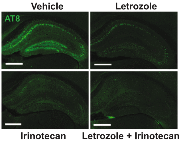
The application of letrozole and irinotecan reduced the level of tau proteins (light green) in mouse brains. (Li et al., Cell, 2025)
Having
picked out letrozole and irinotecan as the best candidates, the
researchers tested them in mouse models of Alzheimer's. When used in
tandem, the drugs were shown to reverse some of the brain changes brought on by the disease.
The harmful clumps of tau protein
that build up in brains affected by Alzheimer's were reduced
significantly, and the mice showed improvements in learning and memory
tasks – two brain capabilities often impaired by Alzheimer's.
By combining the two drugs together, the researchers were
able to target different types of brain cells affected by the disease.
Letorozole seemed to counter Alzheimer's in neurons, while irinotecan
worked in glia.
"Alzheimer's is likely the result of numerous alterations in many genes and proteins that, together, disrupt brain health," says neuroscientist Yadong Huang, from UCSF and Gladstone.
"This makes it very challenging for drug development – which
traditionally produces one drug for a single gene or protein that
drives disease."
It's a promising start, but there's more work to be done:
obviously the drugs have only been directly tested in mice so far, and
these medications also come with side effects
attached. They need to be reconsidered if the drugs are going to be
repurposed for a different disease than what they were originally
approved for.
One of the next steps should be clinical trials for people with Alzheimer's disease.
According to the researchers, this approach could lead to more
personalized and effective treatments, based on how gene expression has
been altered in each case.
It's estimated that more than 55 million people
have Alzheimer's today, and as the world's population ages, that's
expected to more than double in the next 25 years. Finding ways to
prevent the disease and even reverse symptoms would have a huge impact
on global health.
"If completely independent data sources, such as single-cell
expression data and clinical records, guide us to the same pathways and
the same drugs, and then resolve Alzheimer's in a genetic model, then
maybe we're on to something," says Sirota.
"We're hopeful this can be swiftly translated into a real solution for millions of patients with Alzheimer's."
Does your competent? doctor and hospital have enough
functioning neurons to get human testing going and see if this could
restore memory damaged from your stroke?
A groundbreaking study shows that two repurposed cancer drugs, when combined, can correct complex brain cell dysfunction and restore memory in Alzheimer’s mouse models, offering hope for new multi-targeted therapies.
In a recent study published in the journal Cell, a group of researchers investigated whether combining the aromatase inhibitor letrozole and the topoisomerase I inhibitor irinotecan could reverse cell–type–specific transcriptomic disturbances and improve cognition and pathology in Alzheimer’s disease (AD) models.
Background
Every three seconds, someone develops dementia, and more than 50 million people worldwide now live with AD, a figure forecast to triple by 2050. Existing monoclonal antibodies slow amyloid beta (Aβ) accumulation yet leave most patients cognitively impaired, partly because AD involves intertwined neuronal and glial dysfunctions.
Recent single-nucleus RNA sequencing (snRNA-seq) studies have revealed that excitatory neurons, inhibitory neurons, microglia, astrocytes, and oligodendrocyte precursor cells (OPCs) each follow distinct yet interacting degenerative programs.
Repurposing approved drugs offers a faster, safer route to intervention, but single-target candidates rarely tackle this cellular heterogeneity. Therefore, the correction of multi-cell-type networks warrants exploration.
The study identified 25 drugs with predicted multi-cell-type effects, but only five showed a reduced AD risk in human clinical records, including letrozole and irinotecan, which were prioritized for their complementary neuronal and glial targeting. Further research should determine whether dual-action regimens translate into durable clinical benefit and address the sex-specific responses observed in preclinical models.
About the Study
Initial computational screening identified 86 potential drugs targeting individual AD-affected cell types, later refined to 25 candidates predicted to work across multiple cell types.
Investigators first mined integrated human post-mortem snRNA-seq datasets from three independent studies to generate cell-specific AD expression signatures. They then matched these against the Connectivity Map (CMap) compendium, a database of drug perturbations primarily using cancer cell lines, to identify drugs whose perturbation profiles inversely correlated with disease patterns.
Electronic Medical Record (EMR) analytics of 1.4 million adults aged ≥ 65 years across six University of California health systems revealed that exposure to letrozole or irinotecan correlated with a lower AD incidence after propensity-matched adjustment for demographics, comorbidities, and cancer indications.
Notably, cancer patients have lower baseline AD risk, and letrozole’s effects in males were inconclusive due to limited data.
To validate causality, researchers crossed 5xFAD amyloid-overproducing mice with P301S mutant tau transgenic (PS19) mice. They allocated 20 sex-balanced, double-transgenic animals to each of four groups: vehicle, letrozole (1 mg/kg), irinotecan (10 mg/kg), or the combination, administered every other day by intraperitoneal injection for three months, starting at 4–5 months of age.
Morris water maze assessed spatial learning, while brains underwent Sudan Black volumetry, Thioflavin S Aβ staining, AT8 phosphorylated tau (p-tau) immunofluorescence, Iba1 microglial and GFAP astrocytic histology, plus NeuN neuronal counts. Parallel hippocampal snRNA-seq libraries were prepared using 10x Genomics’ Chromium technology and analyzed with UMAP clustering to map drug-induced transcriptomic shifts.
Study Results
During six days of hidden platform training, swim latencies did not differ between groups, confirming equal baseline performance across the two groups. In probe trials, only combination-treated mice spent significantly more time and made more crossings in the target quadrant at both 24 hours and 72 hours, demonstrating recovery of both short- and long-term spatial memory. In contrast, single agents showed partial benefit in males, while females saw negligible improvement. Visual acuity and motility were unchanged, excluding sensorimotor confounds.
Morphologically, all treatments reduced hippocampal atrophy, but the combination achieved the greatest volume preservation. Aβ plaque burden fell across groups, yet p-tau deposition declined only with dual therapy, aligning with its unique cognitive efficacy.
Irinotecan, alone or in combination, reduced microgliosis (Iba1 area), and letrozole monotherapy significantly rescued CA1 neuronal loss, whereas astrocytosis (GFAP) dropped modestly under irinotecan alone. Mechanistically, letrozole preserved neurons while irinotecan tempered glial inflammation – effects that combined additively.
At the transcriptomic level, combination therapy expanded the proportions of CA1 and CA3 pyramidal neurons within hippocampal nuclei. Cell-cell communication analysis showed dampened hyperactive signaling from glia to neurons. Across six major cell types, the regimen counterregulated AD signature genes, notably normalizing APOE expression in microglia, astrocytes, and OPCs.
Gene ontology enrichment tied reversed neuronal genes to estrogen signaling and synaptic plasticity, aligning with letrozole’s aromatase blockade, while glial reversals highlighted oxidative stress mitigation and cholesterol transport, consonant with irinotecan’s anti-inflammatory profile.
Conclusions
To summarize, the letrozole-irinotecan combination delivered a convergent, cell-type-directed therapy that surpassed either monotherapy by restoring memory, shrinking Aβ plaques, lowering p-tau deposition, dampening microgliosis and astrocytosis, and preserving hippocampal neurons. snRNA-seq confirmed the regimen rewired disease-specific gene networks across neurons and glia.
These preclinical results, tempered by methodological caveats (e.g., cancer-cell-derived drug signatures), support repurposed multi-target strategies for AD and justify clinical trials testing this affordable anticancer duo in at-risk populations, with a focus on sex-specific efficacy.
Journal reference:
Li, Y., Serras, C.P., Blumenfeld, J., Xie, M., Hao, Y., Deng, E., Chun, Y. Y., Holtzman, J., An, A., Yoon, S. Y., Tang, X., Rao, A., Woldemariam, S., Tang, A., Zhang, A., Simms, J., Lo, I., Oskotsky, T., Keiser, M. J., Huang, Y., & Sirota, M. (2025). Cell-type-directed network-correcting combination therapy for Alzheimer’s disease. Cell. DOI: 10.1016/j.cell.2025.06.035 https://www.cell.com/cell/fulltext/S0092-8674(25)00737-8
Summary: Blocking the kynurenine pathway, a
regulator of brain metabolism, can restore cognitive function in lab
mice with Alzheimer’s disease. The pathway is overactivated in
Alzheimer’s, disrupting glucose metabolism and starving neurons of
energy.
By inhibiting this pathway, researchers improved memory
and brain plasticity in mice, offering hope for new treatments in
humans. IDO1 inhibitors, currently in cancer trials, could be repurposed
for Alzheimer’s treatment.
Key Facts:
Blocking the kynurenine pathway in mice with Alzheimer’s restored brain metabolism and improved memory.
The kynurenine pathway is overactivated in Alzheimer’s, disrupting glucose metabolism in the brain.
Drugs targeting this pathway, initially developed for cancer, show promise for Alzheimer’s treatment.
Source: Stanford
Among the many ways
neuroscientists think Alzheimer’s disease may strip away brain function
is by disrupting the glucose metabolism needed to fuel the healthy
brain. In essence, declining metabolism robs the brain of energy,
impairing thinking and memory.
Against that backdrop, a
team of neuroscientists at the Knight Initiative for Brain Resilience at
Stanford’s Wu Tsai Neurosciences Institute have zeroed in on a critical
regulator of brain metabolism known as the kynurenine pathway.
Better
yet, this intersection between neuroscience, oncology, and pharmacology
could help speed drugs to market if proved effective in ongoing human
clinical trials for cancer. Credit: Neuroscience News
They
hypothesize that the kynurenine pathway is overactivated as a result of
amyloid plaque and tau proteins that accumulate in the brains of
patients with Alzheimer’s disease.
Now, with support from research and training grants from the Knight
Initiative, they have shown that by blocking the kynurenine pathway in
lab mice with Alzheimer’s Disease, they can improve, or even restore,
cognitive function by reinstating healthy brain metabolism.
“We were surprised that these metabolic improvements were so effective at not just preserving healthy synapses, but in actually rescuing behavior.
The mice performed better in cognitive and memory tests when we gave
them drugs that block the kynurenine pathway,” said senior
author, Katrin Andreasson, a neurologist at the Stanford School of
Medicine and member of the Wu Tsai Neurosciences Institute.
The
study, which included collaborations with researchers at the Salk
Institute for Biological Studies, Penn State University, and others,
appeared August 22, 2024 in the journal Science.
Hungry neurons
In
the brain, kynurenine regulates production of the energy molecule
lactate, which nourishes the brain’s neurons and helps maintain healthy
synapses. Andreasson and her fellow researchers specifically looked at
the enzyme indoleamine-2,3-dioxygenase 1 — or IDO1, for short — which
generates kynurenine.
Their
hypothesis was that increases in IDO1 and kynurenine triggered by
accumulation of amyloid and tau proteins would disrupt healthy brain
metabolism and lead to cognitive decline.
“The kynurenine pathway is over activated in astrocytes, a critical
cell type that metabolically supports neurons. When this happens,
astrocytes cannot produce enough lactate as an energy source for
neurons, and this disrupts healthy brain metabolism and harms synapses”
Andreasson said.
Blocking production of kynurenine by blocking IDO1 restores the ability of astrocytes to nourish neurons with lactate.
Best
of all for Andreasson, and for Alzheimer’s patients, IDO1 is well known
in oncology and there are already drugs in clinical trials to suppress
IDO1 activity and production of kynurenine. That meant Andreasson could
circumvent the time-intensive work of identifying new drugs and to begin
testing in lab mice almost immediately.
In those tests, in which
mice with Alzheimer’s Disease must navigate an obstacle course before
and after drug intervention, Andreasson and team found that the drugs
improved hippocampal glucose metabolism, corrected deficient astrocytic
performance, and improved the mice’s spatial memory.
Promise kept
“We
also can’t overlook the fact that we saw this improvement in brain
plasticity in mice with both amyloid and tau mice models. These are
completely different pathologies, and the drugs appear to work for
both,” Andreasson noted. “That was really exciting to us.”
Better
yet, this intersection between neuroscience, oncology, and pharmacology
could help speed drugs to market if proved effective in ongoing human
clinical trials for cancer.
“We’re hopeful that IDO1 inhibitors developed for cancer could be repurposed for treatment of AD,” Andreasson stressed.
The
next step is to test IDO1 inhibitors in human Alzheimer’s patients to
see if they show similar improvements in cognition and memory. Prior
clinical tests in cancer patients tested the effectiveness of IDO1
inhibitors on cancer but did not anticipate or measure improvements in
cognition and memory. Andreasson is hoping to investigate IDO1
inhibitors in human trials for Alzheimer’s disease in the near future.
About this Alzheimer’s disease and memory research news
Author: Nicholas Weiler Source: Stanford Contact: Nicholas Weiler – Stanford Image: The image is credited to Neuroscience News
Newsweekspoke to a psychiatrist to learn what lifestyle choices can be made at a young age to prevent memory loss and memory-related problems in the years to come.
Minimize Alcohol Consumption or Abstain
Dr. Paul Linde, a board-certified general adult psychiatrist, is the medical director for psychiatry and collaborative care at Ria Health and a clinical professor of psychiatry at theUniversity of California, San Francisco, School of Medicine. He toldNewsweekone thing that can help prevent memory loss later in life is reducing alcohol consumption or keeping it to a minimum.
"Minimizing your alcohol consumption is an easy way to prevent memory loss. A night of heavy drinking can lead to short-term memory loss, aka blacking out, when one appears to be superficially functioning but for which one will have no recollection of your actions or behaviors the next day," he said.
"Severe long-term consequences associated with long-term heavy drinking, chronic alcohol use and alcohol use disorder can also occur," he continued.
"Excessive alcohol use damages multiple parts of the brain, including the cerebral cortex, where new memories are formed and then stored and retrieved," he added.
This can result in conditions like alcohol-induced major neurocognitive disorder, also known as alcoholic dementia or alcohol-induced amnestic disorder. Officially called Wernicke-Korsakoff Syndrome, it is a severe memory disorder caused by thiamine deficiency due to heavy drinking.
"While abstinence is likely the best option to preserve memory and brain function, there are other smarter habits to adopt if one will continue to drink," Linde said.
"Avoid heavy drinking periods and avoid drinking if you're looking to specifically memorize something, as learning and memorizing is impaired by alcohol use," he said. "Lastly, avoid drinking alcohol close to bedtime—good-quality sleep is correlated with high memory function, and alcohol is known to disrupt high-quality sleep, even if one feels sleepy after a drink or two."
The term "sober curious" has trended online as an increasing number of young people dabble in sobriety. Clearly, the brain benefits associated with giving up or minimizing drinking deserve serious consideration.



 Newsweek
 Money Talks News