Semaphorins, originally discovered as guidance cues for
developing axons, are involved in many processes that shape the nervous
system during development, from neuronal proliferation and migration to
neuritogenesis and synapse formation. Interestingly, the expression of
many Semaphorins persists after development. For instance, Semaphorin 3A
is a component of perineuronal nets, the extracellular matrix
structures enwrapping certain types of neurons in the adult CNS, which
contribute to the closure of the critical period for plasticity.
Semaphorin 3G and 4C play a crucial role in the control of adult
hippocampal connectivity and memory processes, and Semaphorin 5A and 7A
regulate adult neurogenesis. This evidence points to a role of
Semaphorins in the regulation of adult neuronal plasticity. In this
review, we address the distribution of Semaphorins in the adult nervous
system and we discuss their function in physiological and pathological
processes.
Introduction
The development of complex tissues depends on
proliferation, differentiation and migration of cells. Cell guidance
cues regulate these events and continue to be essential throughout life
to maintain tissue homeostasis. Semaphorins constitute a large family of
cell guidance cues, which are present in some viruses and conserved
across animal species, from worms and flies to humans. Thirty Semaphorin
proteins have been identified so far. They can be divided into eight
classes (Sema1-7 and the viral Semaphorins, SemaV) on the basis of
phylogenetic relationships and structural features. Sema1, Sema2, and
Sema5C are found in invertebrates, whereas all the other Semaphorin
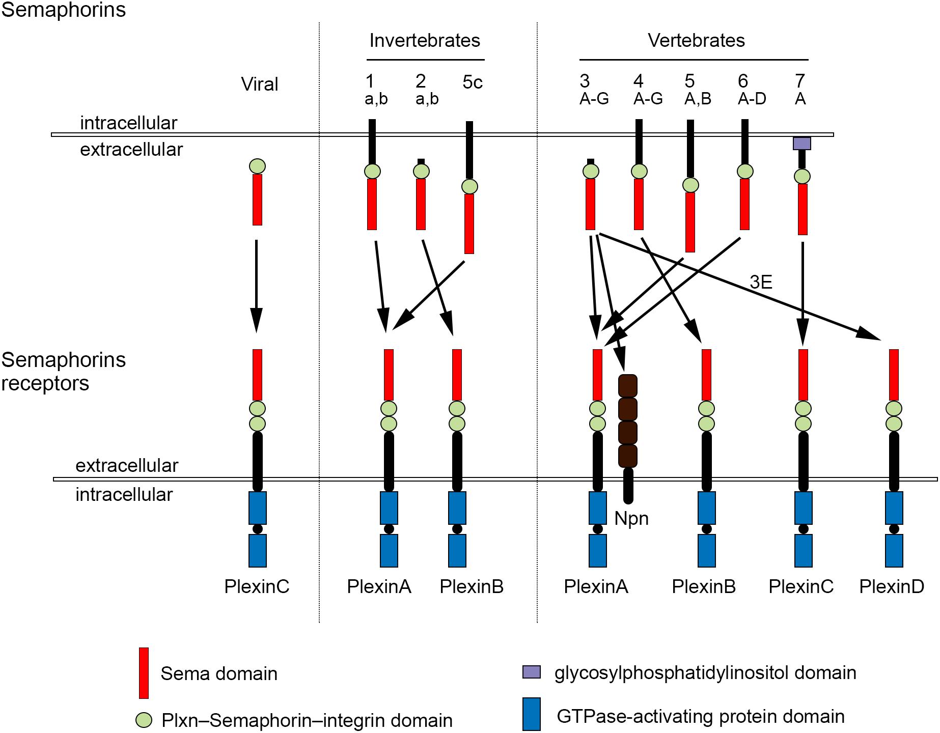
classes are found in vertebrates (Bamberg et al., 1999; Pasterkamp, 2012; Alto and Terman, 2017; Figure 1).
Semaphorins can be secreted (Sema2, Sema3, and SemaV),
membrane-spanning (Sema1, Sema4, Sema5, and Sema6) or
glycosylphosphatidylinositol-anchored (Sema7A). The structural hallmark
of the Semaphorin protein family is an extracellular domain at the
N-terminal region, important for dimerization and interaction
specificity, called Sema domain, which is followed by a
Plxn–Semaphorin–integrin domain and by distinct protein domains that
further define Semaphorins (Zhou et al., 2008; Figure 1).
First characterized by their ability to act as repulsive cues for growing neurites (Kolodkin et al., 1992, 1993; Luo et al., 1993),
Semaphorins are now known to be crucial molecules also for the
development and functioning of the musculoskeletal, cardiovascular,
respiratory, immune, endocrine, reproductive, hepatic, and renal system.
In addition, Semaphorin signaling has been linked to diseases affecting
these systems, as well as to cancer (Roth et al., 2009; Neufeld et al., 2012; Pasterkamp, 2012; Tamagnone, 2012; Giacobini and Prevot, 2013; Kang and Kumanogoh, 2013; Kumanogoh and Kikutani, 2013).
The effects of Semaphorins occur through binding to their receptors, the neuropilin (Npn) and plexin (Plxn) protein families (Figure 1).
Plxns are grouped in four classes, from A to D, with four A-type, three
B-type, one C-type and one D-type. The Plxn extracellular region
contains several sema domains, which are important for binding to
Semaphorins, whereas the intracellular region contains GTPase-activating
protein domains (Takahashi et al., 1999; Tamagnone et al., 1999).
In general, Semaphorins exist as homodimers, both in an unbound state
and when interacting with Plxns. Semaphorin homodimers bring together
two Plxn monomers or disrupt existing Plxn homodimers, relieving Plxn
autoinhibition, which might be caused by an interaction between the sema
domain of Plxn and the rest of the Plxn extracellular domain (Takahashi and Strittmatter, 2001; Kong et al., 2016).
Once activated, Plxn signals through downstream molecules, including
GTPases of the Rho family, protein kinases such as MAPK, and enzymes
such as MICAL (molecule interacting with casL), which induce the
phosphorylation of intracellular proteins of the collapsin responsive
mediator protein (CRMP) family (Vikis et al., 2000; Hu et al., 2001; Liu and Strittmatter, 2001; Terman et al., 2002; Pasterkamp et al., 2003; Hota and Buck, 2012). CRMPs, in turn, affect actin and microtubule dynamics (Hung et al., 2010, 2011; Alto and Terman, 2017).
Membrane-associated Semaphorins can also act as receptors or
co-receptors for Semaphorins located on other cells, a phenomenon known
as reverse signaling (Battistini and Tamagnone, 2016).
Class 3 Semaphorins require Npn as co-receptors (Npn-1
and -2). Npn-1 homodimers function as ligand-binding receptors for
Sema3A and Sema3D; Npn-2 homodimers as receptors for Sema3F; and Npn-1
and Npn-2 heterodimers as receptors for Sema3B, 3C, 3E, and 3G (He et al., 2019; Toledano et al., 2019).
Npn are transmembrane proteins with short intracellular domains that
lack intrinsic enzymatic or signaling activity. They do not seem to act
as a direct bridge between Plxn and Semaphorins but may function in the
presentation of Semaphorin to Plxn. In addition, Npn can bind vascular
endothelial growth factor (VEGF) in co-receptor complexes with VEGF
receptors (Kruger et al., 2005; Pasterkamp, 2012), regulating blood and lymphatic vessel growth (Tammela et al., 2005).
Additional receptors can directly bind Semaphorins, including CD72 (Kumanogoh et al., 2000), Tim2 (Kumanogoh et al., 2002), and integrins (Pasterkamp et al., 2003).
Moreover, co-receptors that associate with Sema binding receptors
affect the signaling outcome of Sema-receptor interactions (Sharma et al., 2012).
Cell adhesion molecules, such as Nr-CAM and L1 CAM can associate with
Npn receptors and can be required for transducing class 3 Sema signals (Castellani et al., 2000, 2004; Falk et al., 2005).
In addition, a number of receptor tyrosine kinases, such as VEGF
receptor 2, Met, ErbB2 and off-track, associate with Plxns and Npns and
become transactivated upon Sema binding (Sharma et al., 2012). Interestingly, Semaphorin function can be modulated by binding to proteoglycans (Kantor et al., 2004; de Wit et al., 2005; Zimmer et al., 2010; Cho et al., 2012; Dick et al., 2013).
For example, class 5 Semaphorins demonstrate axon repulsive properties
on neurites that co-express chondroitin sulfate proteoglycans and Plxns,
while they turn into attractive cues if neurites express heparan
sulfate proteoglycans adjacent to Plxns (Kantor et al., 2004).
Semaphorins have been discovered in the early 1990s as
repulsive axon guidance molecules, enabling axons to find their targets
and thus contributing to nervous system development (Kolodkin et al., 1992, 1993; Luo et al., 1993).
In the peripheral nervous system, Semaphorins of several classes form
molecular boundaries to prevent axons of dorsal root ganglion neurons,
cranial nerves, spinal motoneurons or sympathetic neurons from entering
inappropriate areas (Masuda and Taniguchi, 2016).
Repulsive Semaphorin signaling is also crucial in the control of axon
pathfinding of several classes of central nervous system (CNS) neurons
during development (Sahay et al., 2003; Kolk et al., 2009; Pignata et al., 2016; Alto and Terman, 2017; Okada et al., 2019).
The main mechanism how Semaphorins act as guidance molecules is through
activation of Plxn signaling, which induces cytoskeletal changes in the
growth cone of developing axons, such as depolymerization of actin
filaments, attenuation of microtubule dynamics, and collapse of
microtubule arrays (Goshima et al., 1997; Fritsche et al., 1999; Hung et al., 2010).
In the last three decades, Semaphorins have been shown
to be involved in many other developmental processes that shape the
nervous system, including axon defasciculation (Kolodkin et al., 1992; Tran et al., 2007; Claudepierre et al., 2008; Pecho-Vrieseling et al., 2009; Imai, 2012; Assens et al., 2016), lamina-specific patterning of synaptic connectivity (Skutella and Nitsch, 2001; Pasterkamp, 2012; Xie et al., 2019), axon terminal branching (Bagnard et al., 1998; Bagri et al., 2003; Dent et al., 2004; Cioni et al., 2013; Jung et al., 2019), dendritic morphogenesis and arborization (Polleux et al., 2000; Fenstermaker et al., 2004; Vodrazka et al., 2009; Ng et al., 2013; Cheadle and Biederer, 2014; Yamashita et al., 2014; Danelon et al., 2020), synapse formation (Godenschwege et al., 2002; Morita et al., 2006; Paradis et al., 2007; Yamashita et al., 2007; Tran et al., 2009; Kuzirian et al., 2013; Inoue et al., 2018; McDermott et al., 2018), subcellular target recognition by specific axons (Telley et al., 2016), pruning (Bagri et al., 2003; Sahay et al., 2003; Faulkner et al., 2006; Low et al., 2008; Uesaka et al., 2014), and removal of ectopic synapses (O’Connor et al., 2009; Tran et al., 2009; Mohan et al., 2018, 2021).
Many excellent reviews have addressed the functions of
Semaphorins during nervous system development. Here, we will provide an
overview of the role of Semaphorins in adult CNS physiology and
pathology, including the role of Sema3A in plasticity processes through
its interaction with the extracellular matrix (ECM).
Semaphorins in Adult Nervous System Physiology
Semaphorins are found in the nervous system not only
during development but also in adulthood. Early studies showed that
Sema3A mRNA expression persists in several discrete areas of the adult
CNS and PNS (Luo et al., 1993; Giger et al., 1996, 1998; Pasterkamp et al., 1998; de Wit and Verhaagen, 2003).
Since then, the role of Sema3A and other Semaphorins in the physiology
of the adult nervous system has been progressively unveiled, pointing to
a role of these axon guidance cues in the regulation of
neuroplasticity.


 Daniela Carulli
 Fred de Winter
 Joost Verhaagen