Use the labels in the right column to find what you want. Or you can go thru them one by one, there are only 34,264 posts. Searching is done in the search box in upper left corner. I blog on anything to do with stroke. DO NOT DO ANYTHING SUGGESTED HERE AS I AM NOT MEDICALLY TRAINED, YOUR DOCTOR IS, LISTEN TO THEM. BUT I BET THEY DON'T KNOW HOW TO GET YOU 100% RECOVERED. I DON'T EITHER BUT HAVE PLENTY OF QUESTIONS FOR YOUR DOCTOR TO ANSWER.
Changing stroke rehab and research worldwide now.Time is Brain!trillions and trillions of neuronsthatDIEeach day because there areNOeffective hyperacute therapies besides tPA(only 12% effective). I have 523 posts on hyperacute therapy, enough for researchers to spend decades proving them out. These are my personal ideas and blog on stroke rehabilitation and stroke research. Do not attempt any of these without checking with your medical provider. Unless you join me in agitating, when you need these therapies they won't be there.
What this blog is for:
My blog is not to help survivors recover, it is to have the 10 million yearly stroke survivors light fires underneath their doctors, stroke hospitals and stroke researchers to get stroke solved. 100% recovery. The stroke medical world is completely failing at that goal, they don't even have it as a goal. Shortly after getting out of the hospital and getting NO information on the process or protocols of stroke rehabilitation and recovery I started searching on the internet and found that no other survivor received useful information. This is an attempt to cover all stroke rehabilitation information that should be readily available to survivors so they can talk with informed knowledge to their medical staff. It lays out what needs to be done to get stroke survivors closer to 100% recovery. It's quite disgusting that this information is not available from every stroke association and doctors group.
Of course, your competent? doctor has been enabling research into nanobots for a long time already! NO? So, you don't have a functioning stroke doctor or hospital?
Pharmacotherapy is the core approach for treating various brain diseases. However, the intricate anatomical structure and the blood–brain barrier (BBB) of the brain present challenges for intracerebral drug delivery and therapeutic efficacy. Although systemic administration and surgical interventions can alleviate symptoms, they are limited by low therapeutic effects and potential adverse side effects. Moreover, due to their complex pathogenesis, insidious development, and deep-seated lesions, brain diseases are difficult to diagnose accurately. To address these challenges, there is an urgent need to develop intelligent nanocarriers that can efficiently load drugs and penetrate the BBB for precise therapy of brain diseases. In this connection, micro/nanorobots (MNRs) are multifunctional drug carriers at the micro-nano scale, which possess exceptional penetration and targeting capabilities. Employing externally powered propulsion or chemical self-propulsion, MNRs can navigate in the brain and cross the BBB. This review comprehensively summarizes the recent advances and future outlook of smart MNR drug delivery systems for brain disease treatment. It covers broad topics from nanocarriers to active smart MNRs. Furthermore, it elucidates the therapeutic mechanisms of these smart MNR drug delivery systems in brain diseases based on pathogenesis and pathology. Our aim is to provide a reference for designing and developing novel smart MNRs for drug delivery in the brain, paving the way for their clinical applications in treating brain diseases.
Of course, your competent? doctor has been enabling research into nanobots for a long time already! NO? So, you don't have a functioning stroke doctor or hospital?
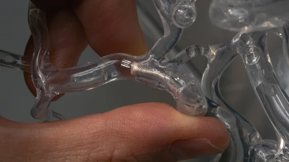
Image courtesy of Andrew Brodhead/Stanford University.
Researchers
with Stanford University have developed tiny robots capable of swimming
through a patient’s vascular system and removing blood clots. The new
technology, called milli-spinner thrombectomy, could potentially be used
to treat strokes, heart attacks, pulmonary embolisms and other
clot-related cardiovascular conditions.
“For most cases, we’re
more than doubling the efficacy of current technology, and for the
toughest clots—which we’re only removing about 11% of the time with
current devices—we’re getting the artery open on the first try 90% of
the time,” Jeremy Heit, MD, PhD, chief of neuroimaging and neurointervention at Stanford and an associate professor of radiology, explained in a statement. “It’s unbelievable. This is a sea-change technology that will drastically improve our ability to help people.”
Heit is the co-author of a new analysis focused on this state-of-the-art technology. Published in Nature, the research explored the impact of these tiny robots in both flow models and animal studies.[1]
“The
milli-spinner thrombectomy directly modifies the clot microstructure to
facilitate clot removal, improving mechanical thrombectomy success
rates compared with current methods that rely on clot rupture or
cutting,” the study’s authors concluded. “This approach offers a
promising new direction for mechanical thrombectomy devices, especially
for treating ischemic stroke, pulmonary embolism and peripheral
thrombosis.”
A brand new way to target blood clots
Milli-spinner thrombectomy is a continuation of the work of Renee Zhao, PhD, an assistant professor of mechanical engineering at Stanford and senior author of the Nature
study. Zhao’s previous research with these robots was primarily focused
on dispensing medicine. The spinning hollow structure of the robots,
with their tiny fins and slits, were originally intended to be used as a
propulsion mechanism. However, Zhao and her team noted that the
structures were also providing localized suction that could be used to
help eliminate blood clots in a fast, effective manner.
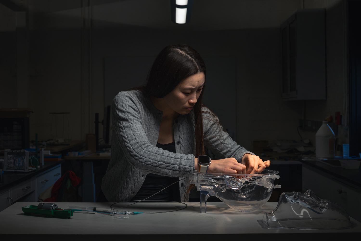
Renee Zhao, PhD, working on her milli-spinner thrombectomy technology. Image courtesy of Andrew Brodhead/Stanford University.
“At
first, we simply wondered whether this suction could help remove a
blood clot,” Zhao said in the same statement. “But when we tested the
spinner on a clot, we observed a striking clot color change, from red to
white, along with a dramatic reduction in volume. Honestly, it felt
like magic. We didn’t fully understand the mechanism at the time.”
For
the current version of this technology, clinicians deliver the tiny
robot to its intended location with a catheter and then it remains in
one location. However, researchers are also exploring the possibility of
an “untethered version” of the milli-spinner that could freely move
through the vascular system.
There is also hope that this same technique could go beyond busting blood clots and be used to remove kidney stone fragments.
“We’re
exploring other biomedical applications for the milli-spinner design,
and even possibilities beyond medicine,” Zhao explained. “There are some
very exciting opportunities ahead.”
This technology is still under development. More research will still be required before it gains FDA approval.
Click here to read the Nature study. In addition, Stanford has produced a video about this breakthrough that is now available online.
Summary: Researchers developed ‘Anthrobots,’
microscopic biological robots made from human tracheal cells,
demonstrating potential in healing and regenerative medicine.
These
self-assembling multicellular robots, ranging from hair-width to
pencil-point size, show remarkable healing effects, particularly in
neuron growth across damaged areas in lab conditions.(Need human testing.)
Building on
earlier Xenobot research, this study reveals that Anthrobots can be
created from adult human cells without genetic modification, offering a
new approach to patient-specific therapeutic tools.
Key Facts:
Anthrobots
are self-assembling biological robots made from human tracheal cells,
capable of movement and encouraging neuron growth.
They can be
created from adult human cells without genetic modifications, making
them a potential patient-specific therapeutic tool.
Anthrobots
represent a significant advancement in regenerative medicine,
potentially aiding in treating a variety of diseases and injuries.
Source: Tufts University
Researchers
at Tufts University and Harvard University’s Wyss Institute have
created tiny biological robots that they call Anthrobots from human
tracheal cells that can move across a surface and have been found to
encourage the growth of neurons across a region of damage in a lab dish.
The
multicellular robots, ranging in size from the width of a human hair to
the point of a sharpened pencil, were made to self-assemble and shown
to have a remarkable healing effect on other cells. The discovery is a
starting point for the researchers’ vision to use patient-derived
biobots as new therapeutic tools for regeneration, healing, and
treatment of disease.
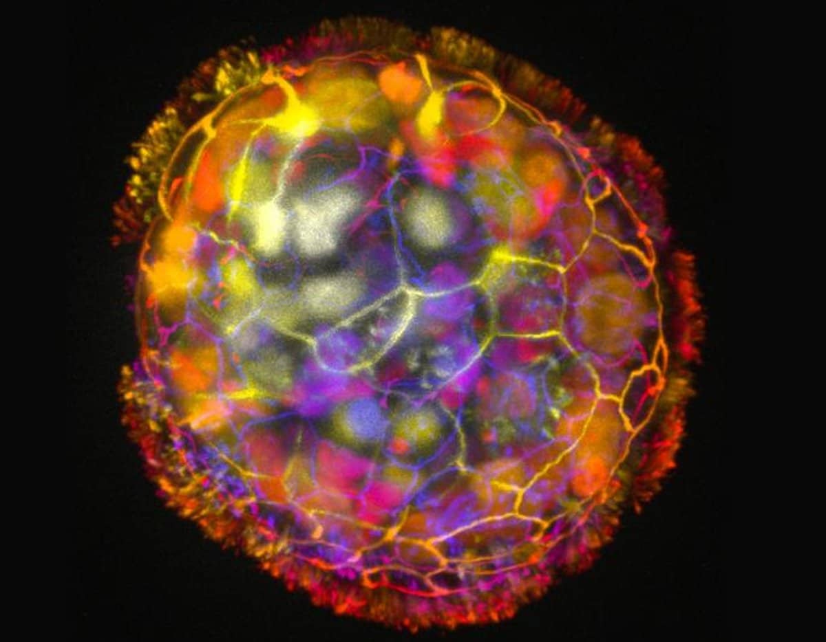
An
Anthrobot is shown, depth colored, with a corona of cilia that provides
locomotion for the bot. Credit: Gizem Gumuskaya, Tufts University
The work follows from earlier research in the laboratories of Michael
Levin, Vannevar Bush Professor of Biology at Tufts University School of
Arts & Sciences, and Josh Bongard at the University of Vermont in
which they created multicellular biological robots from frog embryo
cells called Xenobots, capable of navigating passageways, collecting
material, recording information, healing themselves from injury, and
even replicating for a few cycles on their own.
At the time,
researchers did not know if these capabilities were dependent on their
being derived from an amphibian embryo, or if biobots could be
constructed from cells of other species.
In the current study, published in Advanced Science,
Levin, along with PhD student Gizem Gumuskaya discovered that bots can
in fact be created from adult human cells without any genetic
modification and they are demonstrating some capabilities beyond what
was observed with the Xenobots.
The discovery starts to answer a broader question that the lab has
posed—what are the rules that govern how cells assemble and work
together in the body, and can the cells be taken out of their natural
context and recombined into different “body plans” to carry out other
functions by design?
In this case, researchers gave human cells,
after decades of quiet life in the trachea, a chance to reboot and find
ways of creating new structures and tasks.
“We wanted to probe
what cells can do besides create default features in the body,” said
Gumuskaya, who earned a degree in architecture before coming into
biology.
“By reprogramming interactions between cells, new
multicellular structures can be created, analogous to the way stone and
brick can be arranged into different structural elements like walls,
archways or columns.”
The researchers found that not only could
the cells create new multicellular shapes, but they could move in
different ways over a surface of human neurons grown in a lab dish and
encourage new growth to fill in gaps caused by scratching the layer of
cells.
Exactly how the Anthrobots encourage growth of neurons is
not yet clear, but the researchers confirmed that neurons grew under the
area covered by a clustered assembly of Anthrobots, which they called a
“superbot.”
“The cellular assemblies we construct in the lab can
have capabilities that go beyond what they do in the body,” said Levin,
who also serves as the director of the Allen Discovery Center at Tufts
and is an associate faculty member of the Wyss Institute. “It is
fascinating and completely unexpected that normal patient tracheal
cells, without modifying their DNA, can move on their own and encourage
neuron growth across a region of damage,” said Levin.
“We’re now looking at how the healing mechanism works, and asking what else these constructs can do.”
The
advantages of using human cells include the ability to construct bots
from a patient’s own cells to perform therapeutic work without the risk
of triggering an immune response or requiring immunosuppressants. They
only last a few weeks before breaking down, and so can easily be
re-absorbed into the body after their work is done.
In addition, outside of the body, Anthrobots can only survive in very
specific laboratory conditions, and there is no risk of exposure or
unintended spread outside the lab. Likewise, they do not reproduce, and
they have no genetic edits, additions or deletions, so there is no risk
of their evolving beyond existing safeguards.
How Are Anthrobots Made?
Each
Anthrobot starts out as a single cell, derived from an adult donor. The
cells come from the surface of the trachea and are covered with
hairlike projections called cilia that wave back and forth. The cilia
help the tracheal cells push out tiny particles that find their way into
air passages of the lung.
We all experience the work of ciliated
cells when we take the final step of expelling the particles and excess
fluid by coughing or clearing our throats. Earlier studies by others had
shown that when the cells are grown in the lab, they spontaneously form
tiny multicellular spheres called organoids.
The researchers
developed growth conditions that encouraged the cilia to face outward on
organoids. Within a few days they started moving around, driven by the
cilia acting like oars. They noted different shapes and types of
movement – the first. important feature observed of the biorobotics
platform.
Levin says that if other features could be added to the
Anthrobots (for example, contributed by different cells), they could be
designed to respond to their environment, and travel to and perform
functions in the body, or help build engineered tissues in the lab.
The
team, with the help of Simon Garnier at the New Jersey Institute of
Technology, characterized the different types of Anthrobots that were
produced. They observed that bots fell into a few discrete categories of
shape and movement, ranging in size from 30 to 500 micrometers (from
the thickness of a human hair to the point of a sharpened pencil),
filling an important niche between nanotechnology and larger engineered
devices.
Some were spherical and fully covered in cilia, and some were
irregular or football shaped with more patchy coverage of cilia, or just
covered with cilia on one side. They traveled in straight lines, moved
in tight circles, combined those movements, or just sat around and
wiggled. The spherical ones fully covered with cilia tended to be
wigglers.
The Anthrobots with cilia distributed unevenly tended to
move forward for longer stretches in straight or curved paths. They
usually survived about 45-60 days in laboratory conditions before they
naturally biodegraded.
“Anthrobots self-assemble in the lab dish,”
said Gumuskaya, who created the Anthrobots. “Unlike Xenobots, they
don’t require tweezers or scalpels to give them shape, and we can use
adult cells – even cells from elderly patients – instead of embryonic
cells. It’s fully scalable—we can produce swarms of these bots in
parallel, which is a good start for developing a therapeutic tool.”
Little Healers
Because
Levin and Gumuskaya ultimately plan to make Anthrobots with therapeutic
applications, they created a lab test to see how the bots might heal
wounds. The model involved growing a two-dimensional layer of human
neurons, and simply by scratching the layer with a thin metal rod, they
created an open ‘wound’ devoid of cells.
To ensure the gap would
be exposed to a dense concentration of Anthrobots, they created
“superbots” a cluster that naturally forms when the Anthrobots are
confined to a small space. The superbots were made up primarily of
circlers and wigglers, so they would not wander too far away from the
open wound.
Although it might be expected that genetic
modifications of Anthrobot cells would be needed to help the bots
encourage neural growth, surprisingly the unmodified Anthrobots
triggered substantial regrowth, creating a bridge of neurons as thick as
the rest of the healthy cells on the plate.
Neurons
did not grow in the wound where Anthrobots were absent. At least in the
simplified 2D world of the lab dish, the Anthrobot assemblies
encouraged efficient healing of live neural tissue.
According to the researchers, further development of the bots could
lead to other applications, including clearing plaque buildup in the
arteries of atherosclerosis patients, repairing spinal cord or retinal
nerve damage, recognizing bacteria or cancer cells, or delivering drugs
to targeted tissues. The Anthrobots could in theory assist in healing
tissues, while also laying down pro-regenerative drugs.
Making New Blueprints, Restoring Old Ones
Gumuskaya explained that cells have the innate ability to self-assemble into larger structures in certain fundamental ways.
“The
cells can form layers, fold, make spheres, sort and separate themselves
by type, fuse together, or even move,” Gumuskaya said.
“Two
important differences from inanimate bricks are that cells can
communicate with each other and create these structures dynamically, and
each cell is programmed with many functions, like movement, secretion
of molecules, detection of signals and more. We are just figuring out
how to combine these elements to create new biological body plans and
functions—different than those found in nature.”
Taking advantage
of the inherently flexible rules of cellular assembly helps the
scientists construct the bots, but it can also help them understand how
natural body plans assemble, how the genome and environment work
together to create tissues, organs, and limbs, and how to restore them
with regenerative treatments.
About this neurobotics research news
Author: Mike Silver Source: Tufts University Contact: Mike Silver – Tufts University Image: The image is credited to Gizem Gumuskaya, Tufts University
With this the tPA bolus could be vastly reduced, almost eliminating the chances of a bleed, and for hemorrhages it could deliver clotting material. But our fucking failures of stroke associations don't do a damn thing with stroke research so nothing ever gets better for stroke survivors. There is NO LEADERSHIP AND NO STRATEGY for anything to do with stroke. Prove me wrong.
It’s a computer – inside a cockroach. Nano-sized entities made of DNA
that are able to perform the same kind of logic operations as a
silicon-based computer have been introduced into a living animal.
The DNA computers – known as origami robots because they work by
folding and unfolding strands of DNA – travel around the insect’s body
and interact with each other, as well as the insect’s cells. When they
uncurl, they can dispense drugs carried in their folds.
“DNA nanorobots could potentially carry out complex programs that
could one day be used to diagnose or treat diseases with unprecedented
sophistication,” says Daniel Levner, a bioengineer at the Wyss Institute at Harvard University.
Advertisement
Levner was part of a team that made the nanobots by exploiting the
binding properties of DNA. When it meets a certain kind of protein, DNA
unravels into two complementary strands. By creating particular
sequences, the strands can be made to unravel on contact with specific
molecules – say, those on a diseased cell. When the molecule unravels,
out drops the package wrapped inside.
A bug’s life
The team has now injected various kinds of nanobots into cockroaches.
Because the nanobots are labelled with fluorescent markers, the
researchers can follow them and analyse how different robot combinations
affect where substances are delivered. The team says the accuracy of
delivery and control of the nanobots is equivalent to a computer system.
“This is the first time that biological therapy has been able to match how a computer processor works,” says co-author Ido Bachelet of the Institute of Nanotechnology and Advanced Materials at Bar Ilan University.
“Unlike electronic devices, which are suitable for our watches, our
cars or phones, we can use these robots in life domains, like a living
cockroach,” says Ángel Goñi Moreno of the National Center for Biotechnology in Madrid, Spain. “This opens the door for environmental or health applications.”
DNA has already been used for storing large amounts of information
and circuits for amplifying chemical signals, but these applications
are rudimentary compared with the potential benefits of the origami
robots.
Commodore cockroach
The number of nanobots in the study – more than in previous
experiments – makes it particularly promising, says Bachelet. “The
higher the number of robots present, the more complex the decisions and
actions that can be achieved. If you reach a certain threshold of
capability, you can perform any kind of computation. In this case, we
have gone past that threshold,” he says.
The team says it should be possible to scale up the computing power
in the cockroach to that of an 8-bit computer, equivalent to a Commodore
64 or Atari 800 from the 1980s. Goni-Moreno agrees that this is
feasible. “The mechanism seems easy to scale up so the complexity of the
computations will soon become higher,” he says.
An obvious benefit of this technology would be cancer treatments,
because these must be cell-specific and current treatments are not
well-targeted. But a treatment like this in mammals must overcome the
immune response triggered when a foreign object enters the body.
Bachelet is confident that the team can enhance the robots’ stability
so that they can survive in mammals. “There is no reason why
preliminary trials on humans can’t start within five years,” he says.
I wonder if using this post-stroke would prevent your likely chances of getting dementia. Ask your doctor, s/he won't know a damn thing, not even that you are like to get dementia because they don't read research.
An international team of researchers has shown that a new small-molecule drug can restore brain function and memory in a mouse model of Alzheimer's disease. The drug works by stopping toxic ion flow in the brain that is known to trigger nerve cell death. Scientists envision that this drug could be used to treat Alzheimer's and other neurodegenerative diseases such as Parkinson's and ALS.
"This is the first drug molecule that can regulate memory loss by directly blocking ions from leaking through nerve cell membranes," said Ratnesh Lal, a professor of bioengineering at the University of California San Diego and co-senior author of the study.
Various studies have linked Alzheimer's disease to the accumulation of two particular proteins in the brain called amyloid-beta and tau. One theory is that these protein clusters create pores in nerve cell membranes that allow ions to travel in and out uncontrollably. This would alter ion levels inside the cells and in turn trigger neuronal dysfunction and cell death.
The new drug, a small molecule called anle138b, blocks these pores from moving ions in and out of nerve cells. Anle138b attaches to both amyloid-beta and tau protein clusters and deactivates the pores created by these clusters.
Researchers administered anle138b to mice with a genetic predisposition for developing an Alzheimer's-like condition. The mice had symptoms such as abnormal brain function, impaired memory and high levels of either amyloid-beta or tau proteins in the brain. Treatment with anle138b normalized brain activity and improved learning ability in mice.
The study was led by the German Center for Neurodegenerative Diseases, the University Medical Center Göttingen, the Braunschweig University of Technology, the Max Planck Institute for Biophysical Chemistry, the Center for Nanoscale Microscopy and Molecular Physiology of the Brain in Göttingen, Germany, and the University of California San Diego. Researchers published their findings on Dec. 5 in EMBO Molecular Medicine.
Christian Griesinger, a professor at the Max Planck Institute for Biophysical Chemistry and co-senior author of the study, noted, "The drug is able to reach the brain when taken orally. Therefore, it is easy to administer, and we are currently performing toxicology studies to eventually be able to apply anle138b to humans."
The team cautions that since the drug has so far only been tested in mice, it is unclear how well it would perform in humans. "I would like to emphasize that none of the current animal models fully recapitulate the symptoms seen in Alzheimer's patients. Thus, care has to be taken when interpreting such data. However, our study offers evidence that anle138b has potential for neuroprotection," said André Fischer, a senior researcher at the German Center for Neurodegenerative Diseases and the University Medical Center Göttingen, who is also a co-senior author of the study.
While collaborators in Germany will be pursuing clinical studies in human patients with neurodegenerative diseases, Lal and his research group at the UC San Diego Jacobs School of Engineering are particularly interested in testing anle138b on a variety of other diseases that are linked to toxic ion flow caused by amyloid proteins, including diabetes, tuberculosis and certain types of cancer. Lal's group has performed extensive research on amyloid ion channels and their roles in these diseases. "Blocking the ion leakiness of amyloid channels using anle138b could be an effective therapy for various diseases," Lal said.
Lal serves as co-director for the Center of Excellence for Nanomedicine and Engineering, a subcenter of the Institute of Engineering in Medicine at UC San Diego. His research group will also work on targeted delivery of the drug using their patent pending "nanobowls," which are magnetically guided nanoparticles that can be packed with drugs and diagnostic molecules, deliver them to particular sites in the body and release them on demand. Future studies will focus on using these nanobowls to deliver anle138b to the brain, as well as other diseased tissues and organs affected by toxic amyloid-beta ion channels.