Changing stroke rehab and research worldwide now.Time is Brain! trillions and trillions of neurons that DIE each day because there are NO effective hyperacute therapies besides tPA(only 12% effective). I have 523 posts on hyperacute therapy, enough for researchers to spend decades proving them out. These are my personal ideas and blog on stroke rehabilitation and stroke research. Do not attempt any of these without checking with your medical provider. Unless you join me in agitating, when you need these therapies they won't be there.

What this blog is for:

My blog is not to help survivors recover, it is to have the 10 million yearly stroke survivors light fires underneath their doctors, stroke hospitals and stroke researchers to get stroke solved. 100% recovery. The stroke medical world is completely failing at that goal, they don't even have it as a goal. Shortly after getting out of the hospital and getting NO information on the process or protocols of stroke rehabilitation and recovery I started searching on the internet and found that no other survivor received useful information. This is an attempt to cover all stroke rehabilitation information that should be readily available to survivors so they can talk with informed knowledge to their medical staff. It lays out what needs to be done to get stroke survivors closer to 100% recovery. It's quite disgusting that this information is not available from every stroke association and doctors group.

Showing posts with label astrocytes. Show all posts

Monday, November 18, 2024

Astrocyte Transplantations Offer Hope for Treating Brain Disorders

 Ask your competent? doctor how many astrocytes died during your stroke and the EXACT PROTOCOLS to recover their functionality. NOTHING from your doctor? You don't have a functioning stroke doctor, why are you seeing them? And your competent? doctor needs to get human testing going in stroke survivors. But is your doctor COMPETENT AT ALL? With nothing on 100% recovery, I'd say not competent.

  • astrocytes (101 posts to June 2011)
  • Astrocyte Transplantations Offer Hope for Treating Brain Disorders

    Summary: Astrocytes, critical brain cells, are often lost in neurodegenerative diseases, but recent research highlights the promise of astrocyte transplantation to restore brain function. These transplanted cells integrate into the host brain, forming normal synaptic connections and promoting regeneration, though factors like donor cell type and transplantation timing influence success.

    Studies reveal that transplanted astrocytes can survive for up to a year, adapting to their new environment while retaining features of their original region. This emerging therapy offers a promising avenue for treating conditions like ALS, Parkinson’s, and Alzheimer’s.

    Key Facts:

    • Astrocyte transplantation aids brain regeneration and synaptic function.
    • Donor cell type and timing influence integration into the recipient brain.
    • Transplanted astrocytes can survive up to a year, matching native astrocytes.

    Source: The Conversation

    Astrocytes — named for their star-like shape — are a type of brain cell as abundant as neurons in the central nervous system, but little is known about their role in brain health and disease.

    Many neurological diseases are caused by or result in the loss of cells in the central nervous system. Some diseases are a result of the loss of specific cells, such as the loss of motor neurons in amyotrophic lateral sclerosis (ALS), the loss of dopaminergic neurons in Parkinson’s disease and the loss of GABAergic neurons in Huntington’s disease.
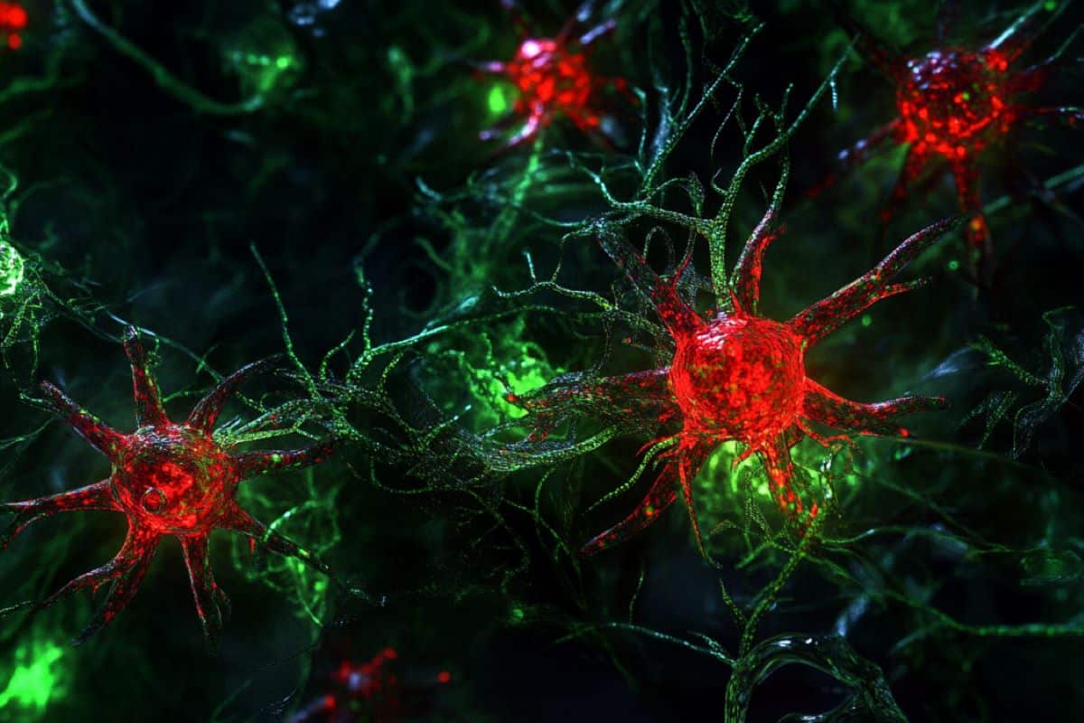
    This shows AI generated representations of astrocytes.
    Cell replacement therapy involves transplanting functional cells in patients. Credit: Neuroscience News

    For other neurodegenerative conditions, like Alzheimer’s disease, a key hallmark is the general loss of cells in brain regions responsible for memory formation.

    Although many brain diseases are marked by the loss of specific cells, a common link among these diseases is the loss of astrocytes. Interestingly, in some animal studies involving cases such as ALS, introducing disease-causing mutations selectively in astrocytes alone produces ALS symptoms and disease progression.

    Transplantation therapy

    Emerging evidence indicates that astrocytes take part in major functions of the brain, including homeostasis and neural network modulation that are essential to everyday cognition. A functioning brain requires healthy astrocytes, and finding strategies to heal or replace damaged astrocytes could help in the treatment of neurological diseases.

    Cell replacement therapy involves transplanting functional cells in patients. In recent years there have been exciting developments in this area with respect to astrocyte transplantation in animal disease models, with one approach even moving to early clinical trial in ALS patients. While there have been some promising outcomes, treatment success varies from one study to the other.

    Our recent study, published in The Journal of Neuroscience, examines how transplanted astrocyte integrate into the recipient central nervous system. We studied the types of transplanted astrocytes, timing of treatment and routes of transplantation.

    Preparing astrocytes

    First, we prepared astrocyte cultures in petri dishes by extracting immature astrocytes from the cerebral cortex of newborn mice and expanding the cell population.

    To track the development of transplanted astrocytes following their delivery to recipient mice, we used astrocytes from genetically modified mice in which astrocytes glow red, and they are transplanted into the brain of mice where astrocytes glow green.

    We found that the transplanted astrocytes could survive for up to one year after transplantation, developing normally and integrating into the recipient brain just like the native astrocytes, with just minor differences.

    Astrocytes depend on their capability to sense signals and exchange materials within the brain environment through molecules such as receptors and ion channels located on their cell surface.

    Transplanted astrocytes displayed comparable numbers of such receptors and channels and possessed similar sizes and complexity when compared to native astrocytes.

    Transplanted astrocytes do appear to take some time to catch up to and perfectly match astrocytes in the recipient mice in terms of the production of these receptors and ion channels.

    Source, type and location

    Intriguingly, we also found that the integration of transplanted astrocytes into the recipient is affected by the age of the mouse, which reflects the maturity of the cellular environment the astrocytes are transplanted into.

    When astrocytes were transplanted to an infant mouse, they could migrate and spread more extensively in the host brain. However, when astrocytes were transplanted into a young adult mouse, they were confined to the site of transplantation.

    Astrocytes in different regions of the brain and the spinal cord display very different features. We were interested in seeing how astrocytes from one region of the brain integrated into a different region. Astrocytes prepared from the cerebral cortex ended up developing into cortical astrocytes even when placed into the cerebellum.

    Therefore, the source and type of astrocytes being transplanted makes a difference, and this intrinsic programming of astrocytes needs to be considered when thinking about astrocyte replacement therapy.

    Exciting potential

    In recent years, increasing studies have been conducted to investigate the potential of astrocyte transplantation. Similar to our findings, transplanted astrocytes have been found to form normal contacts with neuronal synapses and are functioning normally. Astrocyte transplantation has also been shown to promote brain plasticity and regeneration following injury and in different animal models of neurological diseases.

    Therefore, it presents a promising and exciting strategy to treat neurological diseases. By answering principle questions regarding how transplanted astrocytes integrate in the host, our research can support the development of more effective cell therapies that can improve the quality of life of patients.

    About this neurology research news

    Author: Albert HiuKa Fok and Sabrina Chierzi
    Source: The Conversation
    Contact: Albert HiuKa Fok and Sabrina Chierzi – The Conversation
    Image: The image is credited to Neuroscience News

    Thursday, October 10, 2024

    Role of glucose metabolism in Alzheimer’s disease

     Between this earlier research and this, what does your competent? doctor say you should be doing? Not knowing and not answering this is grounds for firing them.

    The latest here:

    Role of glucose metabolism in Alzheimer’s disease

    At a Glance

    • Researchers found that proteins involved in Alzheimer’s disease inhibit glucose metabolism in the brain.
    • Blocking a particular enzyme restored glucose metabolism and cognitive function in mouse models of Alzheimer’s disease.
    • The findings suggest a novel potential approach for Alzheimer’s disease treatment.
    Illustration of an astrocyte with extensions connected to a blood vessel at bottom and a neuron at top. Astrocytes (center) play a crucial role in supporting neurons (top). ART-ur / Shutterstock

    In Alzheimer’s disease (AD), misfolded amyloid β (Aβ) and tau proteins accumulate in the brain. This leads to the progressive loss of connections between neurons. At the same time, glucose metabolism declines in certain types of brain cells, called astrocytes and microglia. One function of astrocytes is to help ensure that neurons have enough energy to support their activity. Astrocytes do this by breaking down glucose into lactate and exporting it to neurons. Neurons can then use the lactate as fuel.

    Recent research has implicated an enzyme in astrocytes, called indoleamine-2,3-dioxygenase 1 (IDO1), in AD. A team of researchers, led by Dr. Katrin Andreasson at Stanford University, examined how IDO1 affects glucose metabolism in astrocytes. They also looked at how IDO1 and glucose metabolism relate to AD pathology and brain function. The study, which was funded in part by NIH, appeared in Science on August 23, 2024.

    The team found that Aβ and tau increased IDO1 levels and activity in astrocytes from both mice and humans. The proteins also suppressed the conversion of glucose to lactate. Inhibiting IDO1 with a drug, or turning off the gene that encodes IDO1, restored lactate production in the presence of Aβ and tau.

    The hippocampus is the brain region responsible for learning and memory. In various mouse models of AD, the team found that lactate production in the hippocampus was suppressed. The mice also had impaired spatial memory and low hippocampal synaptic plasticity (the ability of connections between neurons to strengthen over time). Inhibiting IDO1 restored all three of these to normal levels. But inhibiting IDO1 had no effect on synaptic plasticity when neurons were blocked from importing lactate. This suggests that lactate in the hippocampus is important for spatial memory and plasticity.

    To see if these findings also applied to AD in humans, the team derived stem cells from people with and without late-onset AD. They then induced the stem cells to form astrocytes and neurons. Glucose metabolism and lactate production were reduced in the astrocytes derived from AD patients. The astrocytes also didn’t effectively transfer lactate to neurons. Inhibiting IDO1 restored lactate production in the astrocytes and its uptake by neurons to normal levels.

    The findings suggest that Aβ and tau boost IDO1 activity in astrocytes. This reduces glucose metabolism and lactate production. The loss of lactate, in turn, deprives neurons of an important fuel source.

    Restoring lactate production by inhibiting IDO1 might prevent or even reverse the cognitive effects of AD. IDO1 inhibitors have already been developed for cancer treatment and might be repurposed for AD treatment.

    “We also can’t overlook the fact that we saw this improvement in brain plasticity in mice with both amyloid and tau mice models,” Andreasson notes. “These are completely different pathologies, and the drugs appear to work for both. That was really exciting to us.”

    That suggests that different pathologies may damage neurons via a common mechanism. Thus, this treatment approach could potentially work not only for AD, but for other neurodegenerative diseases as well.

    —by Brian Doctrow, Ph.D.

    Saturday, June 8, 2024

    Brain’s Oxygen Deprivation Mechanism Hinders Memory Formation

     Ask your competent? doctor how long after your stroke this memory problem continues and how EXACTLY your doctor is treating the problem. NO treatment? You don't have a functioning stroke doctor!

    Brain’s Oxygen Deprivation Mechanism Hinders Memory Formation

    Summary: Oxygen deprivation in the brain triggers a feedback loop involving glutamate and nitric oxide, causing anoxia induced long-term potentiation (aLTP). This process disrupts regular memory-enhancing mechanisms, potentially explaining memory loss post-stroke. The study offers insights into treating memory problems in stroke patients.

    Key Facts:

    1. aLTP occurs during temporary oxygen deprivation in the brain, impairing memory.
    2. Glutamate and nitric oxide form a feedback loop that sustains aLTP.
    3. Disrupting this loop may help restore normal memory function after strokes.

    Source: OIST

    When we learn something new, our brain cells (neurons) communicate with each other through electrical and chemical signals. If the same group of neurons communicate together often, the connections between them get stronger. This process helps our brains learn and remember things and is known as long-term potentiation or LTP.  

    Another type of LTP occurs when the brain is deprived of oxygen temporarily – anoxia-induced long-term potentiationor aLTP. aLTP blocks the former process, thereby impairing learning and memory. Therefore, some scientists think that aLTP might be involved in memory problems seen in conditions like stroke. 

    Researchers at the Okinawa Institute of Science and Technology (OIST) and their collaborators have studied the aLTP process in detail. They found that maintaining aLTP requires the amino acid glutamate, which triggers nitric oxide (NO) production in both neurons and brain blood vessels. This process forms a positive glutamate-NO-glutamate feedback loop.

    Their study, published in iScience, indicates that the continuous presence of aLTP could potentially hinder the brain’s memory strengthening processes and explain the memory loss observed in certain patients after experiencing a stroke.  

    The brain’s response to low oxygen 

    When there is a lack of oxygen in the brain, glutamate, a neurotransmitter, is released from neurons in large amounts. This increased glutamate causes the production of NO. NO produced in neurons and brain blood vessels boosts glutamate release from neurons during aLTP. This glutamate-NO-glutamate loop continues even after the brain gets enough oxygen. 

    “We wanted to know how oxygen depletion affects the brain and how these changes occur,” Dr. Han-Ying Wang, a researcher in the former Cellular and Molecular Synaptic Function Unit at OIST and lead author of the study, stated.

    “It’s been known that nitric oxide is involved in releasing glutamate in the brain when there is a shortage of oxygen, but the mechanism was unclear.”  

    During a stroke, when the brain is deprived of oxygen, amnesia – the loss of recent memories – can be one of the symptoms. Investigating the effects of oxygen deficiency on the brain is important because of the potential medicinal benefits.

    “If we can work out what’s going wrong in those neurons when they have no oxygen, it may point in the direction of how to treat stroke patients,” Dr. Patrick Stoney, a scientist in OIST’s Sensory and Behavioral Neuroscience Unit and former member of the Cellular and Molecular Synaptic Function Unit, explained. 

    Brain tissues from mice were placed in a saline solution, mimicking the natural environment in the living brain. Normally, this solution is oxygenated to meet the high oxygen demands of brain tissue. However, replacing the oxygen with nitrogen allowed the researchers to deprive the cells of oxygen for precise lengths of time.  

    The tissues were then examined under a microscope and electrodes were placed on them to record electrical activity of the individual cells. The cells were stimulated in a way that mimics how they would be stimulated in living mice. 

    Stopping memory and learning activity 

    The scientists found that maintaining aLTP requires NO production in both neurons and in blood vessels in the brain. Collaborating scientists from OIST’s Optical Neuroimaging Unit showed that in addition to neurons and blood vessels, aLTP requires the activity of astrocytes, another type of brain cell. Astrocytes connect and support communication between neurons and blood vessels. 

    “Long-term maintenance of aLTP requires continuous synthesis of nitric oxide. NO synthesis is self-sustaining, supported by the NO-glutamate loop, but blocking molecular steps for NO-synthesis or those that trigger glutamate release eventually disrupt the loop and stop aLTP,” Prof. Tomoyuki Takahashi, leader of the former Cellular and Molecular Synaptic Function Unit at OIST, explained.  

    Notably, the cellular processes that support aLTP are shared by those involved in memory strengthening and learning (LTP). When aLTP is present, it hijacks molecular activities required for LTP and removing aLTP can rescue these memory enhancing mechanisms.

    This suggests that long-lasting aLTP may obstruct memory formation, possibly explaining why some patients have memory loss after a short stroke. 

    Prof. Takahashi emphasized that the formation of a positive feedback loop formed between glutamate and NO when the brain is temporarily deprived of oxygen is an important finding. It explains long-lasting aLTP and may offer a solution for memory loss caused by a lack of oxygen.  

    About this memory and neuroscience research news

    Author: Tomomi Okubo
    Source: OIST
    Contact: Tomomi Okubo – OIST
    Image: The image is credited to Neuroscience News

    Original Research: Open access.
    Anoxia-induced hippocampal LTP is regeneratively produced by glutamate and nitric oxide from the neuro-glial-endothelial axis” by Han-Ying Wang et al. iScience

    Tuesday, May 28, 2024

    New Alzheimer’s Breakthrough Targets Plexin-B1 Protein

     You'll want your competent? doctor to make sure followup research occurs because of your increased risk of dementia post stroke. Do you even have a competent doctor?My definition of competence is having EXACT 100% RECOVERY PROTOCOLS. None of this guideline shit!

    Your risk of dementia, has your doctor told you of this?

    1. A documented 33% dementia chance post-stroke from an Australian study?   May 2012.

    2. Then this study came out and seems to have a range from 17-66%. December 2013.`    

    3. A 20% chance in this research.   July 2013.

    4. Dementia Risk Doubled in Patients Following Stroke September 2018

    The latest here:

    New Alzheimer’s Breakthrough Targets Plexin-B1 Protein

    Summary: Researchers identified a novel way to potentially slow or halt Alzheimer’s progression by targeting the plexin-B1 protein. Their study shows how reactive astrocytes and plexin-B1 play crucial roles in clearing amyloid plaques. This discovery opens new pathways for Alzheimer’s treatments and emphasizes the importance of cellular interactions.

    Key Facts:

    • Key Protein: Targeting plexin-B1 protein can enhance the brain’s ability to clear amyloid plaques.
    • Cellular Interactions: Reactive astrocytes help control the clearance of harmful deposits in the brain.
    • Innovative Treatments: The study opens new pathways for developing treatments for Alzheimer’s disease.

    Source: Mount Sinai Hospital

    Researchers at the Icahn School of Medicine at Mount Sinai have made a significant breakthrough in Alzheimer’s disease research by identifying a novel way to potentially slow down or even halt disease progression.

    The study, which focuses on the role of reactive astrocytes and the plexin-B1 protein in Alzheimer’s pathophysiology, provides crucial insights into brain cell communication and opens the door to innovative treatment strategies.

    It was published in Nature Neuroscience on May 27. 

    This shows neurons.
    The research team emphasizes that while their findings mark a significant advance in the fight against Alzheimer’s, more research is needed to translate these discoveries into treatments for human patients. Credit: Neuroscience News

    This groundbreaking work is centered on the manipulation of the plexin-B1 protein to enhance the brain’s ability to clear amyloid plaques, a hallmark of Alzheimer’s disease. Reactive astrocytes, a type of brain cell that becomes activated in response to injury or disease, were found to play a crucial role in this process.

    They help control the spacing around amyloid plaques, affecting how other brain cells can access and clear these harmful deposits.

    “Our findings offer a promising path for developing new treatments by improving how cells interact with these harmful plaques,” said Roland Friedel, PhD, Associate Professor of Neuroscience, and Neurosurgery, at Icahn Mount Sinai and a senior author of the study.

    The research was driven by the analysis of complex data comparing healthy individuals to those with Alzheimer’s, aiming to understand the disease’s molecular and cellular foundations.

    Hongyan Zou, PhD, Professor of Neurosurgery, and Neuroscience, at Icahn Mount Sinai and one of the study’s lead authors, highlighted the broader implications of their findings: “Our study opens new pathways for Alzheimer’s research, emphasizing the importance of cellular interactions in developing neurodegenerative disease treatments.”

    One of the study’s most significant achievements is its validation of multiscale gene network models of Alzheimer’s disease.

    “This study not only confirms one of the most important predictions from our gene network models but also significantly advances our understanding of Alzheimer’s. It lays a solid foundation for developing novel therapeutics targeting such highly predictive network models,” said Bin Zhang, PhD, Willard T.C. Johnson Research Professor of Neurogenetics at Icahn Mount Sinai and one of the study’s lead authors.

    By demonstrating the critical role of plexin-B1 in Alzheimer’s disease, the research underscores the potential of targeted therapies to disrupt the disease’s progression.

    The research team emphasizes that while their findings mark a significant advance in the fight against Alzheimer’s, more research is needed to translate these discoveries into treatments for human patients.

    “Our ultimate goal is to develop treatments that can prevent or slow down Alzheimer’s progression,” Dr. Zhang added, outlining the team’s commitment to further exploring the therapeutic potential of plexin-B1.

    Funding: This study is supported by the NIH National Institute on Aging (NIA) grants U01AG046170 and RF1AG057440 and is part of the NIA-led Accelerating Medicines Partnership – Alzheimer’s Disease (AMP-AD) Target Discovery and Preclinical Validation program.

    This public private partnership aims to shorten the time between the discovery of potential drug targets and the development of new drugs for Alzheimer’s disease treatment and prevention.

    About this Alzheimer’s disease research news

    Author: Jennifer Gutierrez
    Source: Mount Sinai Hospital
    Contact: Jennifer Gutierrez – Mount Sinai Hospital
    Image: The image is credited to Neuroscience News
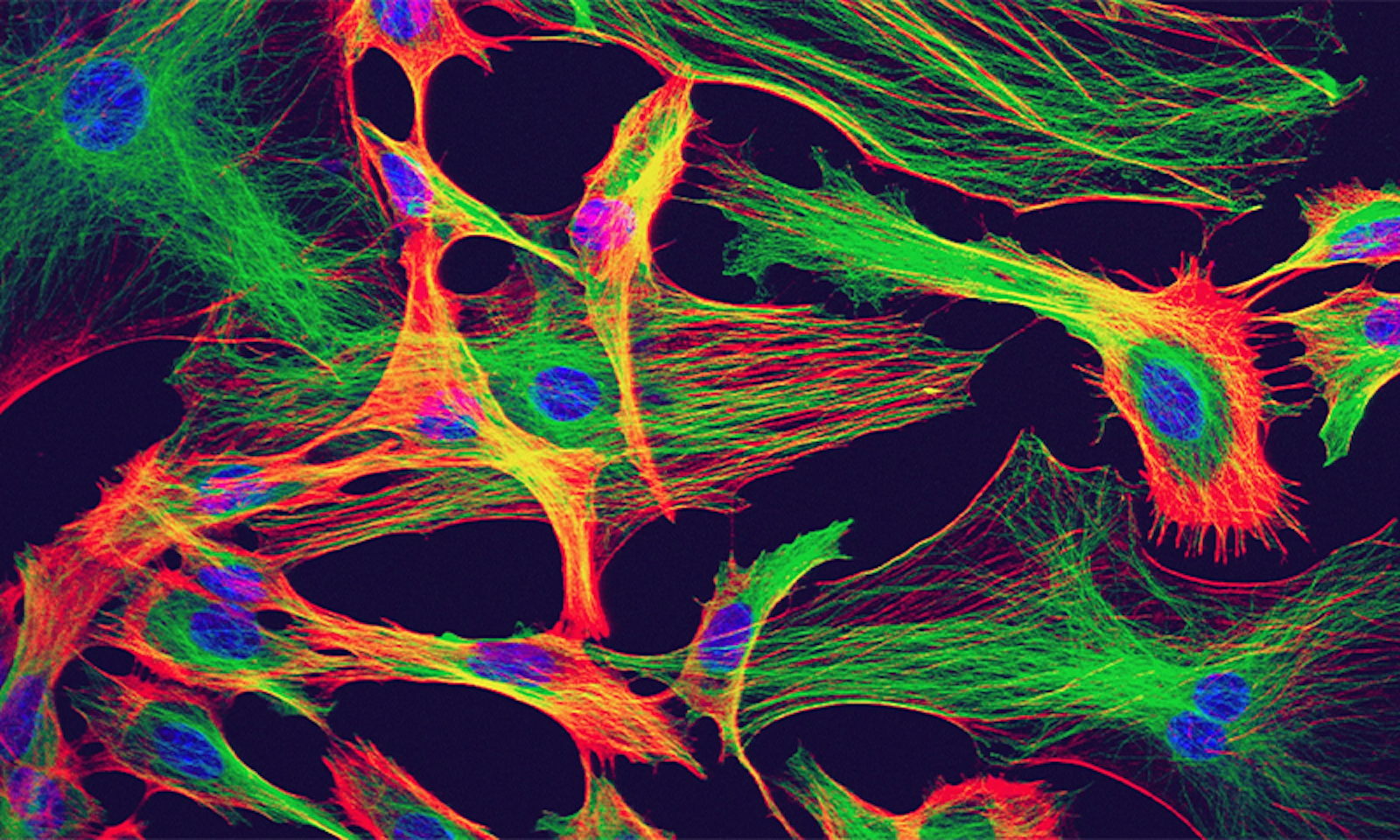
    Wednesday, March 13, 2024

    Omega-3 polyunsaturated fatty acids ameliorate neuroinflammation and mitigate ischemic stroke damage through interactions with astrocytes and microglia


    Did your doctor instruct the dietician to get these into your hospital meals? NO? Then you don't have a functioning stroke doctor!

    What foods provide omega-3s?

    • Fish and other seafood (especially cold-water fatty fish, such as salmon, mackerel, tuna, herring, and sardines)

    • Nuts and seeds (such as flaxseed, chia seeds, and walnuts)

    • Plant oils (such as flaxseed oil, soybean oil, and canola oil)

    Omega-3 polyunsaturated fatty acids ameliorate neuroinflammation and mitigate ischemic stroke damage through interactions with astrocytes and microglia

    , , , , , ,
    https://doi.org/10.1016/j.jneuroim.2014.11.007Get rights and content

    Highlights

    • PUFA n3 reduce stroke damage.

    • PUFA n3 attenuate hypoxia-induced inflammation.

    • PUFA n3 interact with microglia and astroglia.

    Abstract

    Omega-3 polyunsaturated fatty acids (PUFA n3) provide neuroprotection due to their anti-inflammatory and anti-apoptotic properties as well as their regulatory function on growth factors and neuronal plasticity. These qualities enable PUFA n3 to ameliorate stroke outcome and limit neuronal damage. Young adult male rats received transient middle cerebral artery occlusion (tMCAO). PUFA n3 were intravenously administered into the jugular vein immediately after stroke and 12 h later. We analyzed stroke volume and behavioral performance as well as the regulation of functionally-relevant genes in the penumbra. The extent of ischemic damage was reduced and behavioral performance improved subject to applied PUFA n3. Expression of Tau and growth-associated protein-43 genes were likewise restored. Ischemia-induced increase of cytokine mRNA levels was abated by PUFA n3. Using an in vitro approach, we demonstrate that cultured astroglial and microglia directly respond to PUFA n3 administration by preventing ischemia-induced increase of cyclooxygenase 2, hypoxia-inducible factor 1alpha, inducible nitric oxide synthase, and interleukin 1beta. Cultured cortical neurons also appeared as direct targets, since PUFA n3 shifted the Bcl-2-like protein 4 (Bax)/B-cell lymphoma 2 (Bcl 2) ratio towards an anti-apoptotic constellation. Thus, PUFA n3 reveal a high neuroprotective and anti-inflammatory potential in an acute ischemic stroke model by targeting astroglial and microglial function as well as improving neuronal survival strategies. Our findings signify the potential clinical feasibility of PUFA n3 therapeutic treatment in stroke and other acute neurological diseases.

    Introduction

    Stroke is the result of a permanent or transient focal occlusion of major brain arteries or their branches and represents a main cause of death and disability in the industrialized civilization. Brain damage and neuronal cell death following acute ischemia result from a series of complex pathophysiological processes that evolves in time and space beginning a few minutes after stroke onset and lasting for hours and days including secondary damage due to edema spreading even if reperfusion has already been revived. Cell dysfunction and tissue destruction are accompanied by local blood–brain barrier (BBB) breakdown followed by the invasion of peripheral immune cells, i.e. T-lymphocytes, macrophages and polymorph nuclear granulocytes. Beforehand, a massive early disturbance of ion homeostasis, calcium dysregulation, excitotoxicity, mitochondrial impairment together with reactive oxygen species (ROS) formation can be observed (Iadecola and Anrather, 2011, Dirnagl, 2012). The described pathomechanisms coincide with the activation, attraction and proliferation of astroglial and microglial cells. Astrogliosis and microgliosis are prevailing incidents in the penumbra during the initial stage of ischemia. Both glial cell types control and tune early and late neuroinflammatory responses resulting from oxygen and nutrient deprivation soon after the beginning of the ischemic phase (Dang et al., 2011). Although microglia is believed to play the most prominent role in the shaping of inflammatory responses after stroke, latterly astrocytes in the center of ischemic tissue disintegration are considered to actively sense hypoxia and trigger a battery of anti-inflammatory reactions (Ronaldson and Davis, 2012, Habib and Beyer, 2014, Habib et al., 2014). Importantly, both types of glial cells, the adjacent extracellular matrix, the endothelium and neurons form a “neurovascular unit” that represents a dynamic entity which shapes neuroinflammation in the setting of stroke (Dirnagl, 2012).

    Recent studies have shown that omega-3 essential polyunsaturated fatty acids (PUFA n3) and in particular docosahexaenoic acid (DHA, 22:6, n-3) and to a lesser extent eicosapentanaenoic acid (EPA, 20:5, n-3) exert profound anti-inflammatory effects on the brain and protect brain tissue in experimental models of acute stroke in neonatal and adult animals and neuroinflammatory challenges besides being beneficial for brain development and cognitive function (Bazan, 2007, Belayev et al., 2009, Hoffman et al., 2009, Cole et al., 2010, Orr et al., 2013). Following short-term transient middle cerebral artery occlusion (tMCAO), rodents with DHA substitution and higher brain DHA levels revealed reduced infarct areas and cellular inflammatory responses as well as attenuated leukocyte infiltration and concomitantly fewer microglial cells (Belayev et al., 2009, Lalancette-Hebert et al., 2011, Orr et al., 2013). Several hours after stroke, the resident microglial cells become activated, accumulate in the vicinity of the lesion site and in the penumbra region and start proliferating (Kriz and Lalancette-Hebert, 2009, Dang et al., 2011, Lalancette-Hebert et al., 2011). This defines the post-ischemic treatment window with DHA as 3–5 h. There is also good evidence that consumption of fish and fish products (fish oil contains large amounts of DHA) is positively associated with a reduced risk of ischemic events in the CNS and cardiovascular disease (Pascoe et al., 2014). There are several proposed cellular mechanisms which could explain the safeguarding role of PUFA n3 under neuropathological conditions in the brain (Orr et al., 2013). DHA affects growth factor regulation which may be responsible for increased neurite growth and synapse formation (Kim et al., 2011). Coevally, DHA is anti-inflammatory in non-neuronal and neural tissues targeting for instance cyclooxygenases (COX) and cytosolic phospholipase A2 (cPLA2) as well as leukocyte infiltration and nuclear factor kappa-light-chain-enhancer of activated B cells (NF-κB) activation (Marcheselli et al., 2003, Orr et al., 2013). Such effects occur brain-intrinsically but it has also been shown that DHA dampens systemic inflammatory responses (Sijben and Calder, 2007).

    In the present study, we aimed at demonstrating the neuroprotective potency of PUFA n3 in an experimental stroke rat model (transient middle cerebral artery occlusion, tMCAO), its efficacy in restoring motoric and sensory behavioral defects as well as morphological injury, and analyzing its influence on the expression of stroke-associated inflammatory gene markers. By adopting an in vitro hypoxia approach, we intended to curtail cell type-specific effects which might explain neuroprotective mechanisms at the subcellular level.

    More at link.

    Saturday, February 24, 2024

    These Cells Spark Electricity in the Brain. They’re Not Neurons

    What is your doctor doing with this to get you recovered? 

    Do you prefer your  doctor and hospital incompetence NOT KNOWING? OR NOT DOING?

    These Cells Spark Electricity in the Brain. They’re Not Neurons

    For decades, researchers have debated whether brain cells called astrocytes can signal like neurons.

    Article Lead Image
    Explore

    A brain is nothing if not communicative. Neurons are the chatterboxes of this conversational organ, and they speak with one another by exchanging pulses of electricity using chemical messengers called neurotransmitters. By repeating this process billions of times per second, a brain converts clusters of chemicals into coordinated actions, memories, and thoughts.

    Researchers study how the brain works by eavesdropping on that chemical conversation. But neurons talk so loudly and often that if there are other, quieter voices, it might be hard to hear them.

    For most of the 20th century, neuroscientists largely agreed that neurons are the only brain cells that propagate electrical signals. All the other brain cells, called glia, were thought to serve purely supportive roles. Then, in 1990, a curious phenomenon emerged: Researchers observed an astrocyte, a subtype of glial cell, responding to glutamate, the main neurotransmitter that generates electrical activity.

    In the decades since, research teams have come up with conflicting evidence, some reporting that astrocytes signal, and others retorting that they definitely do not. The disagreement played out at conferences and in review after review of the evidence. The two sides seemed irreconcilable.

    A paper published in Nature  in September presents the best proof yet that astrocytes can signal, gathered over eight years by a team co-led by Andrea Volterra, visiting faculty at the Wyss Center for Bio and Neuro Engineering in Geneva, Switzerland. The study includes two key pieces of evidence: images of glutamate flowing from astrocytes, and genetic data suggesting that these cells, dubbed glutamatergic astrocytes, have the cellular machinery to use glutamate the way neurons do.

    The paper also helps explain the decades of contradictory findings. Because only some astrocytes can perform this signaling, both sides of the controversy are, in a sense, right: A researcher’s results depend on which astrocytes they sampled.

    “This study is so cool because it provides an explanation for why both of those pieces of data were out there and conflicting,” said Christopher Dulla, a professor of neuroscience at Tufts University who studies astrocytic signaling and was not involved in the new work. “I tend to buy it.”

    The discovery opens the possibility that some astrocytes form an essential part of the brain’s circuitry. “More and more we come to the idea that there is a participation of all the cell types to the function of the brain,” Volterra said. “It’s much more integrated than it was thought before.”

    A Communicative Web

    The catch-all name “glia”—from the Greek word for “glue”—for all brain cells that aren’t neurons, like astrocytes, conveys scientists’ initial view that their main purpose was to hold neurons together. However, since the first description of astrocytes in 1865, researchers have discovered that . For one thing, they have glutamate receptors, which they use to detect and clean up excess neurotransmitters in the spaces around neurons.

    What’s been less clear is whether they can use glutamate to generate an electrical signal on their own. In 1994, researchers stimulated astrocytes in a dish and saw nearby neurons appear to respond by preparing to send a signal. And in 1997, Volterra and his colleagues observed the reverse: Rat astrocytes answered neurons’ calls with oscillating waves of the signaling molecule calcium. From 2000 to 2012, researchers published more than 100 papers reporting evidence in favor of astrocytes’ ability to communicate via synapses.

    But others questioned how that evidence was gathered and interpreted. In 2014, for example, researchers discovered that a key mouse model was flawed, raising questions about the prior studies that used those mice.

    THERE’S A SPARK: More than 25 years after he first observed astrocytes responding to signals in a dish, the neuroscientist Andrea Volterra from the Wyss Center for Bio and Neuro Engineering in Geneva, Switzerland, has come back with new evidence that some astrocytes actively participate in the brain’s electric conversation. Photo courtesy of Andrea Volterra.

    Meanwhile, the view of astrocytes was evolving, and scientists were beginning to consider them active participants in the brain’s processing of information. While neurons and their branching dendrites are often pictured as trees, astrocytes are more like a fungus, forming a tightly woven mat that blankets the brain and shares information among its constituent parts. In this way, astrocytes seem to form a coordinated web that influences neuronal activity. For example, in 2016, while conducting neuroscience research at the University of California, San Francisco, Kira Poskanzer discovered that mouse astrocytes can prompt nearby neurons to enter a rhythmic sleep state by regulating glutamate.

    “It’s less like an individual cell doing its own thing and more like part of a whole team of cells working together,” said Poskanzer, now at the biotech startup Arcadia Science.

    However, there’s a difference between mopping up glutamate and truly generating signals. Volterra believed that some astrocytes were capable of the latter. But to prove it, he needed evidence that astrocytes can send signals and have the right tools to do so in relevant, meaningful ways.

    A New Class of Brain Cell

    Volterra took advantage of a new approach to studying the brain: single-cell RNA sequencing, which takes a snapshot of the complete suite of genes active in individual cells throughout a tissue. Combing through eight databases of mouse hippocampal cells, he identified nine clusters of astrocytes, distinguished by their gene activity. Astrocytes in one—and only one—of the clusters transcribed proteins known to be involved in neurotransmitter storage, release, and transport using vesicles, as occurs in neurons. The cells were not evenly distributed across the brain region, or even throughout specific circuits.

    To see whether people have these cells, Volterra and his team searched three databases of human hippocampal cells for the same protein signatures they had seen in the mouse astrocytes. The signatures appeared in all three data sets.

    That genetic data, though, was still indirect evidence. Volterra needed to show the signaling in action. He and his team simulated a neuronal signal to astrocytes in slices of mouse brain and took images of the molecules released by the astrocytes. Some—but not all—astrocytes responded with glutamate. And when the researchers prevented astrocytes from using vesicles, the cells could no longer release glutamate.

    To Volterra, the evidence was clear. “We were right. There are astrocytes that release glutamate,” he said. “But we were also wrong, because we thought all astrocytes release glutamate.”

    The findings almost certainly upend the current understanding of the way the brain communicates, said Dmitri Rusakov, a professor of neuroscience at University College London who was not involved in the work. But in what way is an open question.

    Knowing that astrocytes can signal is only the first step. That fact doesn’t answer how synapses respond to astrocytic glutamate. It doesn’t say which functions require astrocyte signaling instead of, or in addition to, neurons. It doesn’t account for why some areas of the brain have more glutamatergic astrocytes than others, or why a subset uses this function while the rest do not.

    Instead, like all new discoveries, it poses new questions for science to answer.

    We’ve got a significant body of evidence,” Rusakov said. “Now you need a theory to put it all together.”

    This article was originally published on the  Quanta Abstractions blog. 

    Lead image: New evidence suggests that some astrocytes can stimulate electrical signals just as neurons do. Credit: David Robertson, ICR / Science Source

    Tuesday, June 27, 2023

    Astrocytes in ischemic stroke: Crosstalk in central nervous system and therapeutic potential

    Astrocyte research has been out there for years. Has your doctor done anything with it? If not, you don't have a functioning stroke doctor or hospital. 

    Do you prefer your doctor and hospital incompetence NOT KNOWING? OR NOT DOING?

    Astrocytes in ischemic stroke: Crosstalk in central nervous system and therapeutic potential

    First published: 21 June 2023

    Jueling Liu and Yuying Guo contributed equally to this work.

    Abstract

    In the central nervous system (CNS), a large group of glial cells called astrocytes play important roles in both physiological and disease conditions. Astrocytes participate in the formation of neurovascular units and interact closely with other cells of the CNS, such as microglia and neurons. Stroke is a global disease with high mortality and disability rate, most of which are ischemic stroke. Significant strides in understanding astrocytes have been made over the past few decades. Astrocytes respond strongly to ischemic stroke through a process known as activation or reactivity. Given the important role played by reactive astrocytes (RAs) in different spatial and temporal aspects of ischemic stroke, there is a growing interest in the potential therapeutic role of astrocytes. Currently, interventions targeting astrocytes, such as mediating astrocyte polarization, reducing edema, regulating glial scar formation, and reprogramming astrocytes, have been proven in modulating the progression of ischemic stroke. The aforementioned potential interventions on astrocytes and the crosstalk between astrocytes and other cells of the CNS will be summarized in this review.

    Tuesday, May 30, 2023

    New research shows astrocytes are key to swaying the pendulum in Alzheimer's disease progression

    It is your doctor's responsibility to understand how to apply this knowledge to prevent MCI and dementia post stroke.

    Your risk of dementia, has your doctor told you of this?  Your doctor is responsible for preventing this!

    1. A documented 33% dementia chance post-stroke from an Australian study?   May 2012.

    2. Then this study came out and seems to have a range from 17-66%. December 2013.`    

    3. A 20% chance in this research.   July 2013.

    4. Dementia Risk Doubled in Patients Following Stroke September 2018 

    The latest here:


    New research shows astrocytes are key to swaying the pendulum in Alzheimer's disease progression

    Why do some people develop Alzheimer's disease while others don't? And, even more puzzlingly, why do many individuals whose brains are chock-full of toxic amyloid aggregates-;a telltale sign of Alzheimer's brain pathology-;never go on to develop Alzheimer's-associated dementias?

    University of Pittsburgh School of Medicine researchers appear to have found the answer. Star-shaped brain cells called astrocytes are key to swaying the pendulum in Alzheimer's disease progression, shows new game-changing research published today in Nature Medicine.

    By testing the blood of more than 1,000 cognitively unimpaired elderly people with and without amyloid pathology, the Pitt-led research team found that only those who had a combination of amyloid burden and blood markers of abnormal astrocyte activation, or reactivity, would progress to symptomatic Alzheimer's in the future, a critical discovery for drug development aimed at halting progression.

    Our study argues that testing for the presence of brain amyloid along with blood biomarkers of astrocyte reactivity is the optimal screening to identify patients who are most at risk for progressing to Alzheimer's disease. This puts astrocytes at the center as key regulators of disease progression, challenging the notion that amyloid is enough to trigger Alzheimer's disease."

    Tharick Pascoal, M.D., Ph.D., senior author, associate professor of psychiatry and neurology at Pitt

    Alzheimer's disease is a neurodegenerative condition that causes progressive memory loss and dementia, robbing patients of many productive years of life. At the tissue level, the hallmark of Alzheimer's disease is an accumulation of amyloid plaques-;protein aggregates lodged between nerve cells of the brain-;and clumps of disordered protein fibers, called tau tangles, forming inside the neurons.

    For many decades brain scientists believed that an accumulation of amyloid plaques and tau tangles is not only a sign of Alzheimer's disease but also its direct culprit. This assumption also led drug manufacturers to heavily invest into molecules targeting amyloid and tau, overlooking the contribution of other brain processes, such as the neuroimmune system.

    Recent discoveries by groups like Pascoal's suggest that the disruption of other brain processes, such as heightened brain inflammation, might be just as important as amyloid burden itself in starting the pathological cascade of neuronal death that causes rapid cognitive decline.

    In his previous research, Pascoal and his group found that brain tissue inflammation triggers the spread of pathologically misfolded proteins in the brain and is a direct cause of eventual cognitive impairment in patients with Alzheimer's disease. Now, almost two years later, researchers revealed that the cognitive impairment can be predicted by a blood test.

    Astrocytes are specialized cells abundant in the brain tissue. Just as other members of the glia-;resident immune cells of the brain-;astrocytes support neuronal cells by supplying them with nutrients and oxygen and protecting them from pathogens. But because glial cells don't conduct electricity and, at first, didn't seem to play a direct role in how neurons communicate with one another, their role in health and disease had been overlooked. The latest research from Pitt changes that.

    "Astrocytes coordinate brain amyloid and tau relationship like a conductor directing the orchestra," said lead author of the study Bruna Bellaver, Ph.D., postdoctoral associate at Pitt. "This can be a game-changer to the field, since glial biomarkers in general are not considered in any main disease model."

    Scientists tested blood samples from participants in three independent studies of cognitively unimpaired elderly people for biomarkers of astrocyte reactivity-;glial fibrillary acidic protein, or GFAP-;along with the presence of pathological tau. The study showed that only those who were positive for both amyloid and astrocyte reactivity showed evidence of progressively developing tau pathology, indicating predisposition to clinical symptoms of Alzheimer's disease.

    The findings have direct implications for future clinical trials for Alzheimer's drug candidates. In aiming to halt disease progression sooner, trials are moving to earlier and earlier stages of pre-symptomatic disease, making correct early diagnosis of Alzheimer's risk critical for success. Because a significant percentage of amyloid-positive individuals will not progress to clinical forms of Alzheimer's, amyloid positivity alone is not enough to determine an individual's eligibility for a therapy.

    Inclusion of astrocyte reactivity markers, such as GFAP, in the panel of diagnostic tests will allow for improved selection of patients who are likely to progress to later stages of Alzheimer's and, therefore, help fine-tune selection of candidates for therapeutic interventions who are more likely to benefit.

    Source:
    Journal reference:

    Bellaver, B., et al. (2023). Astrocyte reactivity influences amyloid-β effects on tau pathology in preclinical Alzheimer’s disease. Nature Medicine. doi.org/10.1038/s41591-023-02380-x.

    Wednesday, April 19, 2023

    Exercise Boosts Brain Health With Chemical Signals

    It is YOUR DOCTOR'S RESPONSIBILITY to get you recovered enough to do this exercise. Don't let them run away from that by using the craptastic saying:'All strokes are different, all stroke recoveries are different'. If you get that from any of your medical personnel, run away, they know nothing useful that will get you recovered, that's just their go to excuse for not getting you recovered..

    Exercise Boosts Brain Health With Chemical Signals

    Summary: Chemical signals released by muscles during exercise promote neural development in the brain, researchers report.

    Source: Beckman Institute

    Physical activity is frequently cited as a means of improving physical and mental health. Researchers at the Beckman Institute for Advanced Science and Technology have shown that it may also improve brain health more directly.

    They studied how the chemical signals released by exercising muscles promote neuronal development in the brain.

    Their work appears in the journal Neuroscience.

    When muscles contract during exercise, like the biceps working to lift a heavy weight, they release a variety of compounds into the bloodstream. These compounds can travel to different parts of the body, including the brain. The researchers were particularly interested in how exercise could benefit a particular part of the brain called the hippocampus.

    “The hippocampus is a crucial area for learning and memory, and therefore cognitive health,” said Ki Yun Lee, a Ph.D. student in mechanical science and engineering at the University of Illinois Urbana-Champaign and the study’s lead author. Understanding how exercise benefits the hippocampus could therefore lead to exercise-based treatments for a variety of conditions including Alzheimer’s disease.

    To isolate the chemicals released by contracting muscles and test them on hippocampal neurons, the researchers collected small muscle cell samples from mice and grew them in cell culture dishes in the lab. When the muscle cells matured, they began to contract on their own, releasing their chemical signals into the cell culture.

    The research team added the culture, which now contained the chemical signals from the mature muscle cells, to another culture containing hippocampal neurons and other support cells known as astrocytes.

    Using several measures, including immunofluorescent and calcium imaging to track cell growth and multi-electrode arrays to record neuronal electrical activity, they examined how exposure to these chemical signals affected the hippocampal cells.

    The results were striking. Exposure to the chemical signals from contracting muscle cells caused hippocampal neurons to generate larger and more frequent electrical signals—a sign of robust growth and health. Within a few days, the neurons started firing these electrical signals more synchronously, suggesting that the neurons were forming a more mature network together and mimicking the organization of neurons in the brain.

    However, the researchers still had questions about how these chemical signals led to growth and development of hippocampal neurons. To uncover more of the pathway linking exercise to better brain health, they next focused on the role of astrocytes in mediating this relationship.

    “Astrocytes are the first responders in the brain before the compounds from muscles reach the neurons,” Lee said. Perhaps, then, they played a role in helping neurons respond to these signals.

    This shows exercise equipment
    When muscles contract during exercise, like the biceps working to lift a heavy weight, they release a variety of compounds into the bloodstream. Image is in the public domain

    The researchers found that removing astrocytes from the cell cultures caused the neurons to fire even more electrical signals, suggesting that without the astrocytes, the neurons continued to grow—perhaps to a point where they might become unmanageable.

    “Astrocytes play a critical role in mediating the effects of exercise,” Lee said. “By regulating neuronal activity and preventing hyperexcitability of neurons, astrocytes contribute to the balance necessary for optimal brain function.”

    Understanding the chemical pathway between muscle contraction and the growth and regulation of hippocampal neurons is just the first step in understanding how exercise helps improve brain health.

    “Ultimately, our research may contribute to the development of more effective exercise regimens for cognitive disorders such as Alzheimer’s disease,” Lee said.

    In addition to Lee, the team also included Beckman faculty members Justin Rhodes, a professor of psychology; and Taher Saif, a professor of mechanical science and engineering.

    About this neuroscience and exercise research news

    Author: Melinh Lai
    Source: Beckman Institute
    Contact: Melinh Lai – Beeckman Institute
    Image: The image is in the public domain

    Original Research: Closed access.
    Astrocyte-mediated Transduction of Muscle Fiber Contractions Synchronizes Hippocampal Neuronal Network Development” by Ki Yun Lee et al. Neuroscience


    Abstract

    Astrocyte-mediated Transduction of Muscle Fiber Contractions Synchronizes Hippocampal Neuronal Network Development

    Exercise supports brain health in part by enhancing hippocampal function. The leading hypothesis is that muscles release factors when they contract (e.g., lactate, myokines, growth factors) that enter circulation and reach the brain where they enhance plasticity (e.g., increase neurogenesis and synaptogenesis). However, it remains unknown how the muscle signals are transduced by the hippocampal cells to modulate network activity and synaptic development.

    Thus, we established an in vitro model in which the media from contracting primary muscle cells (CM) is applied to developing primary hippocampal cell cultures on a microelectrode array.

    We found that the hippocampal neuronal network matures more rapidly (as indicated by synapse development and synchronous neuronal activity) when exposed to CM than regular media (RM).

    This was accompanied by a 4.4- and 1.4-fold increase in the proliferation of astrocytes and neurons, respectively. Further, experiments established that factors released by astrocytes inhibit neuronal hyper-excitability induced by muscle media, and facilitate network development.

    Results provide new insight into how exercise may support hippocampal function by regulating astrocyte proliferation and subsequent taming of neuronal activity into an integrated network.

    Thursday, December 15, 2022

    The role of circadian clock in astrocytes: From cellular functions to ischemic stroke therapeutic targets

     ABSOLUTELY USELESS! You tell us nothing that will help survivors recover.  What the fuck are you doing in stroke if you aren't helping survivors?

    The role of circadian clock in astrocytes: From cellular functions to ischemic stroke therapeutic targets

    Yuxing Zhang1,2, Xin Zhao3, Ying Zhang1,2, Fukang Zeng1,2, Siyang Yan1, Yao Chen1, Zhong Li1, Desheng Zhou1* and Lijuan Liu1*
    • 1Department of Neurology, The First Affiliated Hospital of Hunan University of Chinese Medicine, Changsha, Hunan, China
    • 2The Graduate School, Hunan University of Chinese Medicine, Changsha, Hunan, China
    • 3The Medical School, Hunan University of Chinese Medicine, Changsha, Hunan, China

    Accumulating evidence suggests that astrocytes, the abundant cell type in the central nervous system (CNS), play a critical role in maintaining the immune response after cerebral infarction, regulating the blood-brain barrier (BBB), providing nutrients to the neurons, and reuptake of glutamate. The circadian clock is an endogenous timing system that controls and optimizes biological processes. The central circadian clock and the peripheral clock are consistent, controlled by various circadian components, and participate in the pathophysiological process of astrocytes. Existing evidence shows that circadian rhythm controls the regulation of inflammatory responses by astrocytes in ischemic stroke (IS), regulates the repair of the BBB, and plays an essential role in a series of pathological processes such as neurotoxicity and neuroprotection. In this review, we highlight the importance of astrocytes in IS and discuss the potential role of the circadian clock in influencing astrocyte pathophysiology. A comprehensive understanding of the ability of the circadian clock to regulate astrocytes after stroke will improve our ability to predict the targets and biological functions of the circadian clock and gain insight into the basis of its intervention mechanism.

    Thursday, October 20, 2022

    Adenosine and Astrocytes Control Critical Periods of Neural Plasticity

    I guess you'll have to have your doctor get this article AND write a protocol on neuroplasticity so it can be repeatable on demand. 

    Adenosine and Astrocytes Control Critical Periods of Neural Plasticity

    Abstract

    Windows of plasticity are fundamental for the correct formation of definitive brain circuits; these periods drive sensory and motor learning during development and ultimately learning and memory in adults. However, establishing windows of plasticity also imposes limitations on the central nervous system in terms of its capacity to recover from injury. Recent evidence highlights the important role that astrocytes and adenosine seem to play in controlling the duration of these critical periods of plasticity.

    Get full access to this article