Changing stroke rehab and research worldwide now.Time is Brain! trillions and trillions of neurons that DIE each day because there are NO effective hyperacute therapies besides tPA(only 12% effective). I have 523 posts on hyperacute therapy, enough for researchers to spend decades proving them out. These are my personal ideas and blog on stroke rehabilitation and stroke research. Do not attempt any of these without checking with your medical provider. Unless you join me in agitating, when you need these therapies they won't be there.

What this blog is for:

My blog is not to help survivors recover, it is to have the 10 million yearly stroke survivors light fires underneath their doctors, stroke hospitals and stroke researchers to get stroke solved. 100% recovery. The stroke medical world is completely failing at that goal, they don't even have it as a goal. Shortly after getting out of the hospital and getting NO information on the process or protocols of stroke rehabilitation and recovery I started searching on the internet and found that no other survivor received useful information. This is an attempt to cover all stroke rehabilitation information that should be readily available to survivors so they can talk with informed knowledge to their medical staff. It lays out what needs to be done to get stroke survivors closer to 100% recovery. It's quite disgusting that this information is not available from every stroke association and doctors group.

Showing posts with label neurite outgrowth. Show all posts

Wednesday, August 5, 2026

New Stroke Treatment Turns Brain Cavities Into Repair Hubs

 After your competent? doctor gets human testing going then s/he can create the protocols that fill those cavities with axon pathfinding and dendritic branching! Not understanding any of this IS PURE INCOMPETENCE from your doctor!

New Stroke Treatment Turns Brain Cavities Into Repair Hubs

An injectable biomaterial turned stroke-damaged areas into hubs of repair, helping mouse brains grow new blood vessels and nerve fibers while restoring near-normal movement.

A stroke can leave behind more than damaged brain cells. In severe cases, it creates an empty cavity where living tissue once carried signals, supplied blood, and controlled movement. Duke University researchers are now testing an injectable material designed to turn that biological void into a place where repair can begin.

In mice, the treatment drew immune cells into the stroke cavity and helped organize them into a coordinated healing response. New blood vessels spread through the injured area, nerve fibers became more abundant, and the animals regained motor abilities that approached those of healthy mice.

The findings were published in Cell Biomaterials. The material was injected directly into the damaged region five days after the stroke, meaning it was tested as a repair strategy rather than an emergency treatment.

Why Stroke Damage Is So Hard to Repair

Most strokes occur when a clot cuts off blood flow to part of the brain. Clot-dissolving drugs and procedures that physically remove the blockage can save threatened tissue when delivered quickly. Once brain cells have died, however, restoring circulation cannot bring them back.

A major ischemic stroke may destroy enough tissue to leave a fluid-filled cavity. Rehabilitation can train surviving brain networks to take on new roles, but medicine currently has no established way to reconstruct the missing region itself.

“Once brain tissue has been lost, restoring blood flow is no longer enough,” said Tatiana Segura, the Robert Plonsey Distinguished Professor of Biomedical Engineering at Duke. “Our goal is to engineer the injured space so that immune, vascular, and neural repair processes can begin to work together.”

Tatiana Segura
Tatiana Segura. Credit: Duke University

An Injectable Scaffold for Brain Repair

Rather than trying to manufacture replacement brain tissue, Segura’s team developed a temporary framework that encourages the body to do more of the rebuilding itself.

The treatment is based on MAPS, or microporous annealed particle scaffolds. These injectable materials are assembled from tiny hydrogel particles that connect after delivery while leaving open spaces between them. Unlike a solid gel, the porous structure gives cells room to enter, move, and form new tissue. Microporous scaffolds can support cellular infiltration and blood vessel growth without waiting for the entire material to break down first.

The Duke team had previously investigated similar materials for stroke repair. In the new work, the researchers added biological instructions intended to shape the immune response inside the scaffold.

Astrocyte Signals Guide the Healing Response

Those instructions came from astrocytes, star-shaped cells that support neurons, help regulate the brain’s environment, and react rapidly to injury. Astrocytes communicate partly by releasing extracellular vesicles, or EVs. These nanoscale packages transport proteins, lipids, and genetic material between cells.

Researchers grew astrocytes in the laboratory and exposed them to different signaling molecules. They then collected the EVs produced under those conditions and tested whether the packages could attract immune cells and encourage tissue repair.

Simply releasing EVs into the damaged brain would allow many of them to disperse. To keep the signals where they were needed, the researchers chemically attached the vesicles to the hydrogel particles.

Turning the Scaffold Into a Signaling Hub

This design transformed the scaffold into more than a physical support. It became a localized signaling hub where incoming cells could repeatedly encounter molecular instructions.

“We are not simply placing a material into the brain,” Segura said. “We are engineering a local environment that can coordinate several parts of the repair response.”

EVs produced after astrocytes were exposed to IL-4 and C1q generated the strongest results. The combination attracted macrophages and a surprisingly persistent population of neutrophils into the stroke cavity.
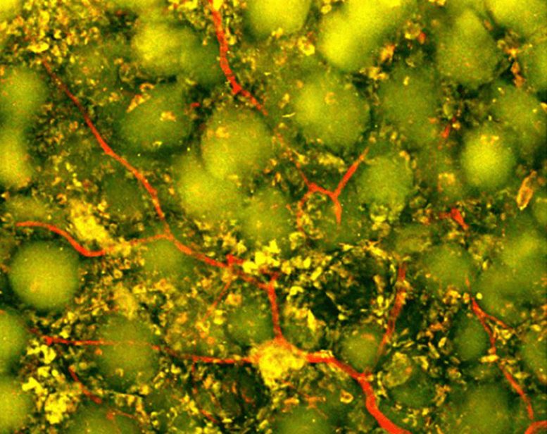
Neutrophil Infiltration
Two-photon imaging at day 27 using Ly6G-green fluorescent protein (GFP) reporter mice to visualize neutrophils. IL-4/C1q-EV + MAPS implants exhibited dense vascularization and focal accumulation of GFP+ cells within scaffold pores. Credit: Duke University

Immune Cells Take on a Surprising Role

Neutrophils are among the immune system’s fastest responders. After a stroke, they are often associated with inflammation and additional tissue damage, especially during the early phase of injury. Yet immune cells do not always have a single fixed role. Their behavior can change depending on timing, location, and the molecular signals surrounding them.

Inside the engineered scaffold, neutrophils appeared to become part of the repair process rather than merely contributing to destruction.

The researchers tested that possibility by depleting the immune-cell population rich in neutrophils. Blood vessel formation dropped sharply, and the scaffold underwent far less remodeling. The experiment showed that these cells were not simply present at the injury site. They were helping drive the response.

“This result changes how we think about neutrophils after stroke,” said Shangjing Xin, lead scientist of the study and a postdoctoral fellow in the Segura Laboratory. “Their role appears to depend on when they arrive, where they are located, and the signals they receive from their surroundings. Our study demonstrates a potential engineering strategy to recruit and retain these cells at the right time.”

New Blood Vessels and Nerve Fibers Emerge

The treatment produced visible changes throughout the damaged region. Blood vessels grew across the cavity, potentially creating the circulation needed to support living tissue. Researchers also detected more axonal fibers within and around the injury. Axons are the long projections neurons use to carry electrical signals to other cells.

Those biological changes were accompanied by improved movement.

During a grid-walking test, scientists measured how often the mice misplaced a front paw while crossing an uneven surface. Animals treated with the optimized scaffold made fewer errors over time. By eight weeks, their performance could not be statistically distinguished from that of healthy control mice, and the improvement continued through the end of the study.

The scaffold itself proved essential. When researchers delivered the EVs without MAPS, they did not observe comparable blood vessel growth. The result suggests that the treatment depended on both components: the biological messages carried by the vesicles and the porous structure that concentrated those messages while giving cells space to organize.

Toward a Scalable Human Stroke Therapy

The study relied on EVs collected from primary rat astrocytes, which would not be a practical source for a widely available human therapy.

Segura’s laboratory is now exploring astrocytes made from human-induced pluripotent stem cells. These cells can be produced from reprogrammed adult cells and expanded in the laboratory, potentially offering a more scalable and clinically relevant source of EVs. Researchers may also be able to adjust the conditions under which the astrocytes grow to better control the messages their vesicles carry.

“You do not restore an ecosystem simply by containing the initial damage,” Segura said. “You have to create the conditions that allow life to return. That is how we think about the stroke cavity. The material is not intended to reproduce the brain itself but to create an environment where the body’s own cells can enter, communicate, and participate in rebuilding vascularized tissue.”

Reference: “IL-4/C1q activated astrocyte-derived extracellular vesicles promote stroke infarct recovery by recruiting peripheral leukocytes” by Shangjing Xin, Lucy Zhang, Nhi V. Phan, Mengying An, Ligen Shi, S. Thomas Carmichael and Tatiana Segura, 21 July 2026, Cell Biomaterials.
DOI: 10.1016/j.celbio.2026.100543


Wednesday, July 1, 2026

New Artificial Neurons Cause Living Brain Cells to Fire

 If your competent? doctor can't figure out how to get dendritic branching/neurite outgrowth and axon pathfinding to work to connect up gray matter again then maybe this could work.

New Artificial Neurons Cause Living Brain Cells to Fire

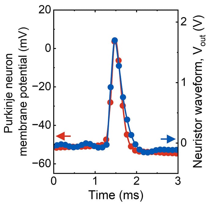
A team at Northwestern University has printed artificial neurons from molybdenum disulfide (MoS₂) — a semiconducting mineral — on flexible plastic that produce spiking waveforms closely matching biological action potentials in shape, width, and timing. When delivered to living Purkinje cells in mouse cerebellar tissue, the artificial spikes drove the cells to fire — the first demonstration that a printed device can produce electrical signals a real brain cell accepts and responds to.

The result, recently published in Nature Nanotechnology, could lay groundwork for a new generation of neural interfaces — prosthetic limbs that deliver realistic sensation, spinal cord bridges that relay motor commands, and benchtop disease models with tunable parameters.

“There’s this white space — organic devices are too slow, metal oxides are too fast — and biology lives in between,” said Mark C. Hersam, PhD, the study’s senior author and Walter P. Murphy Professor of Materials Science and Engineering at Northwestern University in Evanston, Illinois.

photo of Flexible polymide
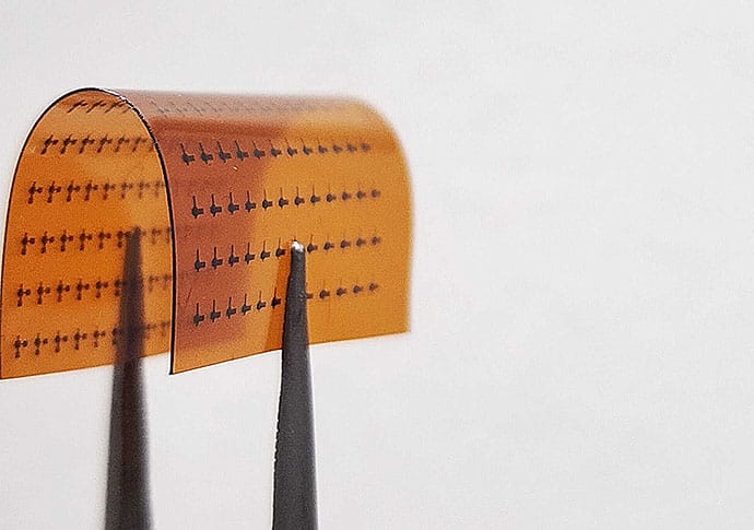
An array of printed artificial neurons on flexible polyimide held by tweezers to show the substrate bending.

“We got these devices working at that timescale,” he said, “and when you have the right timescale and the right spike shape, you can directly interface with living cells.”

Key Points
  • Printed MoS2 artificial neurons generated action-potential-like spikes.
  • Spike shape/timing matched biological APs; duration ≈0.7-2 ms.
  • Mouse Purkinje cells fired to artificial spikes at <200 Hz.
  • 740 Hz output failed; neuronal firing capacity limits response.
  • Potential uses: neural interfaces, spinal bridges, tunable disease models.
How do printed neural interfaces compare with metal oxide devices?
What limits long-term stability of MoS2 neural circuits?
Which cerebellar diseases could benefit from tunable circuit models?

Engineers Interface With Neuroscientists

The collaboration began not in biology but in electrical engineering. Hersam’s National Science Foundation grant aimed to build computing hardware that mimics the brain’s energy efficiency. The human brain runs on about 20 watts, whereas modern AI training runs on megawatts — a millionfold difference — and is extravagantly wasteful and potentially harmful to the environment.

The grant’s challenge: build computing hardware that mimics the brain’s efficiency.

“Most of the artificial neurons in the literature, if you actually look at their spiking profiles, they look more like a sine wave or just an oscillator, not a sharp action potential,” Hersam said. “They don’t achieve things like bursts of spikes, which is one of the things we demonstrate.”

photo of Meghana Holla, PhD
Meghana Holla, PhD

But to mimic the brain, Hersam needed people who study it. He teamed up with Indira M. Raman, PhD, a neurophysiologist in the Department of Neurobiology at Northwestern University, and her lab. Her doctoral students, Spencer Brown, PhD, and Meghana Holla, PhD, visited Hersam’s lab to see what the engineers were up to.

The engineers in Hersam’s lab showed them the device output, with waveforms spiking at 3000 times per second, much too fast to mimic a neural cell. Purkinje cells may be among the fastest-firing neurons in the brain, but they only reach about 100 spikes per second.

“That’s not a neuron,” Brown, an incoming assistant professor of neuroscience at Brandeis University in Waltham, Massachusetts, recalled. “They can’t do that.”

photo of Purkinje neuron Chart
The artificial neuron’s output (blue) overlaid with a living Purkinje cell’s action potential (red). Both spikes match closely in shape and duration, completing within about 2 milliseconds.

Over the following year, both fields discovered they used identical terminology, such as long-term potentiation, memory, and synapse weight, to mean different things.

photo of Spencer T. Brown, PhD
Spencer Brown, PhD

“We thought we were talking about the same things, but we weren’t,” Brown said.

Brown and Holla provided “ground truth”: Each spike had to last between a fraction of a millisecond and a few milliseconds, matching a real action potential.

And the firing rate — the number of spikes per second — had to fall between single digits and low hundreds, not the thousands the engineers’ devices had been producing.

The engineers took the neuroscientists’ advice and successfully reconfigured the circuit to match.

The Glue That Makes Artificial Neurons Fire

The artificial neurons are built from a liquid, a custom ink formulated for a specialized printer. MoS2, a semiconducting mineral, is peeled into flakes that are just a few atoms thick and suspended in ethanol. Without that suspension, the flakes clump together and settle out.

To keep them suspended, the researchers add ethylcellulose, a polymer derived from wood pulp, which coats each MoS2 flake and holds it apart from its neighbors, kind of like glue. The resulting ink is a stable suspension of semiconductor particles in solvent, and it flows through an aerosol jet printer that deposits it as a fine mist onto flexible plastic.

photo of inkjet printer
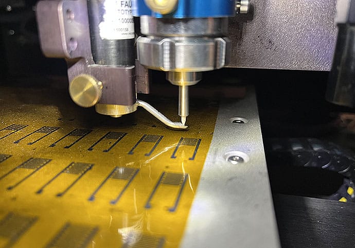
An aerosol jet printer deposits molybdenum disulfide ink onto flexible plastic. The nozzle sprays the ink as a fine mist, printing rows of artificial neurons without a cleanroom.

The ethylcellulose scaffolding is essential for the artificial neurons to communicate like a network. When the printed film is baked at 350 °C, the ethylcellulose partially decomposes into carbon residue that settles into tiny, nanometer-sized gaps between flakes to form conductive bridges. Once fabricated, the device operates at room temperature.

Then comes what’s called electroforming. The first time a large current passes through the device, it doesn’t flow evenly. Some pathways are slightly more conductive, such as wherever carbon residue accumulated more thickly, or wherever flakes overlapped. The more conductive pathways carry more current, and more current generates more heat. That heat decomposes more polymer residue into carbon along the same route, making it more conductive and drawing still more current toward it.

photo of Mark C. Hersam, PhD
Mark C. Hersam, PhD

The result is a single dominant channel — a filament — burned through the thickness of the film. “This occurs in a spatially inhomogeneous manner, leading to the formation of a conductive filament…all the current constricted into a narrow region,” said Hersam.

The filament has two states: hot and conducting, or cool and nonconducting.

On its own, that’s just a switch. What turns it into something that fires like a neuron is the circuit around it.

“This is a random network of flakes with gaps of a few nanometers,” said Vinod K. Sangwan, PhD, co-corresponding author and research associate professor of materials science and engineering at Northwestern University. “You cannot have atoms going from one place to another across that vacuum. The only mechanism left is thermal.”

In the full artificial neuron, the printed switch sits alongside a capacitor, which is a component that stores electrical charge. A steady input current slowly charges the capacitor, the way a biological neuron gradually accumulates signals from its neighbors.

The filament heats up and becomes conductive, and the capacitor rapidly discharges through it. That sudden discharge is the spike — a sharp, fast voltage pulse. Then the filament cools, the switch resets, and the capacitor begins slowly charging again. The cycle repeats: slow accumulation, sudden firing, reset.

And because the filament heats and cools on a millisecond timescale, the spikes fall within the same timing window as a real neuronal action potential.

How Real Brain Cells Respond to Artificial Neurons

Holla, who completed her PhD in Raman’s lab and is now a postdoctoral researcher studying memory at New York University in New York City, designed and ran experiments in mouse cerebellar slices. She positioned a stimulation electrode on the parallel fibers, the main pathway that excites Purkinje cells, and a recording electrode on the Purkinje cells themselves.

She played recordings of the artificial neurons’ waveforms into the tissue through a standard stimulation electrode at four different speeds: 7, 60, 218, and 740 spikes per second.

At every speed below 200 spikes per second, the Purkinje cells fired in response. The strongest results came at 60 spikes per second, where each artificial spike lasted 0.7 milliseconds, which is fast enough to trigger the cell but brief enough to avoid flooding the tissue with unnecessary current.

Above 200 spikes per second, the cells stopped responding. They simply cannot fire that fast. The team included the 740-spikes-per-second condition on purpose to directly challenge the many engineering groups building artificial neurons that operate at those speeds. “We had to show them [740 spikes] wasn’t sufficient,” Brown said. “You can’t work that fast.”

“You can see the living neurons respond to our artificial neuron,” Hersam said. But he is careful to note a caveat: The printed artificial neurons were not touching the brain tissue. The waveforms they generated were recorded and then played back into the slice through standard laboratory stimulation equipment.

The next step is to prove the printed device itself can interface with living tissue.

Clinical Possibilities

Ian Gaudet, PhD, a neuroscientist at Florida Atlantic University in Boca Raton, Florida, who was not involved in the study, sees multiple clinical possibilities from this work.

photo of Ian Gaudet, PhD
Ian Gaudet, PhD

“I’ve been waiting for [work like this] for years,” Gaudet said. “The signals coming off of these devices are the right shape, the right speed, and the right language for real neurons to be properly affected by them.”

The printed artificial neuron, he argues, is like a translator, converting digital information into electrical patterns neurons accept as input. And in prosthetic limbs, it could replace the rectangular pulses that give amputees a buzzing sensation with signals that peripheral nerves evolved to receive. A crude approximation of sensation could become something much closer to the real feeling.

“The idea would be to have this system where you’re controlling your prosthetic limb and you are feeling your prosthetic limb using the existing neuronal systems of your peripheral nervous system,” Gaudet said.

In spinal cord injury, it could convert decoded motor intentions into biologically shaped signals that motor neurons below a lesion treat as natural commands.

“If you can make that signal seamless,” Gaudet said, “people with spinal cord injuries could walk again one day.”

But where these artificial neurons may prove most valuable first is not as replacements for any damaged brain tissue but as test models. Researchers could build a small artificial cerebellar circuit on a benchtop, configure each element to fire like a different cell type, then deliberately break it to change the firing rate, and, in turn, simulate Purkinje cell loss in spinocerebellar ataxia.

Or one could remove an element to model a cerebellar stroke to see what happens — a disease model that could lead to novel treatments.

“You can turn this on, or turn this off,” Gaudet explained. “What happens if we mimic the patterns that we see in people who have a certain disease?”

Gaudet suggests researchers may use these devices as a physical disease model with real electrical dynamics.

What the Artificial Neuron Cannot Do

Hersam’s next goal is a small circuit — perhaps 10 artificial neurons — where each one fires differently, and together they accomplish what would require thousands of conventional transistors.

Silicon achieves complexity by having billions of identical devices,” Hersam said. “The brain is the opposite. It’s heterogeneous. The complexity is at the device level.”

But Gaudet sees a gap no circuit design can yet fill: Biological neurons grow new connections and prune old ones, strengthening pathways that are used and weakening those that aren’t. Hersam’s lab’s printed neurons — or any other neuromorphic technology that mimics neuronal dynamics — can’t achieve that level of complexity yet.

Wednesday, December 3, 2025

Nanomedicine in neurotrauma: A comprehensive review of gold nanoparticles for traumatic brain injury treatment

 I bet your fuckingly incompetent? doctor hasn't implemented gold nanoparticles in the past 4 years? And your board of directors is so incompetent they don't even know how to run a stroke hospital!

Electromagnetized gold nanoparticles improve neurogenesis and cognition in the aged brain October 2011 

Send me personal hate mail on this: oc1dean@gmail.com. I'll print your complete statement with your name(If you can't stand by your name don't bother replying anonymously) and my response in my blog. Or are you afraid to engage with my stroke-addled mind? No excuses are allowed! You're medically trained; it should be simple to precisely state EXACTLY WHY you aren't working on 100% recovery protocols with NO EXCUSES!

The latest here:

Nanomedicine in neurotrauma: A comprehensive review of gold nanoparticles for traumatic brain injury treatment


https://doi.org/10.1016/j.inoche.2025.115922Get rights and content

Highlights

  • TBI treatment using gold nanoparticles (AuNPs) is multi-mechanistic.
  • AuNPs have intrinsic anti-inflammatory and antioxidant properties across the blood-brain barrier.
  • They stimulate neuroregeneration via PI3K/Akt and MAPK/ERK.
  • Drug, growth factor, and genetic material can be targeted via functionalized AuNPs.
  • Preclinical studies demonstrate less lesions, less inflammation, and better cognition.
  • Standardization, toxicity, and manufacturing must be addressed in clinical translation.

Abstract

Traumatic brain injury (TBI) remains an enormous source of mortality and morbidity worldwide with few therapeutic options available today that address the pathophysiology of neuroinflammation, oxidative stress, and disruption of the blood-brain barrier (BBB) effectively. One of the promising classes of nanomedicines being explored extensively today for the treatment of TBI is gold nanoparticles (AuNPs) due to their biocompatibility, ability to target the BBB effectively, and versatility with respect to modification of their surface chemistry. This tutorial review will examine the multifunctional aspects of AuNPs that make them an attractive therapeutic target in the treatment of TBI, with special attention being given to their application in the management of secondary brain injury. AuNPs possess potent anti-inflammatory properties that result from modulating the polarization of microglia toward the restorative M2 subtype combined with the inhibition of the NF-κB signaling pathway. Additionally, AuNPs possess the capacity to counter the disruptive effects of ROS on cells by scavenging them while boosting the levels of the endogenously produced antioxidants. Finally, AuNPs possess the capacity to contribute toward the repair of the neuronal cells of the brain by inducing the growth of neurites with the activation of the PI3K/Akt signaling axis while functioning as vectors of neurotrophins such as NGF/BDNF and siRNA. Preclinical trials utilizing AuNPs illustrate their capacity to inhibit the growth of the lesion area, the activation of glia cells, and the reduction of cognitive impairment combined with the preservation of the integrity of the BBB with enhanced levels of angiogenesis. Despite these breakthroughs made toward the development of AuNP therapeutic agents that target TBI pathologically, there remain issues that must be considered if these therapeutic agents are to emerge safely.