Changing stroke rehab and research worldwide now.Time is Brain! trillions and trillions of neurons that DIE each day because there are NO effective hyperacute therapies besides tPA(only 12% effective). I have 523 posts on hyperacute therapy, enough for researchers to spend decades proving them out. These are my personal ideas and blog on stroke rehabilitation and stroke research. Do not attempt any of these without checking with your medical provider. Unless you join me in agitating, when you need these therapies they won't be there.

What this blog is for:

My blog is not to help survivors recover, it is to have the 10 million yearly stroke survivors light fires underneath their doctors, stroke hospitals and stroke researchers to get stroke solved. 100% recovery. The stroke medical world is completely failing at that goal, they don't even have it as a goal. Shortly after getting out of the hospital and getting NO information on the process or protocols of stroke rehabilitation and recovery I started searching on the internet and found that no other survivor received useful information. This is an attempt to cover all stroke rehabilitation information that should be readily available to survivors so they can talk with informed knowledge to their medical staff. It lays out what needs to be done to get stroke survivors closer to 100% recovery. It's quite disgusting that this information is not available from every stroke association and doctors group.

Showing posts with label repeatable. Show all posts

Thursday, April 25, 2024

New Tool Maps Brain Signals with Unprecedented Clarity


This is where stroke leaders would look at this and mutter to themselves; 'We could use this to detect brain signals occurring during stroke recovery; neurogenesis and neuroplasticity, and thus figure out how to make those signals repeatable'. But we have to do human testing first.

But the leaders would already have started listening to brain signals using one of these already. 

1. Use nanowires to listen in on single neurons

2. Or lay a grid across the cortex to listen in.

But we have NO stroke leaders, nothing will get done until we get survivors in charge.

Leaders solve problems, they don't run away from them.

The latest here:

New Tool Maps Brain Signals with Unprecedented Clarity

Summary: Researchers developed an innovative chemical tool to explore how signals like dopamine and epinephrine interact with neurons via G protein-coupled receptors (GPCRs).

This new tool allows for precise detection of neuromodulators across various brain regions with high spatial resolution. It marks cells with a permanent fluorescent signal, facilitating the study of signal pathways and interactions at a cellular level across the entire brain.

This advancement could significantly enhance our understanding of neuronal signaling and improve targeting GPCRs in drug development.

Key Facts:

  1. The tool enables detailed visualization of GPCR-related signals across the entire brain with high spatial resolution, a balance previously unachievable in neuroscience.
  2. It has been tested on opioids and epinephrine, utilizing both green and red fluorescence to track multiple molecules simultaneously.
  3. While the fluorescence takes several hours to manifest and is not suitable for real-time tracking, the tool provides valuable postmortem insights into neuronal pathways and drug targeting.

Source: University of Michigan

University of Michigan researchers have developed a new tool to better understand how chemicals like dopamine and epinephrine interact with neurons.

These chemicals are among a wide variety of signals that get processed in the brain through G protein-coupled receptors (GPCRs), proteins that sit on the surface of neurons to receive messages—in the forms of proteins, sugars, fats, even light—that inform cellular behavior. 
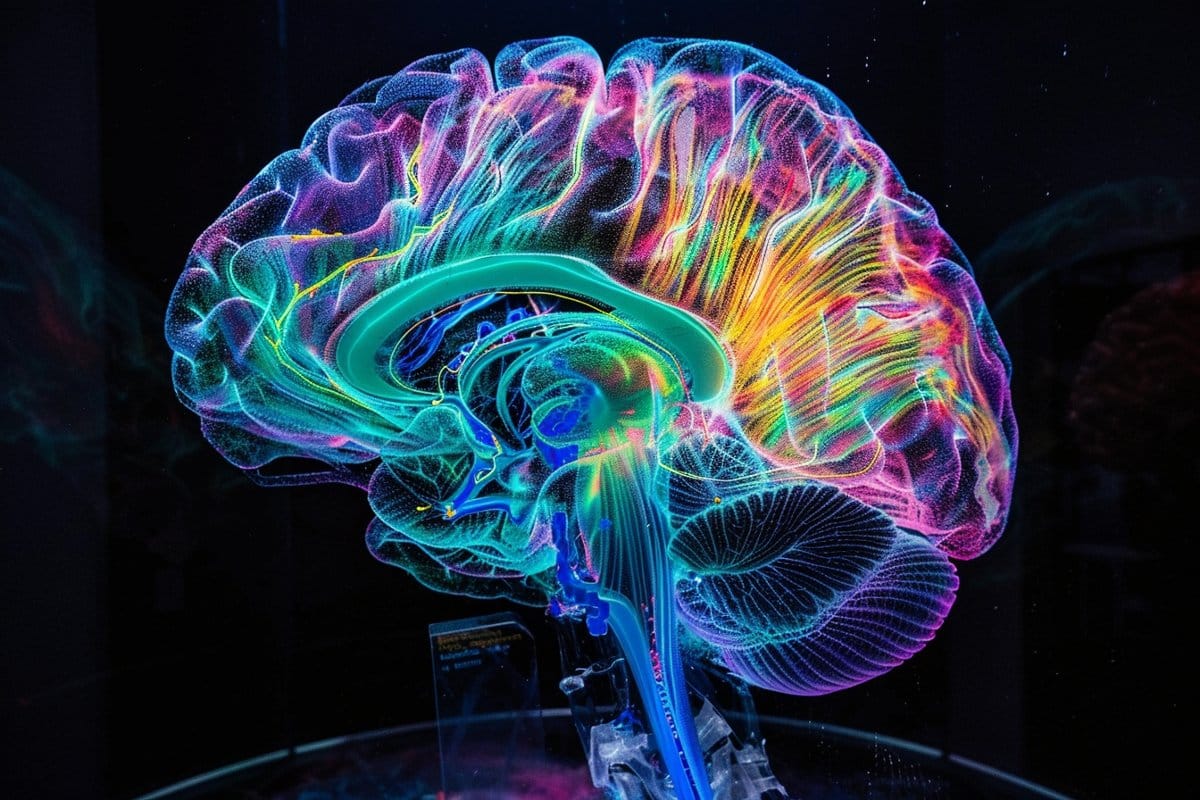
This shows a brain.
Wang’s lab at LSI uses protein engineering to develop technologies that can detect how signaling molecules travel within the brain to reach and interact with specific neurons. Credit: Neuroscience News

GPCRs are involved in an enormous number of biological functions, making them a prime target for treating diseases; more than one-third of FDA-approved drugs target GPCRs. But to fully understand how various molecules interact with GPCRs, researchers need to be able to detect those molecules across the whole brain with high spatial resolution.

“The challenge in our field has been achieving the right balance between a detailed view and the whole picture across the brain,” said Wenjing Wang, a neuroscientist at the U-M Life Sciences Institute. 

LSI faculty member Peng Li said most existing tools can detect a neural modulator either in a small part of the brain with high spatial resolution or in the whole brain with very low resolution.

“But we need to identify the cells that respond to the neuromodulators across various brain regions, in high resolution,” he said.

In a study published in the Proceedings of the National Academy of Sciences, Wang, Li and colleagues unveiled a new chemical tool that achieves both goals for three chemicals that all target GPCRs.

Wang’s lab at LSI uses protein engineering to develop technologies that can detect how signaling molecules travel within the brain to reach and interact with specific neurons. They previously created a tool to reveal the presence of opioids, another GPCR binding partner, at a cellular level.

When the molecule is detected, the tool creates a permanent fluorescent mark in the cells. Thus, researchers can see the specific cells that are highlighted, as well as the whole picture of cells across the brain.

This latest work broadens the utility of that sensor to detect multiple types of GPCR activators, beyond just opioids. So far, the team has tested the tool with opioids and epinephrine in cultured neurons and in mouse models. The team also expanded the tool to use both green and red fluorescence, enabling the tracking of multiple molecules at once.

“Coming from detecting just opioids, we now have a tool that we can begin to easily modulate for various signals that interact with GPCRs,” said Wang, who also is an assistant professor of chemistry at the U-M College of Literature, Science, and the Arts.

“The goal is eventually to even study the interplays of different signaling pathways simultaneously.”

The team cautions that while the tool provides important visualizations of how signals travel across neurons for analysis postmortem, it cannot be used to track chemicals in real time, as it takes several hours for the fluorescence to appear. But it does offer a new path forward for improving understanding of neuronal signaling and the role of GPCRs as drug targets.

“Ideally, we aim to be able to create a brain map for multiple neuromodulators concurrently, offering a comprehensive understanding of the sites of neuromodulation,” said Li, who also is an assistant professor at the U-M School of Dentistry.

About this brain mapping and neurotech research news

Author: Morgan Sherburne
Source: University of Michigan
Contact: Morgan Sherburne – University of Michigan
Image: The image is credited to Neuroscience News

Original Research: Closed access.
“Single-chain fluorescent integrators for mapping G-protein-coupled receptor agonists” by Wenjing Wang et al. PNAS

Saturday, April 6, 2024

Power-integrated, wireless neural recording systems on the cranium using a direct printing method for deep- brain analysis

This is where leaders would look at this and mutter to themselves; 'We could use this to detect brain signals occurring during stroke recovery, neurogenesis and neuroplasticity, and thus figure out how to make those signals repeatable'. But we have to do human testing first.

But the leaders would already have started listening to brain signals using one of these already. 

1. Use nanowires to listen in on single neurons

2. Or lay a grid across the cortex to listen in.

But we have NO stroke leaders, nothing will get done until we get survivors in charge.

Leaders solve problems, they don't run away from them.


The latest here:


Power-integrated, wireless neural recording systems on the cranium using a direct printing method for deep- brain analysis


Yong Won Kwon1,2
†, David B. Ahn 3
†, Young-Geun Park1,2
†, Enji Kim1,2
, Dong Ha Lee1,2
,
Sang-Woo Kim3
, Kwon-Hyung Lee4
, Won-Yeong Kim5
, Yeon- Mi Hong1,2
, Chin Su Koh 6
,
Hyun Ho Jung 6
, Jin Woo Chang7
, Sang-Young Lee5*, Jang- Ung Park1,2,6,8*
Conventional power-integrated wireless neural recording devices suffer from bulky, rigid batteries in head mounted configurations, hindering the precise interpretation of the subject’s natural behaviors. These power sources also pose risks of material leakage and overheating. We present the direct printing of a power-integrated wireless neural recording system that seamlessly conforms to the cranium. A quasi–solid-state Zn-ion microbattery was 3D-printed as a built-in power source geometrically synchronized to the shape of a mouse skull. Soft deep-brain neural probes, interconnections, and auxiliary electronics were also printed using liquid metals on the cranium with high resolutions. In vivo studies using mice demonstrated the reliability and biocompatibility of this wireless neural recording system, enabling the monitoring of neural activities across extensive brain regions without notable heat generation. This all-printed neural interface system revolutionizes brain research, providing bioconformable, customizable configurations for improved data quality and naturalistic experimentation.
INTRODUCTION
The brain constitutes an intricate three-dimensional (3D) network composed of an immense number of neurons continually generating and transmitting signals for communication. These neuronal activities and firing patterns play a pivotal role in governing bodily functions, consciousness, and the formation of memories. Comprehending the electrophysiology of neurons and functional connectivity of network-level neuronal activities is essential for fundamental research in the treatment of numerous neurological diseases, such as Parkinson’s disease (PD), Alzheimer’s disease, epilepsy, and major depressive disorder (1). In response to the challenge, implantable electronic devices known as neural probes have seen notable development. These devices are designed to convert neural signals into electronic signals, allowing for the precise monitoring of neuronal activities within specific brain regions (2–6). In particular, recentadvances in microfabrication technologies and bioelectronics have enabled flexible neural probes for reliable recording by ensuring mechanical and structural compatibility with brain tissues (7–12). A predominant perspective in neuroscience and biomedical engineering is that the activities of a neuronal population are notably influenced by the state of the subject. This perspective allows for a better understanding of neuronal computations related to diverse behavioral and cognitive processes, particularly during unrestricted movement and freely movable states (13, 14). However, the use of numerous neural probes connected to external recording devices via cables and wires limits the subject’s freedom of movement. Consequently, wireless neural recording devices have become indispensable to facilitate more natural movements and behaviors in subjects. This is especially crucial in studies aiming to comprehend how the brain responds to specific environments and tasks. In addition, wireless neural recording holds the potential to enhance data quality by eliminating the possibility of noise and interference caused by wires (15–23). Considering the substantial amount of neural data to be collected, the most applicable technology for wireless neural recording is a battery- powered system with widespread availability of the associated hardware (18, 20–22). Batteries, known for their high energy density and operational stability, make them a promising choice for powering neural interface devices. However, current bulky and rigid battery configurations occupy over 90% of the device volume and more than 60% of its mass (16). To use these batteries on small animals, additional fixtures or suits are required to attach them to the head or back, hindering the free behavior of experimental animals(24, 25). Moreover, the risk of electrolyte leakage and overheating in conventional batteries poses a substantial obstacle to the formation of bio-integrated systems (26–29). Therefore, there is a strong need for batteries that (i) can be shaped to conform to nonplanar biological surfaces and (ii) consist of quasi–solid-state, biocompatible electrolytes to prevent leaks in wireless neural interfaces. In considering the entire neural interface system, various auxiliary electronic components are essential for the collection and processing of raw signals detected by neural probes. While numerous efforts have been made to enhance the long-term stability and signal quality of soft neural probes, the electronics responsible for wireless signal transmission and their electrical connections still typically use flat and rigid printed circuit boards (PCBs) made from solid, fragile materials (30, 31). These structural and material differences
 
Tables at link.
 

Friday, January 29, 2021

Neuroplasticity is a 'Get out of jail free card' for your stroke medical team

 Since there is NO PROTOCOL  on how to make neuroplasticity EXACTLY REPEATABLE, this allows your stroke team to be able to shift blame for not recovering onto the survivor.  I however think ALL BLAME for lack of recovery lies on the doctors, knowing how fucking bad stroke recovery has been for decades and not specifying the needs to researchers to get correct rehab research done.

Friday, November 27, 2020

Big Surprise: The Brain Can Recover Many Years After a Stroke

 This is great, but HOW DO YOU MAKE THIS EXACTLY REPEATABLE? If I were to try swimming now I would drown, so this person is definitely much higher functioning than me in regards to arm use.

Big Surprise: The Brain Can Recover Many Years After a Stroke

Abstract

Most doctors think that after a patient has a stroke, recovery only happens for 6 months to a year; after that there is no point in continuing with rehabilitation therapy. We described a patient who had a severe stroke at age 15 and was left with a completely useless left hand. Then 23 years later, after he started swimming regularly to lose weight, he had some movement in the fingers of his left hand. He began intensive therapy with exercises using a special glove, and now, 37 years after the stroke, he is still improving. The way his brain “rewired” itself all over both sides of the brain is shown with a special imaging method called functional magnetic resonance imaging. This means that intensive physiotherapy and maybe new approaches to brain recovery including stem cell therapy, need to be tried much longer after the stroke than we used to think.

Introduction

What most doctors believe is that after a stroke

, the patient can only recover for a short time, such as 6 months or a year. Young children who have strokes recover much better than people who have strokes as adults.

This is a story of a man who had a stroke at age 15 and had no use of his left hand until he began to recover 23 years later, after taking up swimming to try to lose weight [1]. He is still improving 37 years after the stroke. His story gives hope for recovery much later than we thought possible, and suggests that intensive physiotherapy, and maybe new approaches to brain recovery such as stem cell

therapy might improve recovery long after a stroke [2–5].


Monday, March 23, 2020

New brain implant device could record activity in thousands of neurons

If we are ever going to make neuroplasticity completely repeatable we will need to understand the signals sent between neurons. This might be one way, Your researcher can tell you which of these other ideas would be the best to answer that question. Nothing will ever come of this because we have NO stroke leadership to go to to get a stroke strategy updated.

Right now neuoplasticity is considered the holy grail of stroke rehab but without knowing how to make it repeatable is practically useless.

1. Fast high-resolution miniature two-photon microscopy for brain imaging in freely behaving mice

2. Use nanowires to listen in on single neurons
3. Or lay a grid across the cortex to listen in. 


The latest here:

New brain implant device could record activity in thousands of neurons

A team of Stanford University researchers has created a device that, once implanted in the brain, could help record movies of electrical neural activity in thousands of individual neurons.
The device, described in a paper published March 20 in Science Advances, could be used for research or with prosthetics, and is capable of recording more data while being less intrusive than other options.
"The design of this device is completely different from any existing high-density recording devices, and the shape, size and density of the array can be simply varied during fabrication. This means that we can simultaneously record different brain regions at different depths with virtually any 3D arrangement," Jun Ding, PhD, assistant professor of neurosurgery and neurology and co-author of the paper said in a Stanford News story. "If applied broadly, this technology will greatly excel our understanding of brain function in health and disease states."
At the heart of this invention is a bundle of microwires, each of which is less than half the width of the thinnest human hair. These wires, which are directed into the brain to obtain electrical signals that pass by, are small enough to cause minimal damage but sturdy enough to resist degrading over time.


Abdulmalik Obaid, left, and Nick Melosh with a device they invented that contains a bundle of microwires capable of recording the activity of thousands of neurons in the brain in real time.
The trick was figuring out how to design an orderly array of these super thin wires that is adaptable in terms of size -- some applications of the array may only warrant a few microwires but others would require thousands.
The researchers spent years designing and redesigning the device and the process for making it. Eventually, they found success by encasing each wire in a biologically-safe polymer, then bundling them in a metal collar. Below the collar, the polymer is removed from the wires so they can be inserted into the brain. Topped off with a silicon chip -- like those used in a camera -- the device can begin recording neural activity.
Once the researchers settled on their design, they were able to run tests in living tissues. They began with retinal cells from rats and a 138-wire array.
"We had to take kilometers of microwires and produce large-scale arrays, then directly connect them to silicon chips," Abdulmalik Obaid, a graduate student in materials science and engineering and lead author of the paper told Stanford News. "After years of working on that design, we tested it on the retina for the first time and it worked right away. It was extremely reassuring."
The team has also successfully tested the device in the brains of living mice, using arrays that ranged from 135 to 251 wires, and are continuing these studies so they can better understand the longevity of their invention and the kinds of signals it is able to obtain.
"Electrical activity is one of the highest-resolution ways of looking at brain activity," said Nick Melosh, professor of materials science and engineering and co-senior author of the paper. "With this microwire array, we can see what's happening on the single-neuron level."

Wednesday, February 26, 2020

Research is key to curbing stroke

But only if you have a strategy being followed. If researchers are allowed to willy-nilly choose research on stroke, NOTHING WILL BE ACCOMPLISHED.  So YOU need to educate Ms. McGowan on that.  I'd suggest solving the 5 causes of the neuronal cascade of death in the first week. Or figure out how to make neuroplasticity EXACTLY REPEATABLE.


Research is key to curbing stroke

Sharon McGowan, Chief Executive Officer of the Australian Stroke Foundation, argues that research is key to curbing stroke

Stroke is one of Australia’s biggest killers and a leading cause of disability. It is a devastating disease that strikes the brain, the most complex organ in our bodies, responsible for our movement, thoughts and feelings.
Too many Australian lives are impacted by stroke when we know it can be prevented, treated and beaten. This year there will be more than 56,000 strokes in Australia – that’s one every nine minutes. That number continues to increase as our population grows and ages and lifestyles become more sedentary. Globally, one in four people will have a stroke in their lifetime.

The role of research

Curbing the rate and devastation caused by stroke is an enormous challenge both in Australia and around the world. Medical research is a key component, along with educating the community about stroke prevention.
Recently, the Australian Government announced it is investing $437 million in health and medical research. This included $58 million for cardiovascular disease (stroke and heart disease) research. The funding package builds on a previously announced $220 million Mission for Cardiovascular Health.
It is encouraging to see support from the Australian Government for high-quality research. There is still so much we do not know about the brain and investment is crucial for future breakthroughs in diagnosis, treatment and care.
The true value of research is evident in the significant advances in time-critical acute stroke treatments in the past two decades. This has led to a reduction in the number of lives lost to stroke and a greater number of patients return to independent living.
But we must not stop now, more funding is needed to make further developments a reality. With more people surviving stroke than ever before, we must now find the keys to maximise their recovery, to provide them with the best chance to live long and productive lives.
We know the transition from hospital to home can be challenging. Research in rehabilitation and mental health are important to improve the quality of life in the long term. People with stroke deserve effective treatments underpinned by an evolving understanding of brain recovery and human behaviour.

Stroke Foundation Research and Innovation Program

Research is a core part of our organisation’s mission to prevent stroke, save lives and enhance recovery. Since 2008, almost $5 million in research grants has been awarded to more than 200 researchers.
Historically, our seed grants are awarded to early to mid-career researchers (www.strokefoundation.org.au) for pilot or feasibility studies. This enables researchers to take their first step towards an idea which could provide the next big discovery in stroke. Seed funding the research pipeline is an essential role of funders such as the Stroke Foundation. Our close connection with the stroke community enables us to focus on supporting the research that matters most to those impacted by stroke.
Building capability in the stroke research community is the first link in the chain of discovery and can pave the way for future change in policy, practice and knowledge. It also encourages individuals to enter and stay in the research field.
In awarding the grants, we identify current gaps in research, where it is most needed and where studies can potentially have the biggest impact. A priority in the 2020 grant round was innovation to support the diverse needs of carers, recognising that the impact of stroke is widespread and life-changing for loved ones too.

Stroke researchers look to the skies for the latest breakthrough

Australia has unique needs in stroke research due to its large land mass, dispersed community and indigenous population. In an exciting project currently underway, Australia could become home to the world’s first stroke air ambulance.
Stroke Foundation is part of the Australian Stroke Alliance (ASA), an alliance between 37 research, industry and government entities that wish to develop a suite of World First portable imaging technologies that will radically transform access to early prehospital treatments, and dramatically improve stroke outcomes across Australia.
The ASA was successful in being awarded $1 million in Stage 1 funding from the Commonwealth of Australia under the Medical Research Future Fund – Frontier Health and Medical Research Program (MRFF) to develop a Research Plan for the project, which will form the basis for a MRFF Stage 2 submission which aims to deliver modern prehospital stroke care to Indigenous, rural and metropolitan Australians, including the world’s first stroke capable air ambulance.
This has the potential to transform access to emergency stroke treatment for patients in rural and remote areas and give them the best chance of survival. Sadly, people outside of Australia’s cities are 19% more likely to have a stroke and are also more likely to have a poorer outcome due to their distance to lifesaving, time-critical medical treatments.

Conclusion

Research has the power to save lives, prevent disability and ultimately reduce the burden of stroke on our community. It is more important now than ever as the incidence of stroke grows. By 2050, it is estimated there will be one stroke every four minutes in Australia and one million stroke survivors will be living in our communities unless action is taken.
We know research takes time and perseverance and a great deal of funding, but the outcomes provide hope and can make a difference to the lives of stroke survivors and their loved ones for generations to come.

Monday, December 30, 2019

A review of the progression and future implications of brain-computer interface therapies for restoration of distal upper extremity motor function after stroke

If you were to properly think about this, all rehab therapies would work much better with vastly fewer dead neurons. And you do that by stopping the 5 causes of the neuronal cascade of death in the first week.

If my doctors had done that they would have saved me  5.4 billion neurons. Recovery would have been easy with only 171 million dead neurons.  And sending a bill to my doctor and stroke hospital at $1000 a dead neuron would only cost them 5.4 trillion dollars. That might concentrate their minds.  I don't expect neuroplasticity or neurogenesis to be precisely repeatable for at least 50 years. No one is looking at the signals that occur between neurons such that one neuron drops its current function and takes on a neighbor's function. Knowing that process is the only way to make neuroplasticity EXACTLY REPEATABLE.

A review of the progression and future implications of brain-computer interface therapies for restoration of distal upper extremity motor function after stroke

 Alexander Remsik, Brittany Young, Rebecca Vermilyea, Laura Kiekhoefer, Jessica Abrams, Samantha Evander Elmore,Paige Schultz, Veena Nair, Dorothy Edwards, Justin Williams and Vivek Prabhakaran
Department of Radiology Clinical Science Center, University of Wisconsin Madison School of Medicine and Public Health Ringgold StandardInstitution, Madison, WI, USA

 ABSTRACT

Stroke is a leading cause of acquired disability resulting in distal upper extremity functional motor impairment. Stroke mortality rates continue to decline with advances in healthcare and medical technology. This has led to an increased demand for advanced, personalized rehabilitation. Survivors often experience some level of spontaneous recovery shortly after their stroke event, yet reach a functional plateau after which there is exiguous motor recovery. Nevertheless, studies have demonstrated the potential for recovery beyond this plateau. Non-traditional neurorehabilitation techniques,such as those incorporating the brain-computer interface (BCI), are being investigated for rehabilitation. BCIs may offer a gateway to the brain’s plasticity and revolutionize how humans interact with the world.Non-invasive BCIs work by closing the proprioceptive feedback loop with real-time, multi-sensory feedback allowing for volitional modulation of brain signals to assist hand function. BCI technology potentially promotes neuroplasticity and Hebbian-based motor recovery by rewarding cortical activity associated with sensory-motor rhythms through use with a variety of self-guided and assistive modalities. (So you really know nothing useful?)
 

Monday, May 27, 2019

Wireless Recording in the Peripheral Nervous System with Ultrasonic Neural Dust

With ANY BRAINS AT ALL in stroke leadership, this would have immediately started research using this to listen in on neuron signalling in neuroplasticity. We need to know why and how a neighboring neuron gives up its current task and takes on a neighboring task. Without that knowledge neuroplasticity will never be made repeatable and made into a stroke protocol.  Hell, this has been out since October 2016 so more proof that the stroke medical world is completely incompetent.


Wireless Recording in the Peripheral Nervous System with Ultrasonic Neural Dust


Highlights

  • •
    First in vivo electrophysiological recordings with neural dust motes
  • •
    Passive, wireless, and battery-less EMG and ENG recording with mm-scale devices
  • •
    Recorded signals transmitted via ultrasonic backscatter from implanted neural dust motes
  • •
    Ultrasound as a scalable means of providing wireless power and communication

Summary

The emerging field of bioelectronic medicine seeks methods for deciphering and modulating electrophysiological activity in the body to attain therapeutic effects at target organs. Current approaches to interfacing with peripheral nerves and muscles rely heavily on wires, creating problems for chronic use, while emerging wireless approaches lack the size scalability necessary to interrogate small-diameter nerves. Furthermore, conventional electrode-based technologies lack the capability to record from nerves with high spatial resolution or to record independently from many discrete sites within a nerve bundle. Here, we demonstrate neural dust, a wireless and scalable ultrasonic backscatter system for powering and communicating with implanted bioelectronics. We show that ultrasound is effective at delivering power to mm-scale devices in tissue; likewise, passive, battery-less communication using backscatter enables high-fidelity transmission of electromyogram (EMG) and electroneurogram (ENG) signals from anesthetized rats. These results highlight the potential for an ultrasound-based neural interface system for advancing future bioelectronics-based therapies.

Video Abstract

Flash is missing. Download it from Adobe

Introduction

Recent technological advances (
,
) and fundamental discoveries (
,
,
) have renewed interest in implantable systems for interfacing with the peripheral nervous system. Early clinical successes with peripheral neurostimulation devices, such as those used to treat sleep apnea (
) or control bladder function in paraplegics (
) have led clinicians and researchers to propose new disease targets ranging from diabetes to rheumatoid arthritis (
). A recently proposed roadmap for the field of bioelectronic medicines highlights the need for new electrode-based recording technologies that can detect abnormalities in physiological signals and be used to update stimulation parameters in real time. Key features of such technologies include high-density, stable recordings of up to 100 channels in single nerves, wireless and implantable modules to enable characterization of functionally specific neural and electromyographic signals, and scalable device platforms that can interface with small nerves of 100 μm diameter or less (
) as well as specific muscle fibers. Current approaches to recording peripheral nerve activity fall short of this goal; for example, cuff electrodes provide stable chronic performance but are limited to recording compound activity from the entire nerve. Single-lead intrafascicular electrodes can record from multiple sites within a single fascicle but do not enable high-density recording from discrete sites in multiple fascicles (
). Similarly, surface EMG arrays allow for very-high-density recording (
,
) but do not capture fine details of deep or small muscles. Recently, wireless devices to enable untethered recording in rodents (
,
) and nonhuman primates (
,
,
), as well as mm-scale integrated circuits for neurosensing applications have been developed (
,
,
). However, most wireless systems use electromagnetic (EM) energy coupling and communication, which becomes extremely inefficient in systems smaller than ∼5 mm due to the inefficiency of coupling radio waves at these scales within tissue (
,
; see also Size Scaling and Electromagnetics in the Supplemental Information). Further miniaturization of wireless electronics platforms that can effectively interface with small-diameter nerves will require new approaches.
In contrast to EM, ultrasound offers an attractive alternative for wirelessly powering and communicating with sub-mm implantable devices (
,
,
,
,
). Ultrasound has two advantages. First, the speed of sound is 105 × lower than the speed of light in water, leading to much smaller wavelengths at similar frequencies; this yields excellent spatial resolution at these lower frequencies as compared to radio waves. Second, ultrasonic energy attenuates far less in tissue than EM radiation; this not only results in much higher penetration depths for a given power, but also significantly decreases the amount of unwanted power introduced into tissue due to scattering or absorption. In fact, for most frequencies and power levels, ultrasound is safe in the human body. These limits are well defined, and ultrasound technologies have long been used for diagnostic and therapeutic purposes. As a rough guide, about 72× more power is allowable into the human body when using ultrasound as compared to radio waves (
,
).
We previously introduced the neural dust ultrasonic backscattering concept to harness the potential advantages of ultrasound and showed that, theoretically, such a system could be scaled well below the mm-scale when used for wireless electrophysiological neural recording (
,
). Here, we present the first experimental validation of a neural dust system in vivo in the rat peripheral nervous system (PNS) and skeletal muscle, reporting both electroneurogram (ENG) recordings from the sciatic nerve and electromyographic (EMG) recordings from the gastrocnemius muscle. The neural dust system consists of an external ultrasonic transceiver board which powers and communicates with a millimeter-scale sensor implanted into either a nerve or muscle (Figure 1A). The implanted mote consists of a piezoelectric crystal, a single custom transistor, and a pair of recording electrodes (Figures 1B, 1C, and S1).


Figure thumbnail gr1
Figure 1Neural Dust System Overview

Syringe injectable electronics

With ANY BRAINS AT ALL in stroke leadership, this would have immediately started research using this to listen in on neuron signalling in neuroplasticity. We need to know why and how a neighboring neuron gives up its current task and takes on a neighboring task. Without that knowledge neuroplasticity will never be made repeatable and made into a stroke protocol.  Hell, this has been out since July 2015 so more proof that the stroke medical world is completely incompetent.

Syringe injectable electronics

Associated Data

Supplementary Materials

Abstract

Seamless and minimally-invasive three-dimensional (3D) interpenetration of electronics within artificial or natural structures could allow for continuous monitoring and manipulation of their properties. Flexible electronics provide a means for conforming electronics to non-planar surfaces, yet targeted delivery of flexible electronics to internal regions remains difficult. Here, we overcome this challenge by demonstrating syringe injection and subsequent unfolding of submicrometer-thick, centimeter-scale macroporous mesh electronics through needles with a diameter as small as 100 micrometers. Our results show that electronic components can be injected into man-made and biological cavities, as well as dense gels and tissue, with > 90% device yield. We demonstrate several applications of syringe injectable electronics as a general approach for interpenetrating flexible electronics with 3D structures, including (i) monitoring of internal mechanical strains in polymer cavities, (ii) tight integration and low chronic immunoreactivity with several distinct regions of the brain, and (iii) in vivo multiplexed neural recording. Moreover, syringe injection enables delivery of flexible electronics through a rigid shell, delivery of large volume flexible electronics that can fill internal cavities and co-injection of electronics with other materials into host structures, opening up unique applications for flexible electronics.
The emergence of flexible electronics has significantly extended the applications of electronics by allowing intimate interfaces between electronic units and non-planar surfaces for better monitoring and manipulation of their properties-. A variety of electronic devices- has been integrated on flexible and stretchable substrates to enable applications from foldable display to electronic skin-. 3D interpenetration of flexible electronics within existing structures could further broaden and open up new applications by directly interfacing devices with the internal structures of man-made and biological materials.
Recent work has shown that flexible electronics can be placed into 3D structures through surgical processes- or by being attached to and subsequently released from a rigid delivery substrates- for biological and biomedical applications. However, direct 3D interpenetration of electronics within these structures is limited by the intrinsic thin-film supporting substrates. We have introduced a macroporous mesh paradigm that allow electronics to be combined, for example, with polymer precursors and cells to yield 3D interpenetration, , although controlled delivery and/or non-surgical placement of these ultraflexible open electronic networks into structures with seamless 3D integration and interpenetration has not been possible.
Here, we describe the design and demonstration of macroporous flexible mesh electronics that allow electronics to be precisely delivered into 3D structures by syringe injection and subsequently relax and interpenetrate within the internal space of man-made and biological materials. Distinct from previous reports, , , syringe injection requires complete release of the mesh electronics from a substrate so that the electronics can be driven by solution through a needle. The syringe injectable electronics concept involves (i) loading the mesh electronics into a syringe and needle, (ii) insertion of the needle into the material or internal cavity and initiation of mesh injection (Fig. 1a), (iii) simultaneous mesh injection and needle withdrawal to place the electronics through the targeted region (Fig. 1b), and (iv) delivery of the input/output (I/O) region of the mesh outside of the material (Fig. 1c) for subsequent bonding and measurements.
An external file that holds a picture, illustration, etc.
Object name is nihms-724760-f0001.jpg
Syringe injectable electronics
a to c, Schematics of injectable electronics. The red-orange lines highlight the overall mesh structure and indicate the regions of supporting and passivating polymer mesh layers; the yellow lines indicate metal interconnects between I/O pads (green filled circles) and recording devices (blue filled circles). d, Schematic of the mesh electronics design (upper image), where the orange and red lines represent polymer encapsulated metal interconnects and supporting polymer elements, respectively, and W is the total width of the mesh. The dashed black box (lower image) highlights the structure of one unit cell (white dashed lines), where α is the angle deviation from rectangular. e, Longitudinal mesh bending stiffness, DL, and transverse mesh bending stiffness, DT, as a function of α defined in d. f and g, Images of mesh electronics injection through a glass needle, ID = 95 μm, into 1x PBS solution. Bright-field microscopy image f of the mesh electronics immediately prior to injection into solution; the red arrow indicates the end of the mesh inside the glass needle. 3D reconstructed confocal fluorescence image g recorded following injection of ca. 0.5 cm mesh electronics into 1x PBS solution. The blue and white dashed boxes correspond to regions shown in Supplementary Fig. 3a and b. h, Optical image of an injectable mesh electronics structure unfolded on a glass substrate. W is the total width of the mesh electronics. The red dashed polygon highlights the position of electrochemical devices or FET devices. Green and black dashed boxes highlighted metal interconnect lines and metal I/O pads, respectively. i and j, Yields and change with ±1 standard deviation (±1SD) in properties post-injection for single-terminal electrochemical and two-terminal field-effect transistor (FET) devices. i, Yield (blue) and impedance change (red) of the metal electrodes from the mesh electronics injected through 32, 26 and 22 gauge metal needles. Inset: bright field image of a representative metal electrode on mesh electronics, where the sensing electrode is highlighted by a red arrow. Scale Bar: 20 μm. j, Yield (blue) and conductance change (red) of silicon nanowire FETs following injection through 32, 26, 24, 22 and 20 gauge needles. Inset: scanning electron microscopy (SEM) image of a representative nanowire FET device in the mesh electronics; the nanowire is highlighted by the red arrow. Scale bar: 2 μm.

Design and implementation of electronics for syringe injection

The mechanical properties of the free-standing mesh electronics are important to the injection process. The basic mesh structure (Fig. 1d and Supplementary Fig. 1, a and b) consists of longitudinal polymer/metal/polymer elements, which function as interconnects between exposed electronic devices and I/O pads, and transverse polymer elements. The mesh longitudinal and transverse bending stiffness, DL and DT, are determined by the mesh unit cell and corresponding widths and thickness of the longitudinal and transverse elements, and the angle, α, , . Simulations of DT and DL versus α(Fig. 1e) show that DT (DL) decreases (increases) for increasing α. Hence, increasing α facilitates bending along the transverse direction (reduced DT) and should allow for rolling-up of the mesh electronics within a needle constriction, while at the same time increasing DL, which should reduce bending and potential buckling along the injection direction.
The mesh electronics were fabricated, fully-released from substrates using reported methods, and loaded into glass needles connected to a microinjector (details see, Supplementary Information Sections 2 and 3 and Supplementary Figs. 2 and 3). Images of injection of a 2 mm wide sample through a 95 μm inner diameter (ID) glass needle show the compressed mesh ca. 250 μm from the needle opening (Fig. 1f), and then injected ca. 0.5 cm into 1x phosphate-buffered saline (PBS) solution (Fig. 1g), where the 3D image highlights the unfolding of the mesh structure from the point of the needle constriction (blue dashed box). Higher resolution images (Supplementary Fig. 4, a and b) show that the mesh structure is continuous as it unfolds. Similar results were obtained for injection of a 1.5 cm width sample through a 20 gauge (600 μm ID) metal needle (Supplementary Fig. 4c) demonstrating the generality of this injection through common glass and metal syringe needles.
To test further electrical continuity and functionality of the mesh electronics post-injection, we used anisotropic conductive film (ACF) to connect the I/O pads of theelectronics post-injection to flexible cables that are interfaced to measurement electronics (Supplementary Fig. 5, a-d). Studies of the electrical performance and yield of devices following injection into 1x PBS solution through 100-600 μm ID needles (Fig. 1, i and j) highlight several points. First, metal electrochemical devices had an average device >94% and an average device impedance change, which represents an important characteristic for voltage sensing applications, , of <7% post injection (Fig. 1i). Second, silicon nanowire field-effect transistor (FET) devices had a yield > 90% for needle IDs from 260 to 600 μm, only dropped to 83% for the smallest 100 μm ID needles, and exhibited < 12% conductance change on average post injection (Fig. 1j). Together these results demonstrate the robustness of our mesh electronics design and the capability of maintaining good device performance following injection through a wide-range of needle IDs.
We have characterized the structures of different mesh electronics within glass needle-like constrictions to understand design parameters for successful injection (Fig. 2, a and b). Bright field microscopy images of mesh electronics with different structural parameters recorded from the central region of different ID glass channels (Fig. 2c) highlight two important features. First, mesh electronics with α = 45° and widths substantially larger than the constriction ID can be smoothly injected. Relatively straight longitudinal elements are seen in Fig. 2c, I and II, where the 5 mm 2D mesh widths are 11- and 20-times larger than the respective 450 and 250 μm ID needle constrictions. Second, even 1.5 cm width mesh electronics (Fig. 2c, III) can be injected smoothly through a 33-times smaller ID (450 μm) constriction.

More plus pictures at link.

Thursday, February 7, 2019

Using Virtual Reality, Researchers Get a Closer Look at Autoimmune Disease - Can it be used for stroke?

Our stroke researchers should easily be able to repurpose this for identifying stroke damage and figuring out EXACTLY how neuroplasticity can be made repeatable on demand.  But that won't occur, we have NO STROKE LEADERSHIP. 

Using Virtual Reality, Researchers Get a Closer Look at Autoimmune Disease 

Thu, 02/07/2019 - 11:56am 2 Comments
by Kenny Walter - Digital Reporter -
Adam Lacy-Hulbert and Caroline Stefani utilizing VR tools
Viewing images of diseased cells on a computer screen means limited detail and restricted angles, prohibiting researchers from fully analyzing specimens.
So researchers from Benaroya Research Institute at Virginia Mason (BRI)—a Seattle-based research organization—are taking a different approach.
For more than a year, BRI researchers have used virtual reality (VR) tools to conduct detailed experiments about autoimmune and immune system diseases.
Adam Lacy-Hulbert, PhD, an associate member at BRI, explained in an interview with R&D Magazine how the research lab is utilizing virtual reality platforms to both speed up and enhance the research process.
“So instead of viewing [cell images] as a three-dimensional model on a flat computer screen we could actually project them into a VR space and directly interact with the three-dimensional images of these cells in virtual reality,” Lacy-Hulbert said. “That’s really been a game-changer for how we initially analyze some of our data. Now we can very rapidly go from capturing the images on the microscope to actually imaging them directly with VR. It’s really become a key part in how we interact with our imaging data.”
Much of the work at BRI is focused on imaging cells using a confocal microscope and fluorescent tags, where they are able to image four colors at once at a high resolution. The confocal builds up individual optical slides of a cell or of multiple cells that are interacting.
 “We actually put those slices together to make a three-dimension model of the cell that we can look at and try to interpret how the cell is working and what goes wrong in a cell with an autoimmune disease like lupus compared to a healthy individual,” said Lacy-Hulbert.
The lab uses the ConfocalVR system provided by Immersive Science, which is also based in Seattle. This tool stacks confocal microscope images in fully immersive VR, allowing researchers to see never-before-seen details of cell structures in the images. One of the major advantages of using VR tools in the lab is it gives researchers free reign to change the cells to try to learn more about their internal structure.
“It’s really intuitive to spin cells around in 3D, to get the right orientation so you can see how different structures inside the cell fit together,” Lacy-Hulbert said. “You can very quickly manipulate using the wands, the hand held controllers, to expand and contract your image. You have the ability to feel like you are holding something and just turn it maybe two degrees in a few different directions and you can immediately see how the internal structures interact with each other.”
Caroline Stefani, PhD, a senior postdoctoral research associate at BRI, said that from a researcher’s standpoint the ability to look at a cell image from multiple angles, coupled with the increased speed at which this takes place, makes VR a useful research tool.
“It really has been helpful for us to go faster and it is important for us to have the 3D effect,” she said in an interview with R&D Magazine.
According to Lacy-Hulbert, BRI initially brought in the VR tools to supplement how they present data at the end of experiments. However, he quickly discovered that these tools are useful from the beginning of a research experiment to the end.
“Initially when this started we thought this might be a way to eventually visualize sort of polished data or a presentation of our data,” Lacy-Hulbert said. “What was certainly a surprise to me as the lab head was how many people were using this right in the beginning as an integral part of the research process, rather than just something they might tack on at the end. There are ways in which using VR changes the way you interact with data in ways in which you wouldn’t have expected.”
Lacy-Hulbert said when researchers and patients suffering from an autoimmune disease visit the institute they are often amazed at the details they can see in the diseased cells using VR goggles.
According to the BRI, one out of every 15 people in the U.S. suffer from an autoimmune disease, including type 1 diabetes, multiple sclerosis, Crohn’s disease and rheumatoid arthritis. There are currently many different causes of these diseases with many people suffering from multiple autoimmune diseases.
BRI researchers have collaborated with other research entities to conduct clinical trials and translate lab discoveries into real-life applications. BRI boasts some success stories in this field, including breakthroughs in disease risk prediction applications, treatments and decreasing the progression while making related therapies safer and more effective.
Lacy-Hulbert said the research institute is currently working with Immersive Science to develop new VR tools. He said one of the ideas they are trying to work into new platforms is the ability to view cells using five to 10 fluorescent channels and the ability to image more complex objects, including whole tissues or even entire organisms like fish embryos.

Sunday, February 3, 2019

Cerebral Plasticity as the Basis for Upper Limb Recovery following Brain Damage

Well shit, we've known about neuroplasticity for years. We just need an EXACT REPEATABLE PROTOCOL to make it work for us. When the fuck will you get there? Just saying the word does nothing for getting us 100% recovered

Cerebral Plasticity as the Basis for Upper Limb Recovery following Brain Damage


Highlights

•
Neural basis for training and plasticity in healthy volunteers.
•
Non-invasive methods to observe processes of neural plasticity.
•
Postlesional plasticity and training in animals and in humans.
•
Biomarkers for prediction of motor outcome and therapy planning.
None of these highlights are of any use

Abstract

Neural plasticity is the basis for an adaptation process of functional and structural characteristics of the nervous system in response to a changing environment. However, changes during training in healthy volunteers are only partially comparable to that observed in patients with circumscribed lesions. Pathologies can even be associated with maladaptive plasticity. We first introduce basic processes underlying brain plasticity with respect to the sensorimotor system and outline their limitations. A number of methods showing potential in the evaluation of these processes are compared before literature on postlesional plasticity is reviewed. Approaches in monitoring plasticity processes of the healthy sensorimotor system are partially applicable after brain damage and for the documentation of recovery processes. Some of these techniques can further be used for outcome prediction or therapy selection and optimization. Extreme examples from athletes or professional musicians illustrate the amount of plastic changes the human brain can achieve. Profound understanding of neural plasticity in health and disease will help to modify and individually optimize therapy strategies in neurorehabilitation.

Tuesday, January 29, 2019

How to Heal the Brain with Neuroplasticity After Injury

I know this is trying to help but we never get specifics. Without specifics we can't make it repeatable and write a protocol on it. If I could brush my teeth with my left hand I would but there is way too damn many spastic muscles preventing that. Solve spasticity first and I could easily recover.

How to Heal the Brain with Neuroplasticity After Injury

Saturday, January 26, 2019

How to Rapidly Image Entire Brains at Nanoscale Resolution

How fucking long will it take before stroke leadership looks at this and says; 'Maybe we could repurpose this to identify stroke damage and processes that fix such stroke damage.' Like exactly how neuroplasticity and neurogenesis can be made repeatable on demand.  

But nothing will occur since we have NO STROKE LEADERSHIP.

How to Rapidly Image Entire Brains at Nanoscale Resolution


Summary

A powerful new technique combines expansion microscopy with lattice light-sheet microscopy for nanoscale imaging of fly and mouse neuronal circuits and their molecular constituents that’s roughly 1,000 times faster than other methods.
Scientists mapped the location of all synapses – over 40 million – across an adult fruit fly brain. A half-million colored balls represent synapses associated with dopaminergic neurons. Credit: Gao et al./ Science 2019
Eric Betzig didn’t expect the experiment to work.
Two scientists, Ruixuan Gao and Shoh Asano, wanted to use his team’s microscope on brain samples expanded to four times their usual size – blown up like balloons. The duo, part of Ed Boyden’s lab at the Massachusetts Institute of Technology (MIT), uses a chemical technique to make small specimens bigger so scientists can more easily see molecular details.
Their technique, called expansion microscopy, worked well on single cells or thin tissue sections imaged in conventional light microscopes, but Boyden’s team wanted to image vastly larger chunks of tissue. They wanted to see complete neural circuits spanning millimeters or more. The scientists needed a microscope that was high-speed, high resolution, and relatively gentle – something that didn’t destroy a sample before they could finish imaging it.
So, they turned to Betzig. His team at the Howard Hughes Medical Institute’s Janelia Research Campus had used their lattice light-sheet microscope to image the rapid subcellular dynamics of sensitive living cells in 3-D. Combining the two microscopy techniques could potentially offer rapid, detailed images of wide swaths of brain tissue.
“I thought they were full of it,” Betzig remembers. “The idea does sound a bit crude,” Gao says. “We’re stretching tissues apart.” But Betzig invited Gao and Asano to try the lattice scope out.
“I was going to show them,” Betzig laughs. Instead, he was blown away. “I couldn’t believe the quality of the data I was seeing. You could have knocked me over with a feather.”
A forest of dendritic spines protrudes from the branches of neurons in the mouse cortex. Credit: Gao et al./ Science 2019
Now, he and his Janelia colleagues have teamed up with Boyden’s group and imaged the entire fruit fly brain and sections of mouse brain the thickness of the cortex. Their combined method offers high resolution with the ability to visualize any desired protein – and it’s fast, too. Imaging the fly brain in multiple colors took just 62.5 hours, compared to the years it would take using an electron microscope, Boyden, Betzig, and their colleagues report January 17, 2018, in the journal, Science.
“I can see us getting to the point of imaging at least 10 fly brains per day,” says Betzig, now an HHMI investigator at the University of California, Berkeley. Such speed and resolution will let scientists ask new questions, he says, like how brains differ between males and females, or how brain circuits vary between flies of the same type.
Boyden’s group dreams of making a map of the brain so detailed you can simulate it in a computer. “We’ve crossed a threshold in imaging performance,” says Boyden, who was selected as an HHMI investigator in 2018. “That’s why we’re so excited. We’re not just scanning incrementally more brain tissue, we’re scanning entire brains.”

Expanding the brain

Making detailed maps of the brain requires charting its activity and wiring – in humans, the thousands of connections made by each of more than 80 billion neurons. Such maps could help scientists spot where brain disease begins, build better artificial intelligence, or even explain behavior. “That’s like the holy grail for neuroscience,” Boyden says.
Years ago, his group had an idea to figure out how everything was organized: What if they could actually make the brain bigger – big enough to look inside? By infusing samples with swellable gels – like the stuff in baby diapers – the team invented a way to expand tissues, making the molecules inside less crowded and easier to see under a microscope. Molecules lock into a gel scaffold, keeping the same relative positions even after expansion.
After expanding the fruit fly brain to four times its usual size, scientists used lattice light-sheet microscopy to image all of the dopaminergic neurons (green). Credit: Gao et al./ Science 2019
But it wasn’t easy to image large tissue volumes. The thicker a specimen gets, the harder it is to illuminate only the parts you want to see. Shining too much light on samples can photobleach them, burning out the fluorescent “bulbs” scientists use to light up cells.
Expanding a sample just four-fold increases its volume 64-fold, so imaging speed also becomes paramount, Gao says. “We needed something that was fast and didn’t have much photobleaching, and we knew there was a fantastic microscope at Janelia.”
The lattice light-sheet microscope sweeps an ultrathin sheet of light through a specimen, illuminating only that part in the microscope’s plane of focus. That helps out-of-focus areas stay dark, keeping a specimen’s fluorescence from being extinguished.
Inside the mouse cortex, myelin sheaths insulate nerve cells. Scientists can measure how these sheaths vary along the length of a nerve cell’s axon. Credit: Gao et al./ Science 2019
When Gao and Asano first tested their expanded mouse tissues on the lattice scope, they saw a thicket of glowing nubs protruding from neurons’ branches. These nubs, called dendritic spines, often look like mushrooms, with bulbous heads on skinny necks that can be hard to measure. But the scientists were able to see even “the smallest necks possible,” Asano says, while simultaneously imaging synaptic proteins nearby.
“It was incredibly impressive,” says Betzig. The team was convinced that they should explore the combined technique further. “And that’s what we’ve been doing ever since,” he says.