Changing stroke rehab and research worldwide now.Time is Brain! trillions and trillions of neurons that DIE each day because there are NO effective hyperacute therapies besides tPA(only 12% effective). I have 523 posts on hyperacute therapy, enough for researchers to spend decades proving them out. These are my personal ideas and blog on stroke rehabilitation and stroke research. Do not attempt any of these without checking with your medical provider. Unless you join me in agitating, when you need these therapies they won't be there.

What this blog is for:

My blog is not to help survivors recover, it is to have the 10 million yearly stroke survivors light fires underneath their doctors, stroke hospitals and stroke researchers to get stroke solved. 100% recovery. The stroke medical world is completely failing at that goal, they don't even have it as a goal. Shortly after getting out of the hospital and getting NO information on the process or protocols of stroke rehabilitation and recovery I started searching on the internet and found that no other survivor received useful information. This is an attempt to cover all stroke rehabilitation information that should be readily available to survivors so they can talk with informed knowledge to their medical staff. It lays out what needs to be done to get stroke survivors closer to 100% recovery. It's quite disgusting that this information is not available from every stroke association and doctors group.

Showing posts with label near infrared light. Show all posts

Saturday, May 31, 2025

Q&A: Open-Motion offers rapid, reliable detection of large vessel occlusion stroke

 Your competent? doctor and hospital have been using near infrared light for your recovery for years, right? NO? So, you have complete incompetency in your stroke hospital? Have you fired the board of directors yet?

  • near infrared light (10 posts to December 2014)
  • Do you prefer your doctor, hospital and board of director's incompetence NOT KNOWING? OR NOT DOING? Your choice; let them be incompetent or demand action!

    The latest here:

    Q&A: Open-Motion offers rapid, reliable detection of large vessel occlusion stroke

                   ByRobert Herpen, MA
    Fact checked byShenaz Bagha

    Key takeaways:

    • Rapid, reliable detection in a pre-hospital setting is critical ahead of treating large vessel occlusion stroke.
    • The Open-Motion device will undergo a future validation study to further determine its viability.

    Large vessel occlusion-type strokes are urgent enough that a single clinical scale utilized for detection in a pre-hospital setting may lead to misdiagnosis that endangers the patient, according to new research.

    Open-Motion 3.0, a mobile evaluation device manufactured by Openwater, quickly and easily detected large vessel occlusions in a study published in the Journal of Stroke and Cerebrovascular Diseases.

    NEURO0525Favilla_Graphic_01

    The device correctly identified 79% of patients who had large vessel occlusions (LVO) while correctly excluding 84% of those patients who did not, outperforming stroke scales used prior to arrival at the hospital.

    Healio spoke to Christopher G. Favilla, MD, assistant professor of neurology at the Hospital of the University of Pennsylvania and lead author of the study, to learn more about OpenMotion and its application in rapid stroke detection.

    Healio: What is the Open-Motion 3.0 device, how does it detect LVOs, and how is that better than current detection techniques?

    Favilla: Open-Motion is a diagnostic device and platform that uses low intensity near-infrared light to non-invasively measure blood flow and volume in tissue. Changes in the absorption and scattering of this light can identify abnormalities in anterior circulation of the brain. Current clinical examination-based assessments are not sufficiently sensitive for LVO. Too often, patients are brought to hospitals that are not equipped to perform the endovascular procedure used to most effectively treat LVO stroke. Open-Motion is a portable device that could be used in the field to improve prehospital stroke triage to ensure patients are taken to the hospital that can best treat them.

    Healio: What makes LVO strokes more urgent than other types of stroke?

    Favilla: If untreated, LVO result in the largest and most disabling strokes for patients. A large vessel occlusion refers to a blockage in a major artery in the base of the brain which cuts off critical blood flow and leads to extensive brain injury if not treated quickly. Fortunately, endovascular thrombectomy (EVT) is a revolutionary treatment opportunity for this patient population that dramatically increases the likelihood of a favorable outcome.

    However, approximately 2 million neurons die every minute as the stroke evolves, and the benefits of EVT are time dependent. Rapid identification and triage are critical in ensuring that patients can reduce treatment times and thereby maximize the benefit of EVT.

    Healio: How does the OpenMotion device cut down on diagnostic times and head off the possibility of false negatives?

    Favilla: Only about 10 percent of hospitals can perform endovascular thrombectomy. This leads to the majority of patients with an LVO presenting to the emergency department of a hospital that is unable to provide the care they need. After the occlusion is identified on a scan, the patients require transfer to an endovascular-capable hospital, which unfortunately can take hours and further delay treatment.

    OpenMotion device provides the opportunity to quickly and easily detect an LVO. If EMS providers could harness this technology in the back of the ambulance, they could reduce stroke treatment times by bypassing the nearest hospital and driving directly to an endovascular-capable hospital.

    Minimizing false negatives is key to maximizing the impact of this diagnostic approach and ensuring patients receive the appropriate care. It is also important to consider the impact of false positives, given the implications for EMS workflow and emergency department patient volumes. Minimizing these false positives ensures that critical resources are allocated to the patients most in need. The OpenMotion device provides the opportunity to titrate the threshold, such that the balance of false negatives and false positives can be optimized.

    Christopher Favilla

    Healio: Based on existing study data, what form would an independent validation take?

    Favilla: We are preparing for a multicenter validation study in which patients with suspected stroke will complete an OpenMotion scan during the clinical stroke alert workflow. By enrolling a broad population of patients from multiple centers, we will not only provide an opportunity to validate the device performance by comparing the OpenMotion results with gold standard clinical neurovascular imaging.

    We expect enrollment to begin before the end of 2025 and complete enrollment before the end of 2027.

    Healio: Would the device solely be used for EMS personnel, or would it have applications in the home or the clinic?

    Favilla: Our current approach focuses on EMS utilization. One could imagine potential use in the home setting, but that would require an entirely different scientific approach. Other environments, such as nursing homes or long-term care facilities, could similarly be a consideration in future work.

    Healio: Could OpenMotion be applied to other forms of stroke that would or would not be addressed by EVT?

    Favilla: The current algorithm is designed to differentiate LVO, but ongoing work is evaluating the potential to differentiate hemorrhagic and ischemic stroke. It is too early to know, but we will certainly report findings in that space as soon as they become available. There are several potential future applications, including a range of neurologic and cardiac disease states in which optimizing brain perfusion is a pillar of clinical care.

    Reference:

    Favilla CG, et al. J NeuroInterventional Surg. 2025;doi:10.1136/jnis-2024-021536.

    Favilla CG, et al. J Stroke Cerebrovasc Dis. 2025;doi:10.1016/j.jstrokecerebrovasdis.2025.108323.

    Researchers using Openwater monitor report accurate detection of large vessel occlusion in suspected stroke. https://www.einpresswire.com/article/810706727/researchers-using-openwater-monitor-report-accurate-detection-of-large-vessel-occlusion-in-suspected-stroke. Published May 8, 2025. Accessed May 22, 2025.

    For more information:

    Christopher Favilla, MD, can be reached at neurology@healio.com.

    Friday, May 31, 2024

    Light Therapy Increases Brain Connectivity Following Injury

    Didn't your competent doctor start using this years ago? Oh, you don't have a competent doctor, do you?

    Augmentation of cognitive brain functions with transcranial lasers December 2014 

    Exposure to near-infrared light before bed linked to better sleep, daytime function June 2023 

    Near-Infrared Light Regenerates Damage From Traumatic Brain Injury, Latest of Five Studies Show May 2016

    Light Therapy Increases Brain Connectivity Following Injury

    Low-level light therapy appears to affect healing in the brains of individuals who suffered from a moderate traumatic brain injury (TBI), according to a study published in Radiology.

    Previous studies have shown that low-level light therapy can modulate recovery in patients with TBI. However, the impact of this treatment on the functional connectivity

    click to scroll down to continue reading the article
    article continues here
    of the brain when at rest has not been well studied.

    For the current study, Suk-tak Chan, PhD, Massachusetts General Hospital, Boston, Massachusetts, and colleagues used functional magnetic resonance imaging to assess the effect of low-level light therapy on whole-brain resting-state functional connectivity in patients with moderate TBI at acute (within 1 week), subacute (2-3 weeks), and late-subacute (3 months) recovery phases.

    The researchers evaluated 17 patients treated with low-level light therapy, 21 treated with sham, and 23 healthy controls.  

    Seven brain region pairs exhibited a greater change in connectivity in patients treated with low-level light therapy than in those treated with sham between the acute and subacute phases (range of z differences, 0.37; 95% confidence interval [CI], 0.20-0.53 to 0.45; 95% CI, 0.24-0.67; false discovery rate (FDR)-adjusted P value range, .010-.047).

    There was an increase in connectivity in 13 different brain regions among patients treated with sham between the subacute and late-subacute phases (range of z differences, 0.17; 95% CI, 0.09- 0.25 to 0.26; 95% CI, 0.14-0.39; FDR-adjusted P value range, .020-.047).

    When measured according to Rivermead Postconcussion Symptoms Questionnaire scores, there was no evidence of a difference in clinical outcomes between patients treated with low-level light therapy and those treated with sham (range of differences in medians, -3.54; 95% CI, -12.65 to 5.57 to -0.59; 95% CI, -7.31 to 8.49; P value range, .44-.99).

    “There was increased connectivity in those receiving light treatment, primarily within the first 2 weeks,” said Nathaniel Mercaldo, PhD, Massachusetts General Hospital. “We were unable to detect differences in connectivity between the 2 treatment groups long term, so although the treatment appears to increase the brain connectivity initially, its long-term effects are still to be determined.”

    “There is still a lot of work to be done to understand the exact physiological mechanism behind these effects,” added Dr. Chan.

    Additional studies with larger cohorts of patients and correlative imaging beyond 3 months may help determine the therapeutic role of light in TBI.

    “There are lots of disorders of connectivity, mostly in psychiatry, where this intervention may have a role,” concluded Rajiv Gupta, MD, Massachusetts General Hospital.

    Reference: https://pubs.rsna.org/doi/10.1148/radiol.230999

    SOURCE: Radiological Society of North America

    Wednesday, May 29, 2024

    Light Therapy Boosts Brain Healing in TBI Patients

     Didn't your competent doctor start using this years ago? Oh, you DON'T have a competent doctor, do you?

    Augmentation of cognitive brain functions with transcranial lasers December 2014 

    Exposure to near-infrared light before bed linked to better sleep, daytime function June 2023 

    Near-Infrared Light Regenerates Damage From Traumatic Brain Injury, Latest of Five Studies Show May 2016

    Light Therapy Boosts Brain Healing in TBI Patients

    Summary: Low-level light therapy aids brain healing in patients with moderate traumatic brain injury (TBI). Using a helmet that emits near-infrared light, researchers observed increased brain connectivity within two weeks of treatment.

    While the long-term effects are still unknown, the therapy shows promise for treating various neurological conditions. The study highlights the potential of light therapy as a non-invasive treatment.

    Key Facts:

    • Study Findings: Light therapy increased brain connectivity in TBI patients within two weeks.
    • Research Method: 38 patients received therapy through a near-infrared light helmet.
    • Potential Applications: Light therapy could also treat PTSD, depression, and autism.

    Source: RSNA

    Low-level light therapy appears to affect healing in the brains of people who suffered significant brain injuries, according to a study published today in Radiology.

    Lights of different wavelengths have been studied for years for their wound-healing properties. Researchers at Massachusetts General Hospital (MGH) conducted low-level light therapy on 38 patients who had suffered moderate traumatic brain injury, an injury to the head serious enough to alter cognition and/or be visible on a brain scan.

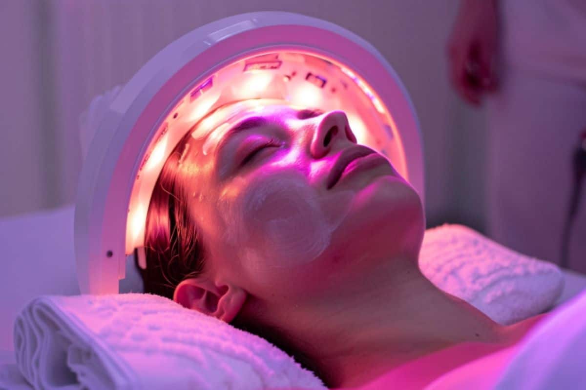
    This shows a woman undergoing light therapy.
    Patients who received low-level light therapy showed a greater change in resting-state connectivity in seven brain region pairs during the acute-to-subacute recovery phase compared to the control participants. Credit: Neuroscience News

    Patients received light therapy within 72 hours of their injuries through a helmet that emits near-infrared light.

    “The skull is quite transparent to near-infrared light,” said study co-lead author Rajiv Gupta, M.D., Ph.D., from the Department of Radiology at MGH. “Once you put the helmet on, your whole brain is bathing in this light.”

    The researchers used an imaging technique called functional MRI to gauge the effects of the light therapy. They focused on the brain’s resting-state functional connectivity, the communication between brain regions that occurs when a person is at rest and not engaged in a specific task.

    The researchers compared MRI results during three recovery phases: the acute phase of within one week after injury, the subacute phase of two to three weeks post-injury and the late-subacute phase of three months after injury.

    Of the 38 patients in the trial, 21 did not receive light therapy while wearing the helmet. This was done to serve as a control to minimize bias due to patient characteristics and to avoid potential placebo effects.

    Patients who received low-level light therapy showed a greater change in resting-state connectivity in seven brain region pairs during the acute-to-subacute recovery phase compared to the control participants.

    “There was increased connectivity in those receiving light treatment, primarily within the first two weeks,” said study coauthor Nathaniel Mercaldo, Ph.D., a statistician with MGH.

    “We were unable to detect differences in connectivity between the two treatment groups long term, so although the treatment appears to increase the brain connectivity initially, its long-term effects are still to be determined.”

    The precise mechanism of the light therapy’s effects on the brain is also still to be determined. Previous research points to the alteration of an enzyme in the cell’s mitochondria (often referred to as the “powerhouse” of a cell), Dr. Gupta said.

    This leads to more production of adenosine triphosphate, a molecule that stores and transfers energy in the cells. Light therapy has also been linked with blood vessel dilation and anti-inflammatory effects.

    “There is still a lot of work to be done to understand the exact physiological mechanism behind these effects,” said study coauthor Suk-tak Chan, Ph.D., a biomedical engineer at MGH.

    While connectivity increased for the light therapy-treated patients during the acute to subacute phases, there was no evidence of a difference in clinical outcomes between the treated and control participants.

    Additional studies with larger cohorts of patients and correlative imaging beyond three months may help determine the therapeutic role of light in traumatic brain injury.

    The researchers expect the role of light therapy to expand as more study results come in. The 810-nanometer-wavelength light used in the study is already employed in various therapeutic applications. It’s safe, easy to administer and does not require surgery or medications.

    The helmet’s portability means it can be delivered in settings outside of the hospital. It may have applications in treating many other neurological conditions, according to Dr. Gupta.

    “There are lots of disorders of connectivity, mostly in psychiatry, where this intervention may have a role,” he said. “PTSD, depression, autism: these are all promising areas for light therapy.”

    About this TBI and neurotech research news

    Author: Linda Brooks
    Source: RSNA
    Contact: Linda Brooks – RSNA
    Image: The image is credited to Neuroscience News

    Original Research: The findings will appear in Radiology.

    Saturday, May 11, 2024

    Near-Infrared II Photobiomodulation Preconditioning Ameliorates Stroke Injury via Phosphorylation of eNOS

     Didn't your competent? doctor figure out a protocol on this 5 years ago? NO? So you DON'T have a functioning stroke doctor? Why are you seeing them?

    I guess a major problem with this it has to be performed before your stroke. You'll need time travel or precognitive dreams about your upcoming stroke.

    Near-Infrared II Photobiomodulation Preconditioning Ameliorates Stroke Injury via Phosphorylation of eNOS

    eNOS

    Originally publishedhttps://doi.org/10.1161/STROKEAHA.123.045358Stroke. 2024;0

    BACKGROUND:

    The current management of patients with stroke with intravenous thrombolysis and endovascular thrombectomy is effective only when it is timely performed on an appropriately selected but minor fraction of patients. The development of novel adjunctive therapy is highly desired to reduce morbidity and mortality with stroke. Since endothelial dysfunction is implicated in the pathogenesis of stroke and is featured with suppressed endothelial nitric oxide synthase (eNOS) with concomitant nitric oxide deficiency, restoring endothelial nitric oxide represents a promising approach to treating stroke injury.

    METHODS:

    This is a preclinical proof-of-concept study to determine the therapeutic effect of transcranial treatment with a low-power near-infrared laser in a mouse model of ischemic stroke. The laser treatment was performed before the middle cerebral artery occlusion with a filament. To determine the involvement of eNOS phosphorylation, unphosphorylatable eNOS S1176A knock-in mice were used. Each measurement was analyzed by a 2-way ANOVA to assess the effect of the treatment on cerebral blood flow with laser Doppler flowmetry, eNOS phosphorylation by immunoblot analysis, and stroke outcomes by infarct volumes and neurological deficits.

    RESULTS:

    Pretreatment with a 1064-nm laser at an irradiance of 50 mW/cm2 improved cerebral blood flow, eNOS phosphorylation, and stroke outcomes.

    CONCLUSIONS:

    Near-infrared II photobiomodulation could offer a noninvasive and low-risk adjunctive therapy for stroke injury. This new modality using a physical parameter merits further consideration to develop innovative therapies to prevent and treat a wide array of cardiovascular diseases.

    Saturday, June 17, 2023

    Exposure to near-infrared light before bed linked to better sleep, daytime function

    Well fuck, we really need someone to write and distribute a protocol on this. All this previous research; OR ARE WE WAITING FOR SOMEONE ELSE TO SOLVE THE PROBLEM?     


    A new treatment protocol using photobiomodulation and muscle/bone/joint recovery techniques having a dramatic effect on a stroke patient's recovery: a new weapon for clinicians Sept. 2012      


    Interplay between up-regulation of cytochrome-c-oxidase and hemoglobin oxygenation induced by near-infrared laser June 2017      


    Photobiomodulation therapy promotes neurogenesis by improving post-stroke local microenvironment and stimulating neuroprogenitor cells Oct. 2017    


    Increased Functional Connectivity Within Intrinsic Neural Networks in Chronic Stroke Following Treatment With Red/Near-Infrared Transcranial Photobiomodulation: Case Series With Improved Naming in Aphasia November 2019

    The latest here which of course NOTHING will happen with.  Your stroke hospital is completely fucking incompetent. 

    The latest here:


    Exposure to near-infrared light before bed linked to better sleep, daytime function

    Key takeaways:

    • Thirty adults were randomized to wear near-infrared light-emitting cervical collar or sham device every other night for 5 weeks.
    • Patients with NIR collar self-reported improvement in sleep-related symptoms.

    INDIANAPOLIS — Transdermal exposure to near-infrared light before bed via a wearable device was linked to improved sleep, relaxation and next-day functionality compared with sham, according to research presented at SLEEP 2023.

    “Given the emerging field of photobiomodulation and its potential neuroprotective and vasodilating effects, this red-light and near-infrared emitting device may be useful if milliwatt power level, dosage and frequency of use are refined,” Kathryn E.R. Kennedy, BS, lead study author and PhD candidate in the department of psychiatry at the University of Arizona, said in a related release.

    Sleep duration, sleep timing, social jetlag and shift work were not associated with fecundability or live birth. Source: Shutterstock
    Results of a randomized, sham-controlled clinical trial featuring a cervical near-infrared light device found that exposure was linked to better sleep, daytime function. Image: Adobe Stock

    Noting therapeutic effects, such as relaxation, with delivery of transdermal near-infrared (NIR) light, Kennedy and colleagues examined the effects of NIR exposure on sleep and daytime functioning when given before bed.

    Their randomized, sham-controlled, 5-week study included 30 individuals, aged 30 to 60 years, who self-reported complaints about sleep but were not diagnosed with a sleep disorder. Following a 2-week baseline period, participants wore a NIR-emitting cervical collar (combined 660 nm, 740 nm, 810 nm and 870 nm) or sham device every other night before bed for 3 weeks.

    Researchers assessed physical symptoms and sleep-related experiences using the Systematic Assessment of Treatment Emergent Effects (SAFTEE) questionnaire. They also examined differences between groups with the Insomnia Severity Index (ISI), daily sleep diaries, Oura rings and daily participant ratings of sleep quality and feeling refreshed, daytime function, energy levels, relaxation and performance.

    According to results, significant differences between active and sham groups were reported in SAFTEE total score (–9.4 vs. +13.8) and sleep-related symptoms (–2.9 vs. +4.1), while no differences were reported in ISI scores, Oura recordings or sleep diary data.

    While most changes were consistent between groups, the active group had decreased sleep latency (–6 mins) and REM% (–0.9%), increased average sleep quality (+0.6 pts) and feeling refreshed (+0.7 pts), and perceived increase in relaxation (17.5 pts) and daytime function (17.5 pts). Conversely, the sham group had decreases in perceived overall performance (18.7 pts) and energy level (26 pts).

    “This novel phototherapy device — while still being explored and in need of further research — appeared to be generally well-tolerated by a small group of participants,” Kennedy said in the release. “Those with active, light-emitting devices — as opposed to the inert sham devices — self-reported an increase in relaxation and better sleep with use.”

    Reference:

    Wednesday, November 13, 2019

    Increased Functional Connectivity Within Intrinsic Neural Networks in Chronic Stroke Following Treatment With Red/Near-Infrared Transcranial Photobiomodulation: Case Series With Improved Naming in Aphasia

    Well fuck, we really need someone to write and distribute a protocol on this. All this previous research; OR ARE WE WAITING FOR SOMEONE ELSE TO SOLVE THE PROBLEM?     


    A new treatment protocol using photobiomodulation and muscle/bone/joint recovery techniques having a dramatic effect on a stroke patient's recovery: a new weapon for clinicians Sept. 2012      


    Interplay between up-regulation of cytochrome-c-oxidase and hemoglobin oxygenation induced by near-infrared laser June 2017      


    Photobiomodulation therapy promotes neurogenesis by improving post-stroke local microenvironment and stimulating neuroprogenitor cells Oct. 2017    

    The latest here which of course NOTHING will happen with.  Your stroke hospital is completely fucking incompetent.

    Increased Functional Connectivity Within Intrinsic Neural Networks in Chronic Stroke Following Treatment With Red/Near-Infrared Transcranial Photobiomodulation: Case Series With Improved Naming in Aphasia November 2019

    Published Online:https://doi.org/10.1089/photob.2019.4630

    Abstract

    Objective: To examine effects of four different transcranial, red/near-infrared (NIR), light-emitting diode (tLED) protocols on naming ability in persons with aphasia (PWA) due to left hemisphere (LH) stroke. This is the first study to report beneficial effects from tLED therapy in chronic stroke, and parallel changes on functional magnetic resonance imaging (fMRI).
    Materials and methods: Six PWA, 2–18 years poststroke, in whom 18 tLED treatments were applied (3 × /week, 6 weeks) using LED cluster heads: 500 mW, red (633 nm) and NIR (870 nm), 22.48 cm2, 22.2 mW/cm2.
    Results: After Protocol A with bilateral LED placements, including midline, at scalp vertex over left and right supplementary motor areas (L and R SMAs), picture naming was not improved. P1 underwent pre-/postovert, picture-naming task-fMRI scans; P2 could not. After Protocol A, P1 showed increased activation in LH and right hemisphere, including L and R SMAs. After Protocol B with LEDs only on ipsilesional, LH side, naming ability significantly improved for P1 and P2; the fMRI scans for P1 then showed activation only on the ipsilesional LH side. After Protocol C with LED placements on ipsilesional LH side, plus one midline placement over mesial prefrontal cortex (mPFC) at front hairline, a cortical node of the default mode network (DMN), P3 and P4 had only moderate/poor response, and no increase in functional connectivity on resting-state functional-connectivity MRI. After Protocol D, however, with LED placements on ipsilesional LH side, plus over two midline nodes of DMN, mPFC, and precuneus (high parietal) simultaneously, P5 and P6 each had good response with significant increase in functional connectivity within DMN, p < 0.0005; salience network, p < 0.0005; and central executive network, p < 0.05.
    Conclusions: NIR photons can affect surface brain cortex areas subjacent to where LEDs are applied on the scalp. Improved naming ability was present with optimal Protocol D. Transcranial photobiomodulation may be an additional noninvasive therapy for stroke.

    Thursday, June 27, 2019

    Interpreting Prefrontal Recruitment During Walking After Stroke: Influence of Individual Differences in Mobility and Cognitive Function

    I would prefer that you interpret this in a useful way. Creating stroke rehab protocols. 

    Interpreting Prefrontal Recruitment During Walking After Stroke: Influence of Individual Differences in Mobility and Cognitive Function

    • 1Brain Rehabilitation Research Center (BRRC), Malcom Randall VA Medical Center, Gainesville, FL, United States
    • 2Department of Physical Therapy, University of Florida, Gainesville, FL, United States
    • 3Brooks Rehabilitation, Jacksonville, FL, United States
    • 4Department of Neurology, University of Florida, Gainesville, FL, United States
    • 5Department of Biostatistics, University of Florida, Gainesville, FL, United States
    • 6Department of Applied Physiology and Kinesiology, University of Florida, Gainesville, FL, United States
    • 7Department of Aging and Geriatric Research, University of Florida, Gainesville, FL, United States
    • 8Geriatric Research, Education and Clinical Center, Malcom Randall VA Medical Center, Gainesville, FL, United States
    Background: Functional near-infrared spectroscopy (fNIRS) is a valuable neuroimaging approach for studying cortical contributions to walking function. Recruitment of prefrontal cortex during walking has been a particular area of focus in the literature. The present study investigated whether task-related change in prefrontal recruitment measured by fNIRS is affected by individual differences in people post-stroke. The primary hypotheses were that poor mobility function would contribute to prefrontal over-recruitment during typical walking, and that poor cognitive function would contribute to a ceiling in prefrontal recruitment during dual-task walking (i.e., walking with a cognitive task).
    Methods: Thirty-three adults with chronic post-stroke hemiparesis performed three tasks: typical walking at preferred speed (Walk), serial-7 subtraction (Serial7), and walking combined with serial-7 subtraction (Dual-Task). Prefrontal recruitment was measured with fNIRS and quantified as the change in oxygenated hemoglobin concentration (ΔO2Hb) between resting and active periods for each task. Spatiotemporal gait parameters were measured on an electronic walkway. Stepwise regression was used to assess how prefrontal recruitment was affected by individual differences including age, sex, stroke region, injured hemisphere, stroke chronicity, 10-meter walking speed, balance confidence measured by Activities-specific Balance Confidence (ABC) Scale, sensorimotor impairment measured by Fugl-Meyer Assessment, and cognitive function measured by Mini-Mental State Examination (MMSE).
    Results: For Walk, poor balance confidence (ABC Scale score) significantly predicted greater prefrontal recruitment (ΔO2Hb; R2 = 0.25, p = 0.003). For Dual-Task, poor cognitive function (MMSE score) significantly predicted lower prefrontal recruitment (ΔO2Hb; R2 = 0.25, p = 0.002).
    Conclusions: Poor mobility function predicted higher prefrontal recruitment during typical walking, consistent with compensatory over-recruitment. Poor cognitive function predicted lower prefrontal recruitment during dual-task walking, consistent with a recruitment ceiling effect. These findings indicate that interpretation of prefrontal recruitment should carefully consider the characteristics of the person and demands of the task.

    Thursday, November 23, 2017

    Blue-Light Therapy following Mild Traumatic Brain Injury: Effects on White Matter Water Diffusion in the Brain

    Would this help stroke patients? I don't think they are referring to the blue light of your smartphones or iPads. Anything would have been helpful to counteract the massive fatigue I had post-stroke.  Or you can ask your doctor about the effectiveness of near-infrared light since it is only two years old and your doctor and stroke hospital are so efficient they already have incorporated infrared into their stroke protocols. (sarcasm)

    New Study Discovers Near-Infrared Light Therapy (NILT) Effectively Treats Traumatic Brain Injury (TBI) Patients  Aug. 2015

    Blue-Light Therapy following Mild Traumatic Brain Injury: Effects on White Matter Water Diffusion in the Brain

    • Social, Cognitive and Affective Neuroscience Laboratory (SCAN Lab), Department of Psychiatry, College of Medicine, University of Arizona, Tucson, AZ, United States
    Mild traumatic brain injury (mTBI) is a common and often inconspicuous wound that is frequently associated with chronic low-grade symptoms and cognitive dysfunction. Previous evidence suggests that daily blue wavelength light therapy may be effective at reducing fatigue and improving sleep in patients recovering from mTBI. However, the effects of light therapy on recovering brain structure remain unexplored. In this study, we analyzed white matter diffusion properties, including generalized fractional anisotropy, and the quantity of water diffusion in isotropic (i.e., isotropic diffusion) and anisotropic fashion (i.e., quantitative anisotropy, QA) for fibers crossing 11 brain areas known to be significantly affected following mTBI. Specifically, we investigated how 6 weeks of daily morning blue light exposure therapy (compared to an amber-light placebo condition) impacted changes in white matter diffusion in individuals with mTBI. We observed a significant impact of the blue light treatment (relative to the placebo) on the amount of water diffusion (QA) for multiple brain areas, including the corpus callosum, anterior corona radiata, and thalamus. Moreover, many of these changes were associated with improvements in sleep latency and delayed memory. These findings suggest that blue wavelength light exposure may serve as one of the potential non-pharmacological treatments for facilitating structural and functional recovery following mTBI; they also support the use of QA as a reliable neuro-biomarker for mTBI therapies.


    Introduction

    Mild traumatic brain injury (mTBI) is a common and often unobtrusive wound that occurs when kinetic energy is transferred to the brain through some form of traumatic event, such as a fall, blow to the head, or blast wave. While there are typically no exceptionally conspicuous physical or neuroimaging signs of mTBI, the mechanical trauma to the brain leads to a mild temporary disruption of consciousness or other alteration of ongoing cognition. Also commonly known as “concussion,” mTBI can further lead to persistent alterations in neuropsychological functions, including changes in mood (e.g., depression), poor attention and concentration, and memory problems (1, 2). Importantly, sleep deprivation is also known to produce many of these same symptoms (3, 4). It is therefore possible that sleep disturbances following mTBI may cause, or at least exacerbate, ongoing post-concussion symptoms. However, the nature of these complaints and their contribution to the experience of daytime sleepiness is not well understood (5). An objective measure of daytime sleepiness is the multiple sleep latency test (MSLT), which is used to determine the time it takes an individual to fall asleep (sleep onset latency) when given the opportunity to take a nap. Following a head trauma, symptoms are believed to result from neuronal damage in the form of diffuse axonal injury (6, 7), leading to the release of specific proteins that in turn promote maladaptive functional and structural changes within the brain (8). Identifying neuro-markers of these changes remains an important challenge in ongoing attempts to understand and treat mTBI and post-concussive symptoms.
    A very limited number of treatment options for mTBI have been proposed and experimentally validated. Available treatments include cognitive behavior therapy (9), neuropsychological rehabilitation (10), educational intervention (11), and pharmacological intervention (12). Although the effects are small, some intervention studies report reliable reductions in post-concussion symptoms, including sleep problems, following successful treatment (13). Considering a range of post-concussion symptoms can also occur as the result of sleep loss, it is likely that improving sleep quality in particular would also lead to improvements of other post-concussion symptoms, such as attention, concentration, memory, and mood disturbances. While improving sleep makes sense, this is often easier said than done. A natural and potentially powerful method for regulating the sleep–wake cycle is through targeted exposure to bright light in the morning hours. Exposure to short wavelength light (~430–475 nm; blue wavelength light) has been demonstrated as an alternative to pharmacological treatment methods that focus on improving alertness, concentration, daytime sleepiness, as well as sleep quality (14, 15). Intrinsically photosensitive retinal ganglion cells are particularly responsive to light within the short wavelengths. These cells transmit signals to hypothalamic nuclei, which in turn regulate the production of melatonin (16, 17). Morning exposure to blue wavelength light leads to a suppression of melatonin production, which contributes to a phase delay and stabilization of the circadian rhythm (18), increases daytime alertness and vigilance, and earlier onset of evening sleep (19, 20). Interestingly, a recent clinical trial showed that 4 weeks of 45 min of morning blue-light therapy (BLT) in comparison to longer wavelength placebo light was effective at reducing self-rated fatigue and daytime sleepiness among individuals recovering from TBI (21). However, the extent to which these behavioral changes correspond to structural changes within the brain has not been explored.
    When considering the potential influences of BLT on mTBI, it is important to consider that mTBI is associated with microscopic changes in brain structure, particularly within the white matter axonal tracts. Abnormalities in fractional anisotropy (FA) in the brain following an mTBI have been studied extensively using diffusion tensor imaging (DTI), a method that allows high-resolution imaging of the directional movement of water molecules along axonal fiber tracts (i.e., how fast water molecules move along fiber tracts). Abnormalities in FA in individuals with an mTBI are reported in areas such as uncinate fasciculus (UF) (22), superior longitudinal fasciculus (SLF) (23), anterior corona radiata (ACR) (22), corpus callosum (CC) (24), and thalamus (25). Alterations in FA within (a) UF are reported to be associated with changes in Mini-Mental State Examination (MMSE) scores (cognitive function) and specifically, memory performance (22, 26); (b) SLF and CC are reported to be associated with executive function (attention and memory) (27); (c) ACR changes are correlated with changes in attention (22); and (d) anterior thalamic nucleus changes are also linked to changes in executive function, memory, and attention (25). In addition, studies have found that individuals with mTBI show alterations in white matter within the frontal lobe (frontal cortex/dorsolateral prefrontal cortex, DLPFC), and that these alterations are correlated with lower executive control and related cognitive functions (28). Also, compared to healthy controls (HCs), there are multiple studies that have reported abnormally high FA values in individuals with mTBI within several areas, including the genu and splenium of CC, ACR (bilaterally), lUF, and internal capsule (IC; bilaterally) (29, 30). Recently, new diffusion measures—quantitative anisotropy (QA), isotropic diffusion (ISO), and generalized fractional anisotropy (GFA)—were introduced to the field of DTI for the analysis of diffusion properties of white matter (31). QA and ISO represent how much water diffuses (i.e., density) in a specific/restricted direction and in an isotropic fashion (i.e., total isotropic component), respectively. In contrast, GFA, which is calculated from an orientation distribution function, is a measure of how fast water diffuses (i.e., diffusivity) in an anisotropic fashion, i.e., it represents degree to which diffusion is anisotropic (31, 32). Highly significant correlations between FA and GFA were reported in the past (33). In addition, the difference between QA and GFA pertains to the fact that QA is a measure of water diffusion along each fiber orientation, whereas GFA/FA is defined for each voxel. Compared to GFA/FA, QA is also reported to have lower susceptibility to partial volume effects of crossing-fibers, free-water diffusion in ventricles, and non-diffusive particles (31). Moreover, normalization of QA helps to stabilize the spin-density measurement across subjects. In this study, we investigated multiple diffusion measures (i.e., diffusivity as well as density measures) simultaneously to better characterize the white matter properties; therefore, in conjunction with GFA, we also estimated normalized QA (NQA) and ISO measures. To the best of our knowledge, no study to date has used these metrics simultaneously to examine the effect of light exposure treatment on the brain following mTBI.
    In individuals with mTBI, how changes in post-concussion symptoms following an exposure to BLT may correspond to structural changes within the brain has not yet been explored. Recent evidence suggests that sleep is important for clearing the neurotoxins that build up throughout the day (34) and increases the production of oligodendrocyte progenitor cells that contribute to myelin formation (35), which could conceivably facilitate repair of axonal damage. Based on this, we hypothesized that 6 weeks of daily morning BLT, compared to a placebo condition with an amber-light therapy (ALT) device, would improve sleep and, consequently, lead to changes in white matter water diffusion, improvements in cognitive abilities such as attention and memory, and daytime sleepiness. To this end, we investigated whether individuals in the BLT and ALT groups showed significant changes in diffusion (i.e., GFA, NQA, and ISO), cognitive, and sleep measures. Furthermore, we examined the correlations between changes in diffusion measures from pre- to post-treatment and changes in neuropsychological performance and sleep onset latency.

    Friday, June 2, 2017

    Interplay between up-regulation of cytochrome-c-oxidase and hemoglobin oxygenation induced by near-infrared laser

    Sounds interesting, talking about oxygen supply and blood volume, although I could see significant problems getting through the skull to actually affect the brain itself. But that is for your doctor and researchers to solve. 

    Interplay between up-regulation of cytochrome-c-oxidase and hemoglobin oxygenation induced by near-infrared laser


    Abstract

    Photobiomodulation, also known as low-level laser/light therapy (LLLT), refers to the use of red-to-near-infrared light to stimulate cellular functions for physiological or clinical benefits. The mechanism of LLLT is assumed to rely on photon absorption by cytochrome c oxidase (CCO), the terminal enzyme in the mitochondrial respiratory chain that catalyzes the reduction of oxygen for energy metabolism. In this study, we used broadband near-infrared spectroscopy (NIRS) to measure the LLLT-induced changes in CCO and hemoglobin concentrations in human forearms in vivo. Eleven healthy participants were administered with 1064-nm laser and placebo treatments on their right forearms. The spectroscopic data were analyzed and fitted with wavelength-dependent, modified Beer-Lambert Law. We found that LLLT induced significant increases of CCO concentration (Δ[CCO]) and oxygenated hemoglobin concentration (Δ[HbO]) on the treated site as the laser energy dose accumulated over time. A strong linear interplay between Δ[CCO] and Δ[HbO] was observed for the first time during LLLT, indicating a hemodynamic response of oxygen supply and blood volume closely coupled to the up-regulation of CCO induced by photobiomodulation. These results demonstrate the tremendous potential of broadband NIRS as a non-invasive, in vivo means to study mechanisms of photobiomodulation and perform treatment evaluations of LLLT.
    PMID:
    27484673
    PMCID:
    PMC4971496
    DOI:
    10.1038/srep30540

    Wednesday, May 11, 2016

    Near-Infrared Light Regenerates Damage From Traumatic Brain Injury, Latest of Five Studies Show

    But does it actually work or just a placebo?

    Stroke Rounds: Light Therapy Flopped in Acute Stroke

    near-infrared laser energy to treat ischemic stroke

    Wavelength-dependent penetration depth of near infrared radiation into cartilage


    http://www.prnewswire.com/news-releases/near-infrared-light-regenerates-damage-from-traumatic-brain-injury-latest-of-five-studies-show-300264268.html
    A revolutionary non-invasive brain treatment invokes a powerful restorative impact on TBI, the latest science published this month demonstrates. This new science harnesses the power of light - near-infrared light. High powered or multi-watt coherent infrared light, or "neuro-laser" safely reaches into the brain and activates numerous brain reparative processes, including the production of brain-derived neurotrophic factor, the brain's own repair system.
    Theodore Henderson, MD, PhD, authored the study Multi-watt near-infrared light therapy as a neuroregenerative treatment for traumatic brain injury, which was published May 3, 2016 in the Neural Regeneration Research, and is cited in the National Library of Congress (Pubmed).
    "Only multi-watt neuro-laser has sufficient energy to penetrate skin and skull, reaching the deep parts of the brain where injuries are, with no irritation to the skin or other side effects," said Dr. Henderson, who has co-authored five related studies in the last year outlining the efficacy of the procedure and the biological mechanisms of the light (see 5 Recent Studies Show NILT Helps TBI).  "The patients are responding very positively, and our data is consistently showing robust responses. This treatment is really helping turn lives around. The stories we're hearing are amazing."
    The new treatment is spearheaded by Drs. Henderson and Larry Morries, co-Founders of the Neuro-Laser Foundation. They are on the forefront of studying the effectiveness of treating TBI patients with a specialized method referred to as NILT.
    With millions of TBI survivors, and various other possible applications for NILT, this is one of the hottest topics in neuro-science today, according to Dr. Henderson. Their recent work has also revealed NILT may have promise for treating depression.
    Former police officer Jennifer Fortezzo is one example. After retiring from the police force due to injury, the after-effects of numerous traumatic brain injuries began taking a serious toll on her life. Beleaguered with daily suicidal thoughts, she sought solutions to what is largely considered to be an "untreatable" condition. She began NILT treatments with Dr. Henderson in March. Just a few months later, today Fortezzo has joined the "Say Goodbye TBI" campaign and reports, "It's been very freeing for me. I felt for so many years like I was in bondage. And this is the first time I feel free."
    "The need is tremendous. We suspect there are many other ways NILT could help the brain heal," said Dr. Morries, who together with Dr. Henderson, is seeking $2.5 million through the "Say Goodbye TBI" campaign for research and for financial aid to provide treatment for military veterans and first-responders.
    The study also highlighted that the required equipment and low number of applications shown to be effective point to potential for wide dissemination, as it has also been found to be safe in the hands of properly trained professionals. The method, currently in the patent process, utilizes unique infrared laser instruments developed to Drs. Morries and Henderson's specifications.
    The Neuro-Laser Foundation (NLF) is a non-profit organization, based in Denver, Colorado, dedicated to transforming lives of those distraught with various psychiatric and neurological conditions, some resulting from traumatic brain injury (TBI). Building on more than 30 years of studying the effects of near-infrared light on cells and tissues, the Foundation is advancing technology and treatment approaches that will increase quality of life factors for people impacted by traumatic brain injury.
    More information is available at TBI.care or calling (720) 493-1101.
    Media Contact: David Jahr, (949) 874-2667, Email
    SOURCE Neuro-Laser Foundation