Use the labels in the right column to find what you want. Or you can go thru them one by one, there are only 33,991 posts. Searching is done in the search box in upper left corner. I blog on anything to do with stroke. DO NOT DO ANYTHING SUGGESTED HERE AS I AM NOT MEDICALLY TRAINED, YOUR DOCTOR IS, LISTEN TO THEM. BUT I BET THEY DON'T KNOW HOW TO GET YOU 100% RECOVERED. I DON'T EITHER BUT HAVE PLENTY OF QUESTIONS FOR YOUR DOCTOR TO ANSWER.
Changing stroke rehab and research worldwide now.Time is Brain!trillions and trillions of neuronsthatDIEeach day because there areNOeffective hyperacute therapies besides tPA(only 12% effective). I have 523 posts on hyperacute therapy, enough for researchers to spend decades proving them out. These are my personal ideas and blog on stroke rehabilitation and stroke research. Do not attempt any of these without checking with your medical provider. Unless you join me in agitating, when you need these therapies they won't be there.
What this blog is for:
My blog is not to help survivors recover, it is to have the 10 million yearly stroke survivors light fires underneath their doctors, stroke hospitals and stroke researchers to get stroke solved. 100% recovery. The stroke medical world is completely failing at that goal, they don't even have it as a goal. Shortly after getting out of the hospital and getting NO information on the process or protocols of stroke rehabilitation and recovery I started searching on the internet and found that no other survivor received useful information. This is an attempt to cover all stroke rehabilitation information that should be readily available to survivors so they can talk with informed knowledge to their medical staff. It lays out what needs to be done to get stroke survivors closer to 100% recovery. It's quite disgusting that this information is not available from every stroke association and doctors group.
Showing posts with label Montreal Cognitive Assessment. Show all posts
Showing posts with label Montreal Cognitive Assessment. Show all posts
Once again, useless predictions of impairment rather than delivering EXACT RECOVERY PROTOCOLS that get survivors recovered! I'd have you all fired for incompetence!
Let's see how long you've been incompetent in not solving this post stroke cognitive impairment problem! Which also proves the board of directors' incompetence for every hospital that isn't solving this problem!
Post-stroke cognitive impairment (PSCI) significantly affects stroke survivors. Identifying modifiable risk factors for PSCI is essential. Serum multi-trace elements are crucial for neurological function but vary in concentration among older adults. It remains unclear whether increasing multi-trace elements can reduce the incidence of PSCI. We investigated the associations between baseline serum multi-trace elements and PSCI. The Montreal Cognitive Assessment defined PSCI. We used logistic regression analyses to evaluate the association between serum multi-trace elements and PSCI. Subsequently, we assessed the associations between serum multi-trace elements and three different cognitive domains using the Kruskal–Wallis test. We further evaluated improvements in the predictive ability of serum multi-trace elements. Finally, 626 patients (mean age: 62.85 ± 7.54 years) were followed up for a median of 1.2 years. Lower concentrations of serum iron (odds ratio [OR] = 2.498, 95% confidence interval [CI]: 1.505–4.145) and zinc (OR = 2.015, 95% CI: 1.233–3.293) were associated with a higher PSCI risk. Higher concentrations of serum iron (OR = 0.368, 95% CI: 0.227–0.595) and magnesium (OR = 0.273, 95% CI: 0.164–0.454), along with lower concentrations of serum copper (OR = 0.544, 95% CI: 0.34–0.872), were significantly correlated with a lower PSCI risk. Cognitive impairments varied across multi-trace elements. Serum iron affected wider cognition, while magnesium and copper levels were strongly associated with language and executive function. Adding serum multi-trace elements to the conventional model improved PSCI risk reclassification (area under curve: 0.676–0.718). Multi-trace elements may influence PSCI progression. This study was registered with the Chinese Clinical Trial Registry (URL: https://www.chictr.org.cn/; unique identifier: ChiCTR1900022675).
It it your doctor's COMPLETE RESPONSIBILITY TO GET YOU RECOVERED so you don't do this. You doctor needs to solve the problem instead of just telling you not to lie in bed. That would be a sign of an incompetent doctor! NO solutions!
After a mild stroke or transient ischaemic attack (TIA), longer in-bed time and sleep duration were linked to greater small vessel disease burden and poorer cognitive performance,
underscoring the potential role of sleep patterns as modifiable risk
factors for brain health after a stroke, according to a study published
in the journal Neurology.
“These results show that
disturbed sleep may be a marker of adverse brain health, even for people
with mild strokes or TIAs,” said Joanna M. Wardlaw, MD, University of
Edinburgh, Edinburgh, United Kingdom. “While many people know that a
lack of sleep can lead to health issues, less is known about the effects
of sleeping longer at night or spending a long time in bed trying to
make up for having trouble sleeping --whether people are doing this
consciously or not.”
The study involved 422 people (65.6 ± 11.8
years; 67% male) from Edinburgh and Hong Kong with an average age of 66
years who had a mild stroke or a TIA (NIHSS <7). Within 1 to 3 months
after the stroke, cerebral small vessel disease
was assessed on MRI, cognitive performance was assessed using Montreal
Cognitive Assessment (MoCA), and sleep quality was analysed using a
structured sleep questionnaire at baseline visit.
Longer in-bed time was independently associated with greater global small vessel disease and Fazekas periventricular white matter hyperintensity
burden, and with lower total MoCA score after covariate adjustment.
Longer sleep duration was independently associated with presence of cerebral microbleeds.
“More
research is needed to confirm these findings and also to look at
whether prolonged sleep has negative effects on people who have never
had a stroke or TIA,” Dr. Wardlaw said. “Of course, research is also
needed on whether improving people’s sleep patterns after stroke could
ward off some of these possible detrimental effects.”
The
researchers noted that their results should be interpreted with caution
because they only examined baseline cross-sectional data, which cannot
establish causal relationships.
“Some relationships between sleep,
small vessel disease, and cognition may differ in a longitudinal
context and at different time points after stroke,” they stated.
Nothing here even remotely suggests how to make neuroplasticity repeatable on demand.
We don't SPECIFICALLY know why a neuron gives up its' current job and takes on a neighbors.
Thus nothing on neuroplasticity is scientifically repeatable on demand. So, DEMAND your doctor give you EXACT PROTOCOLS to use. Don't allow your doctor to give you generalities or guidelines.
Department of Physical Therapy, Akhmim Hospital, Sohag 82749, Egypt
2
Department of Physical Therapy for Neurology, Faculty of Physical Therapy, Cairo University, Giza 12613, Egypt
3
Department of
Rehabilitation Sciences, College of Health and Rehabilitation Sciences,
Princess Nourah bint Abdulrahman University, P.O. Box 84428, Riyadh
11671, Saudi Arabia
4
Physical Therapy Program, Batterjee Medical College, Jeddah 21442, Saudi Arabia
5
Basic Science for Physical Therapy, Faculty of Physical Therapy, Cairo University, Giza 12613, Egypt
6
Department of Neurology and Clinical Neurophysiology, Faculty of Medicine, Cairo University, Giza 12613, Egypt
*
Author to whom correspondence should be addressed.
Background: Stroke is a primary
cause of adult disability and often causes cognitive impairment.
Rehabilitation interventions aim to enhance patients’ cognitive
abilities, thereby addressing care needs, improving quality of life, and
optimizing performance in compromised functions.
Objective: To
evaluate the impact of incorporating cognitive–behavioral training (CBT)
into a selected exercise program on cortical reorganization and
cognitive recovery in post-stroke patients.
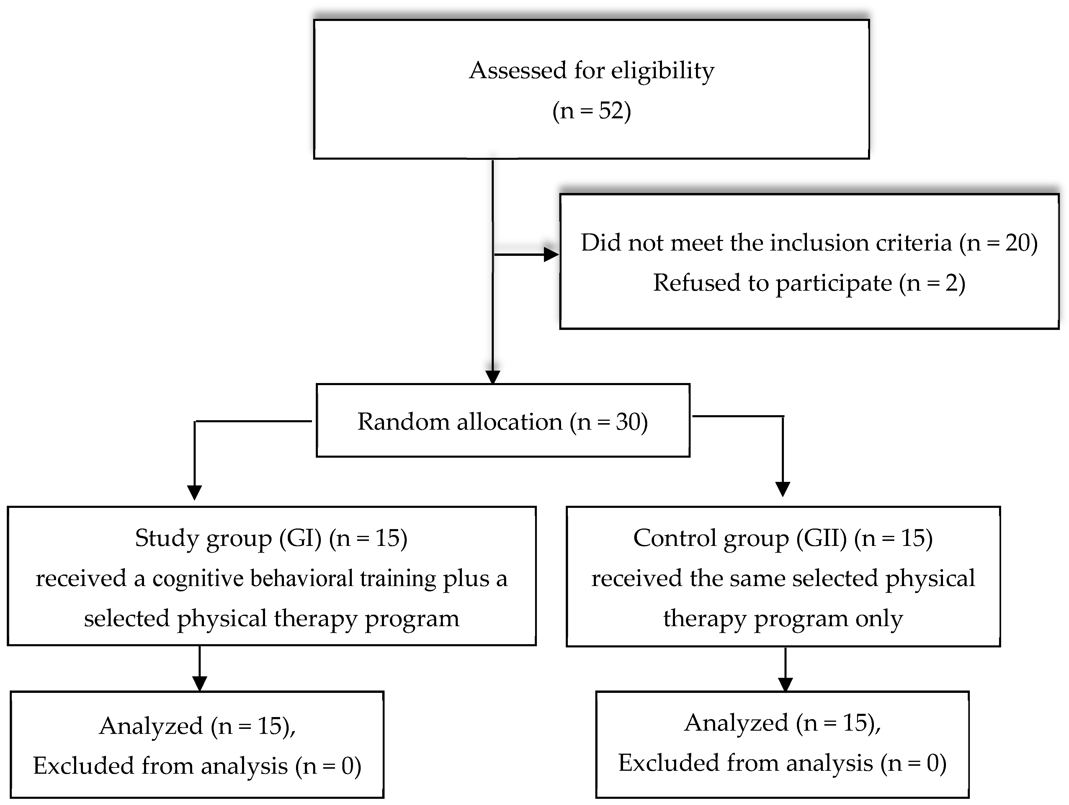
Methods: Thirty
post-stroke patients of both sexes (27 male and 3 female) aged from 40
to 65 years were randomly divided into two groups: the study group (n =
15) received CBT combined with a selected exercise program including
weight-bearing, balance, and aerobic exercises, while the control group
(n = 15) underwent the selected exercise program only. All participants
engaged in an 8-week intervention with three sessions per week. Cortical
reorganization was measured using quantitative electroencephalography
(QEEG) at electrode sites F3, F4, T5, and T6, and cognitive function was
assessed using the Montreal Cognitive Assessment (MoCA) and RehaCom,
focusing on memory, attention, concentration, logical reasoning, and
reaction behavior. Assessments were carried out for all patients before
and after the 8-week treatment program.
Results: Improvements
were assessed through three key measures: QEEG, the MoCA, and RehaCom.
Post-intervention, the study group demonstrated a significantly higher
(alpha + beta)/(delta + theta) ratio at F3, F4, T5, and T6 (p
< 0.01), indicative of enhanced cortical reorganization. MoCA scores
increased by 16.98% in the study group compared to 7.40% in the control
group (p < 0.01). Additionally,
RehaCom assessments revealed marked improvements in memory, attention,
logical reasoning, and reaction behavior in the study group (p < 0.01).
Conclusions:
Integrating cognitive–behavioral training with a selected exercise
program significantly enhances cortical reorganization and cognitive
recovery in post-stroke patients. These findings suggest that adding CBT
to rehabilitation protocols can effectively address deficits in memory
and attention, ultimately improving functional outcomes.
Because
of the rising incidence and decreased mortality associated with stroke,
post-stroke cognitive impairment (PSCI) is becoming more common in
people after stroke [1].
Cognition encompasses the brain’s core functions for processing,
storing, retrieving, and manipulating the information necessary for
problem-solving. After a stroke, up to 55% of patients experience
deficits in episodic memory, 40% experience executive function
impairment, 23% show deficits in language, and 70% suffer some cognitive
decline, all of which affect their functional abilities, work
performance, and capacity for independent living [2]. PSCI significantly affects independence and the ability to return to work [3].
Memory,
learning, and attention problems can have a substantial impact on a
stroke survivor’s functional independence, and multiple studies have
found that higher levels of cognitive impairment are linked to lower
self-reported quality of life [4].
These associations have prompted significant efforts to identify
effective treatments to improve cognitive function following a stroke [5].
Computerized cognitive training consists of organized exercises on standardized, mentally stimulating tasks [6],
offering several benefits compared to traditional drill-and-practice
approaches. These advantages include engaging visual interfaces,
efficient and scalable delivery, and the ability to continuously adjust
training content and difficulty based on individual performance [7].
The
RehaCom software package offers a comprehensive approach to cognitive
assessment and rehabilitation. This evidence-based tool integrates three
core therapeutic strategies: enhancing patients’ understanding of
cognitive processes, boosting motivational aspects, and developing
compensatory techniques and adaptive skills to manage cognitive deficits
[8].
Neurorehabilitation
aims to directly quantify brain damage healing through the use of
trustworthy, objective, and interpretable measurements of
neuroplasticity or changes in brain function [9].
Because it measures cortical activity and reflects the brain’s
spatiotemporal information, quantitative electroencephalography (QEEG)
is a popular tool for developing assistive rehabilitation devices and
evaluating neurophysiological responses to rehabilitation interventions.
QEEG is also a non-invasive and easy way to record brain activity. QEEG
signals are recorded from four standard frequency bands, alpha (8–12
Hz), beta (12–30 Hz), theta (4–8 Hz), and delta (1–4 Hz) waves,
providing valuable insights into cortical brain activity [10].
It is cheap, easy, and nearly risk-free when compared to other brain
imaging methods. It offers electrophysiological information that is not
available from other imaging modalities or clinical evaluations, and it
has a high temporal resolution. Additionally, without requiring the
patient to cooperate, QEEG allows doctors to objectively measure brain
function and conduct real-time brain evaluations [11].
Several
studies have demonstrated the efficacy of cognitive–behavioral training
(CBT) in improving cognitive functions such as memory, attention, and
executive function in stroke patients. CBT facilitates neuroplasticity
by modulating neural oscillations and strengthening synaptic
connections, thereby enhancing cortical reorganization. Research has
also shown that cognitive training interventions improve functional
brain connectivity and contribute to cognitive recovery in stroke
populations [12,13,14].
Compared to other cognitive rehabilitation approaches, CBT offers a
structured and adaptive method that targets specific cognitive domains
essential for post-stroke recovery [15,16].
Despite
promising outcomes reported in prior studies, the neurophysiological
mechanisms by which cognitive–behavioral training (CBT) enhances
cognitive recovery in post-stroke patients remain underexplored. In
particular, there is limited evidence regarding how CBT influences
cortical reorganization using objective neurophysiological measures.
This study addresses this gap by combining CBT with a selected exercise
to elucidate its impact on neural oscillatory activity and cognitive
function in chronic post-stroke patients. Based on the existing
literature, the aims of the study are to evaluate the additive effect of
CBT when integrated with a selected exercise program in improving
post-stroke cognitive impairment (PSCI) and investigate the neuroplastic
changes associated with these interventions using
electroencephalography (EEG). We hypothesize that adding
cognitive–behavioral training using computerized cognitive training to a
selected exercise program will lead to greater improvements for
post-stroke patients in terms of cortical reorganization, as evidenced
by enhanced QEEG indices, and superior cognitive recovery, as reflected
in increased MoCA and RehaCom scores, compared to the selected physical
therapy program alone.
Remotely supervised (RS) transcranial direct current stimulation
(tDCS) appears to be viable and safe for post-stroke cognitive
rehabilitation in chronic stroke patients with moderate cognitive
decline, as reported in a study.
The study included 26 patients with chronic stroke and cognitive
impairment (Korean version of the Montreal Cognitive Assessment [K-MoCA]
score <26). They were randomly assigned to groups that received real
or sham RS-tDCS groups with concurrent computerized cognitive training.
TDCS was applied by patients and caregivers themselves. A training
was conducted beforehand to ensure correct application. Treatment was
conducted 5 days a week for 4 weeks.
Researchers evaluated several cognition tests including K-MoCA,
Korean version of the Dementia Rating Scale-2, Korean-Boston Naming
Test, Trail Making Test, Go/No Go, and Controlled Oral Word Association
Test at the end of the training sessions and 1 month later. They used
repeated-measures ANOVA to compare the outcomes between the groups and
within each group. The adherence rate of the appropriate RS-tDCS session
was also assessed.
Within-group comparisons showed that the real group, but not the sham
group, achieved significant improvement in K-MoCA (p=0.004 and p=0.132,
respectively). The treatment effect of RS-tDCS was more pronounced in
patients with lower baseline K-MoCA (K-MoCA 10–17: p=0.001 in the real
group vs p=0.835 in the sham group; K-MoCA 18–25: p=0.060 vs p=0.064,
respectively), as well as those with left hemispheric lesions (left:
p=0.010 vs p=0.454; right: p=0.106 vs p=0.128).
Comparison between groups likewise revealed a significant
between-group difference in K-MoCA in the lower baseline K-MoCA
subgroup, in favour of the real RS-tDCS intervention (K-MoCA 10–17: ptime×group=0.048). This difference was not seen in other cognitive tests.
Successful application of RS-tDCS had a high adherence rate at 98.4 percent, and no serious adverse effects were documented.
Well shit, at age 50, when I stroked, my right carotid artery was 80% blocked, then 3 years later had completely closed up. 13 years post stroke collateral arteries went around it. I am 100% certain that I have no cognitive impairment 14 years post stroke. Arrogance is not a cognitive impairment on my part.
Researchers
explored the long-term predictive as well as interaction impacts of
structural and functional carotid atherosclerosis markers on cognitive
decline in future. They analyzed 528 middle-aged individuals, who got
enrolled in the carotid atherosclerosis examination in Kaohsiung
Atherosclerosis Longitudinal Study between 2006-2009. These individuals
were examined for cognition between 2016-2019. Low 10-year Montreal
Cognitive Assessment (MoCA) scores with p < 0.001 and p = 0.03,
respectively, were noted in relation to each case of advanced structural
atherosclerosis and advanced functional atherosclerosis. Experts found
an interaction impact between structural and functional atherosclerosis
on the MoCA score 10 years later. They concluded that carotid
atherosclerosis in middle-aged persons can serve as a predictor of their
cognitive function in 10 years. An improved predictive power for
cognitive decline could be achieved with the help of combined
information about both arterial wall and stiffness.
And what EXACTLY are you doing so ALL GROUPS get functional gains to 100% recovery? Yes, that will be difficult, but leaders tackle and solve difficult problems. Are you a leader or a mouse? Leave no survivor behind. I absolutely hate recovery predictions because they mean useful research was not done and they don't help one whit in getting survivors recovered. Until stroke leadership understands that survivors will be screwed.
Study validated subgroups of cognitive impairment on the Montreal
Cognitive Assessment (MoCA)-defined as normal (score of 25-30), mildly
impaired (score of 20-24), and moderately impaired (score less than
19)-by determining whether they differ in rehabilitation gain during
inpatient stroke rehabilitation. Linear regression models were conducted
and predictors included MoCA subgroups and relevant baseline
demographic and clinical covariates. Separate models included the
cognitive subscale of the Functional Independence Measure (FIM)
instrument as a predictor. Participants were 334 patients with
mild-moderate strokes who were administered the MoCA on admission to the
inpatient rehabilitation facility of an urban, academic medical center.
Outcome variables included the mean relative FIM gain (mRFG), which
quantifies the amount of functional gain achieved as a percentage of the
total functional gain possible, and mean relative functional efficiency
(mRFE), which adjusts for length of stay) on the FIM total. MoCA
subgroups significantly predicted mRFG and mRFE after accounting for
age, sex, education, stroke severity, and recurrent vs first stroke. The
normal group exhibited greater mRFG and mRFE than the mildly impaired
group, while the moderately impaired group had significantly worse mRFG
and mRFE than the mildly impaired group. The moderately impaired group
had a significantly smaller proportion of individuals who made a
clinically meaningful change on the total-FIM than the mildly impaired
and normal groups. MoCA subgroups better accounted for mRFG and mRFE
than a standard-of-care cognitive assessment (cognitive-FIM). Use of
MoCA-defined subgroups can assist providers in predicting(There is that useless term again.) functional
gain in survivors of stroke being treated in inpatient rehabilitation.
Descriptor Terms:
CLIENT CHARACTERISTICS, COGNITIVE DISABILITIES, DEMOGRAPHICS,
FUNCTIONAL EVALUATION, MEASUREMENTS, OUTCOMES, PERFORMANCE STANDARDS,
REHABILITATION, STROKE
Citation: Jaywant, Abhishek, Toglia, Joan, Gunning, Faith M., O'Dell, Michael W.. (2020). Subgroups defined by the Montreal cognitive assessment differ in functional gain during acute inpatient stroke rehabilitation.
Archives of Physical Medicine and Rehabilitation, 101(2), Pgs. 220-226. Retrieved 4/17/2020, from REHABDATA database.
What are you doing to change that trajectory to full recovery? Or is this your 'get out of jail free' card to allow you to do nothing? I care absolutely nothing about these disability predictions and you should scream in their faces for anyone using them. It means they have given up on your recovery.
1Institute of Neuroscience and Physiology, Rehabilitation Medicine, University of Gothenburg, Gothenburg, Sweden
2Centre for Person-Centred Care (GPCC), University of Gothenburg, Gothenburg, Sweden
Introduction: After a stroke, cognitive
impairment is commonly associated with poor functional outcomes. The
primary aim of this study was to investigate if cognitive function,
assessed with the Montreal Cognitive Assessment (MoCA) 36–48 h after
stroke, could predict functional dependence 3 months later. The
secondary aim was to identify an optimal threshold for the MoCA score
that could predict functional dependence.
Materials and Methods: This was a
longitudinal cohort study. The research database from a stroke unit at
the Sahlgrenska University Hospital was linked with the Swedish Stroke
Register—Riksstroke. Cognitive function and activities of daily living
(ADL) were assessed with the MoCA and the Barthel Index (BI),
respectively, 36–48 h after stroke. Functional outcome 3 months after
stroke was studied with the modified Rankin Scale. The predictive
characteristics of the MoCA were investigated using logistic regression
analyses. Receiver operating characteristic curves (AUC) were used for
identifying the optimal cutoff score on the MoCA for predicting
functional dependence. The MoCA score that had equal sensitivity and
specificity was chosen as the optimal score for predicting functional
dependence.
Results: A total of 305 participants were included in the study (mean age: 68.8 years, n
= 179 men). The MoCA quartiles were a significant predictor of
functional dependence 3 months after stroke as an individual variable (p
< 0.001, AUC = 0.72) and when adjusted for covariates such as age at
stroke onset, living arrangement prior to stroke, and ADL measured with
BI within 36–48 h after stroke (p = 0.01, AUC = 0.84). The MoCA
score of ≤ 23 for impaired cognition had equal sensitivity and
specificity for predicting functional dependence 3 months after stroke.
Discussion and Conclusion: Cognitive
function assessed with the MoCA within 36–48 h after stroke could
predict functional dependence 3 months later. The participants with MoCA
scores ≤ 23 for impaired cognition were more likely to be functionally
dependent.
In the initial study data establishing the MoCA, normal controls had an
average score of 27.4, compared with 22.1 in people with mild cognitive
impairment (MCI) and 16.2 in people with Alzheimer's disease.
NO, NO, NO you blithering lazy bastards. We want protocols that get us 100% recovered, NOT this prediction crap that allows you to not even try for better rehab. DAMN YOU ALL TO HELL.
You need to scream at your doctor for being incompetent if you get this test. Your doctor should be giving you protocols for recovery not this, 'Well, according to this test, you'll only recover so much. No point in giving you much rehab.' Hell, why not just figure out the 10% of survivors who fully recover and throw up your hands in defeat for that other 90%? It would be faster and you could save the insurance companies vast amounts of money. .
A
simple test taken within 1 week of a stroke may help predict how well
people will have recovered up to 3 years later, according to a study
published in Neurology.
“We found that this test, which takes less than 10 minutes, can help
predict whether people will have impaired thinking skills, problems that
keep them from performing daily tasks such as bathing and dressing, and
even whether they will be more likely to die,” said Martin Dichgans,
MD, Ludwig-Maximilians University, Munich, Germany. “This test should be
used to screen people with stroke and to counsel them and their
families about long-term prognosis and also to identify those who would
most benefit from interventions that could improve their outcomes.”
For the study, 274 people in Germany and France who had a stroke were
given the Montreal Cognitive Assessment within 1 week of the stroke.
They were then divided into 2 groups: those with no problems with
thinking and memory skills and those with cognitive impairment. The
participants were tested for their thinking and memory skills, motor
functioning, and ability to complete daily living tasks 6 months later
and then at 1 and 3 years after the stroke.
The study found that those who had thinking problems within 1 week of
the stroke were 7 times more like to die during the 3 years of the
study than those who did not have thinking problems. The survival rate
for those with thinking problems after 3 years was 83%, while the rate
was 97% for those with no thinking problems early on.
Those with thinking problems on the first test were also 5 times more
likely to have problems with their motor skills than those who did not
have thinking problems early on. By 3 years after the stroke, 29% of
those with thinking problems on the first test had problems with their
motor skills, compared with 5% of those who did not have thinking
problems early on.
Those with cognitive impairment were more than twice as likely to
have problems completing their daily activities such as bathing and
dressing, with 42% having problems compared with 13% 3 years after the
stroke.
Those with cognitive impairment were 5 times more likely to continue
having thinking problems 3 years after the stroke than the other group.
Dr. Dichgans noted that the test helped predict outcomes even when
other factors such as the severity of the stroke were taken into
account.
A limitation of the study was that most of the people involved had
relatively mild strokes, so more research is needed to determine whether
the results apply to people with more severe strokes.
Reference: http://n.neurology.org/lookup/doi/10.1212/WNL.0000000000006506
SOURCE: American Academy of Neurology
You'll have to aggressively go after your doctor to get you back up to your baseline cognition rather than accepting the tyranny of low expectations your doctor is trying to get you to accept. Don't settle for anything less than 100% recovery. Screaming may be required, your doctor should feel embarrassed that they KNOW NOTHING on how to get you back to 100%.
When I was given the orientation portion(date, month, year, day, place, and city.) after spending 3 days in the ER and step down units, I failed completely.
Low MoCA scores post-stroke tied to cognitive and functional impairment 3 years later
by Judy George, Contributing Writer, MedPage Today
This article is a collaboration between MedPage Today® and:
Action Points
Early administration of
the Montreal Cognitive Assessment (MoCA) to patients with stroke
predicted cognitive and functional outcome out to 3 years in two
prospective European cohort studies.
While the populations
studied tended to have milder strokes, the authors concluded that the
ease of administering this assessment tool could help to identify stroke
patients that might benefit from rehabilitation strategies.
Early
post-stroke test scores on the Montreal Cognitive Assessment (MoCA)
predicted long-term cognitive and functional outcomes, a pooled analysis
of two European studies found.
Patients assessed with a MoCA score of <26 within 7 days of stroke
were five times more likely to have cognitive impairment in at least
one cognitive domain and functional impairment 3 years later, reported
Martin Dichgans, MD, of Ludwig-Maximilians University in Munich,
Germany, and co-authors, writing online in Neurology.
"This
test should be used to screen people with stroke and to counsel them
and their families about long-term prognosis and also to identify those
who would most benefit from interventions that could improve their
outcomes," Dichgans said in a statement.(Fucking tyranny of low expectations here.)
While MoCA has been used to evaluate cognitive function
in different stroke settings, this study set out to investigate whether
administering the test within 7 days after stroke predicted long-term
outcomes independently from pre-morbid cognitive status, demographic
characteristics, or stroke severity.
The analysis leveraged data from two prospective hospital-based
studies that were planned in parallel. In total, researchers studied 274
stroke patients: 125 patients from the DEDEMAS (Determinants of Dementia After Stroke) study in Germany and 149 patients from STROKDEM
(Study of Factors Influencing Post Stroke Dementia) in France. Patients
were enrolled from 2010 to 2014, and over 95% had ischemic strokes. The
median admission NIH Stroke Scale (NIHSS) score in the cohorts was 2,
indicating a large proportion of patients with minor stroke (only 75
patients had NIHSS scores >3).
MoCA was administered within 7 days after stroke symptom onset, with
cognitive impairment defined by a MoCA score <26. All patients had a
comprehensive evaluation of cognitive and functional outcome in
face-to-face interviews during follow-up visits at 6, 12, and 36 months
after stroke. Analyses were adjusted for age, sex, education, history of
hypertension and diabetes mellitus, baseline Informant Questionnaire on
Cognitive Decline in the Elderly (IQCODE) score, and NIHSS score at
admission.
About 43% of patients in DEDEMAS and 44% in STROKDEM had a MoCA score
<26. Pooled analyses showed that a baseline MoCA score <26 was
associated with cognitive impairment at 3-year follow-up, defined by
neuropsychological testing (OR 5.30, 95% CI 2.75–10.22) and by a
Clinical Dementia Rating (CDR) score ≥0.5 (OR 2.53, 95% CI 1.53–4.18).
Baseline
MoCA score <26 also was linked to functional impairment at 3 years,
defined by modified Rankin Scale score >2 (OR 5.03, 95% CI
2.20–11.51) and by Instrumental Activities of Daily Living score <8
(OR 2.48, 95% CI 1.40–4.38). Baseline MoCA score <26 was also tied to
mortality (HR 7.24, 95% CI 1.99–26.35) across the follow-up period.
MoCA increased the area under the curve for predicting cognitive
impairment (defined by neuropsychological testing; 0.81 versus 0.72, P=0.01) and functional impairment (defined by modified Rankin score >2; 0.88 versus 0.84, P=0.047). Applicability to Other Populations?
While these results are promising, it's not clear they can be applied
to other patient populations, observed Elisabeth Marsh, MD, of Johns
Hopkins University School of Medicine, and Franz Fazekas, MD, of Medical
University of Graz, Austria, writing in an accompanying editorial.
"The mean admission NIHSS score, a measure of stroke severity, was
low for the cohort: only 2," they wrote. "The authors attempt to address
this by stratifying their analysis (by NIHSS score <3 versus
greater) without finding significant differences; however, given that
the majority of their population had low scores, results may be
different for a patient presenting initially with a more severe stroke."
Applying and interpreting MoCA also will be more difficult in patients with severe strokes, the editorial added.
The analysis also excluded patients with a known history of dementia
before infarction and did not take into account possible consequences of
post-stroke rehabilitation or cognitive therapy. "Therefore, despite
the temptation to use the MoCA to identify patients for aggressive
rehabilitation paradigms meant to reduce long-term morbidity, it remains
unknown whether such interventions will be effective in modifying
outcome or whether in these cases the MoCA is merely a tool for
stratification of poor prognosis," Marsh and Fazekas noted.
Dichgans and co-authors also listed other study limitations,
including attrition bias -- i.e., that people not examined by
face-to-face follow-up visits may be more likely to have dementia. In
addition, pre-stroke cognitive function was assessed only by the IQCODE
questionnaire and residual confounding is possible.
Despite these limitations, "our sample is representative of patients
who are most likely to benefit from targeted prevention," the
researchers wrote. "Given the brevity of the test and its feasibility in
the setting of acute stroke, our findings support the use of the MoCA
as a routine clinical tool to identify high-risk patients who might
benefit from close monitoring."
The German
portion of the study was funded by the German Research Foundation,
German Center for Neurodegenerative Diseases, European Union, and
Vascular Dementia Research Foundation. The French portion of the study
was funded by the French Health Ministry and French Foundation for the
Head and Arteries.
Dichgans and co-authors reported having no disclosures relevant to the manuscript.
Marsh
and Fazekas reported financial relationships with Biogen Idec, Genzyme,
Merck, Novartis, Perceptive Informatics, Roche, Teva-Ratiopharm, and
Actelion.
Reviewed by
Dori F. Zaleznik, MD
Associate Clinical Professor of Medicine (Retired), Harvard Medical
School, Boston and Dorothy
Caputo, MA, BSN, RN, Nurse Planner
Will you stop with the assessments and recovery predictions and just deliver stroke rehab protocols. Survivors want 100% recovery. Why are you doing these wastes of time?
1Institute of Neuroscience and Physiology, Rehabilitation medicine, University of Gothenburg, Gothenburg, Sweden
2Department of Occupational Therapy and Physiotherapy, Sahlgrenska University Hospital, Gothenburg, Sweden
3Centre for Person-Centred Care (GPCC), University of Gothenburg, Gothenburg, Sweden
Objective: To investigate the
feasibility of assessing cognitive function using the Montreal Cognitive
Assessment (MoCA) given 36–48 h post stroke to explain dependence in
activities of daily living (ADL).
Methods: This is a cross-sectional,
exploratory study. Cognitive function and basic ADL were assessed with
the MoCA and the Barthel Index (BI), respectively, within 36–48 h of
admission. Neurological functions were assessed with the National
Institute of Health Stroke Scale (NIHSS) upon admittance to the
hospital. Binary logistic regression analyses were performed to assess
the feasibility of the MoCA in explaining ADL dependence.
Results: Data were available for 550 patients (42% females, mean age 69 years). Moderate correlations (rs > +0.30, p
< 0.001) were found between the total score on the BI, MoCA, and
visuospatial/executive functions. The regression analysis model
including only MoCA as an independent variable had a high sensitivity
for explaining ADL dependence. However, the model with independent
variables of MoCA, NIHSS, and age had the best area under the curve
value (0.74).
Conclusions: Cognitive functions
assessed with the MoCA partly explain ADL dependence 36–48 h post
stroke. Stroke-related neurological deficits and age should be
additional considerations.
Introduction
Cognitive functions play an important role in patients' rehabilitation setting management (1)
and safe discharge. Few studies have investigated the utility of
assessing patients' cognitive functions during the early stages of
stroke onset to explain activity-related outcomes. As even mild stroke
can lead to cognitive impairments and influence patients' everyday
functioning (2), it is important to identify these difficulties.
The Montreal Cognitive Assessment (MoCA) is a recommended tool for assessing cognitive functions in patients with acute stroke (3, 4). Good validity and reliability were reported for those with mild to moderate stroke (4).
Studies performed on a subacute stroke population showed a positive
association between impaired cognitive function assessed with the MoCA
and a high level of global disability (5). Poor executive and memory functions were positively associated with dependence in activities of daily living (ADL) (6). However, explanatory factors for favorable ADL outcomes 3 to 12 month post stroke were stroke localization (7), younger age, less severity of neurological deficits, and good function in the upper extremities (7, 8).
The length of stay after the stroke has decreased
substantially and particularly, the patients with very mild to mild
neurological deficits, face very short hospital stay (9).
The clinicians often have only couple of days to identify stroke
related difficulties. Thus, there is increased need of very early
assessments of cognitive functions. The MoCA and basic ADL with the
Barthel Index (BI) are both commonly used instruments for this, in order
to plan discharge. Whether cognitive functions assessed by the MoCA can
explain patients' ADL ability at the early stage of stroke onset
remains unknown. The aim of this study was therefore to investigate the
feasibility of the MoCA to explain ADL dependence 36–48 h post stroke.
First Published December 26, 2016
research-article
Abstract
The
diagnostic accuracy of the short Montreal Cognitive Assessment
(s-MoCA), a cognitive screening instrument recently derived by item
response theory and computerized adaptive testing from the original
MoCA, for the diagnosis of dementia and mild cognitive impairment (MCI)
was assessed in 2 patient cohorts referred to a dedicated memory clinic
in order to examine the validity and reproducibility of s-MoCA.
Diagnosis used standard clinical diagnostic criteria for dementia and
MCI as reference standard (prevalence of cognitive impairment = 0.43 and
0.46 in each cohort, respectively). There were significant differences
in s-MoCA test scores for dementia, MCI, and subjective memory
impairment (P ≤ .01), and s-MoCA effect sizes (Cohen d)
were medium to large (range: 0.65-1.42) for the diagnosis of dementia
and MCI. Using the cut-off for s-MoCA specified in the index study, it
proved highly sensitive (>0.9) for diagnosis of dementia but with
poor specificity (≤0.25), with moderate sensitivity (≥0.75) and
specificity (≥0.60) for diagnosis of MCI. In conclusion, in these
pragmatic diagnostic test accuracy studies, s-MoCA proved acceptable and
sensitive for the diagnosis of cognitive impairment in a memory clinic
setting, with a performance similar to that of the original MoCA.



 Tamar Abzhandadze
 Lena Rafsten
 Åsa Lundgren Nilsson
 Annie Palstam1 and
  Katharina S. Sunnerhagen
 Tamar Abzhandadze