Use the labels in the right column to find what you want. Or you can go thru them one by one, there are only 34,245 posts. Searching is done in the search box in upper left corner. I blog on anything to do with stroke. DO NOT DO ANYTHING SUGGESTED HERE AS I AM NOT MEDICALLY TRAINED, YOUR DOCTOR IS, LISTEN TO THEM. BUT I BET THEY DON'T KNOW HOW TO GET YOU 100% RECOVERED. I DON'T EITHER BUT HAVE PLENTY OF QUESTIONS FOR YOUR DOCTOR TO ANSWER.
Changing stroke rehab and research worldwide now.Time is Brain!trillions and trillions of neuronsthatDIEeach day because there areNOeffective hyperacute therapies besides tPA(only 12% effective). I have 523 posts on hyperacute therapy, enough for researchers to spend decades proving them out. These are my personal ideas and blog on stroke rehabilitation and stroke research. Do not attempt any of these without checking with your medical provider. Unless you join me in agitating, when you need these therapies they won't be there.
What this blog is for:
My blog is not to help survivors recover, it is to have the 10 million yearly stroke survivors light fires underneath their doctors, stroke hospitals and stroke researchers to get stroke solved. 100% recovery. The stroke medical world is completely failing at that goal, they don't even have it as a goal. Shortly after getting out of the hospital and getting NO information on the process or protocols of stroke rehabilitation and recovery I started searching on the internet and found that no other survivor received useful information. This is an attempt to cover all stroke rehabilitation information that should be readily available to survivors so they can talk with informed knowledge to their medical staff. It lays out what needs to be done to get stroke survivors closer to 100% recovery. It's quite disgusting that this information is not available from every stroke association and doctors group.
Have your competent? doctor give you EXACT PROTOCOLS TO BUILD BRAIN RESERVE and thus prevent dementia post stroke! Oh, can't or won't do that? INCOMPETENCE PERSONIFIED! Fire them!
(My cognitive reserve was obviously high
enough to withstand the stroke quite well. But NO one can tell me how or
if I can build it up again to prevent dementia.)
Greater brain reserve and higher socioeconomic status were associated with reduced cognitive vulnerability to Alzheimer disease pathology among cognitively unimpaired older adults.
Greater cognitive and brain reserve may buffer the cognitive consequences of Alzheimer disease (AD) pathology, according to a study published in Neurology.
Researchers from AdventHealth Research Institute in the United States evaluated whether measures of cognitive and brain reserve modified the relationship between AD pathology and cognitive performance using baseline data from the Investigating Gains in Neurocognition in an Intervention Trial of Exercise (IGNITE) study (ClinicalTrials.gov Identifier: NCT02875301).
Adults (N=621) aged 65 to 80 years who were cognitively unimpaired and physically inactive between 2017 and 2020 underwent cognitive assessment, magnetic resonance imaging (MRI), and plasma sampling for phosphorylated tau (p-tau) 217. A subset of participants (n=355) also underwent positron emission tomography (PET). The primary outcome was the effect of brain-predicted age difference (brain-PAD) and volumetric AD phenotype on cognitive performance. Objective socioeconomic status (SES) was calculated as a composite of annual family income, total savings, debt-adjusted savings, and duration of standard of living maintenance if current income was lost.
[W]e found some evidence that higher SES weakens the relationship between AD pathology and cognitive function, highlighting the need for further research in this area and to address socioeconomic inequalities…
The study population was 71% women and 75% White, with a mean (SD) age of 69.9 (3.8) years. The participants had completed a mean (SD) of 16.3 (2.2) years of education, and mean (SD) objective SES was 0.0 (0.91). In addition, 27% were apolipoprotein E (APOE) ε4 carriers, mean (SD) Montreal Cognitive Assessment (MoCA) score was 25.8 (2.6), and mean (SD) p-tau217 level was 0.43 (0.28) pg/mL.
The cohort had a mean (SD) brain-PAD of -4.05 (6.7) years and a mean (SD) AD signature of 64,958.6 (5387.4) mm3.
The AD signature was negatively correlated with brain-PAD (r, -0.204; P ≤.001) and positively correlated with executive function/attentional control (r, 0.217; P ≤.001), working memory (r, 0.214; P ≤.001), processing speed (r, 0.197; P ≤.001), visuospatial function (r, 0.193; P ≤.001), and episodic memory (r, 0.132; P ≤.001).
Years of education (β range, 0.18-0.30; all P <.001) and objective SES (β range, 0.11-0.21; all P <.004) were significantly associated with cognitive performance across domains. However, years of education did not moderate the relationship between p-tau217 and cognitive outcomes.
Significant brain-PAD-by-p-tau217 interactions were observed for working memory (β, -0.10;P<.01), episodic memory (β, -0.09;P<.05), processing speed (β, -0.08;P<.05), and executive function/attentional control (β, -0.08;P<.05). When AD signature was included in the brain-PAD moderation analysis, brain-PAD continued to interact with p-tau217 levels for working memory (β, -0.10;P=.008), episodic memory (β, -0.08;P=.025), and executive function/attentional control (β, -0.07;P=.044).
Volumetric AD signature did not moderate the relationship between p-tau17 or PET centiloids and cognitive outcomes.
Study limitations include the cross-sectional design, lack of PET data from the full cohort, and limited generalizability because the population was relatively well educated and predominantly non-Hispanic White.
The study authors concluded, “These results suggest that greater brain reserve may help buffer the cognitive consequences of AD pathology.” They continued, “In addition, we found some evidence that higher SES weakens the relationship between AD pathology and cognitive function, highlighting the need for further research in this area and to address socioeconomic inequalities as contributors to improving brain health.”
Disclosures: One study author declared affiliations with biotech, pharmaceutical, and/or device companies. Please see the original reference for a full list of authors’ disclosures.
Have your competent? doctor give you EXACT PROTOCOLS TO BUILD BRAIN RESERVE and thus prevent dementia post stroke! Oh, can't or won't do that? INCOMPETENCE PERSONIFIED! Fire them!
Summary: A collaborative study reveals that maintaining robust overall brain health can protect memory and thinking skills from the early, destructive effects of Alzheimer’s disease.
Investigating why certain older adults remain cognitively sharp despite carrying early Alzheimer’s-related brain changes, researchers found that physical brain integrity acts as a powerful buffer. The findings highlight a critical window for targeted, preventative lifestyle strategies before noticeable cognitive decline or dementia sets in.
Key Facts
The Pathological Paradox: Some older adults harbor early Alzheimer’s-related brain pathology but show absolutely no noticeable cognitive problems or memory impairment.
Brain Health as a Shield: The primary finding indicates that maintaining good overall brain health significantly reduces the negative impact of early Alzheimer’s-related biological changes on cognitive function.
Socioeconomic Influence: The study observed early evidence suggesting that individuals with a higher socioeconomic status (measured by education, income, savings, and financial security) may be less affected by early Alzheimer’s pathology when it comes to memory performance.
The Clinical Cohort: Researchers analyzed data from more than 600 independently living older adults in the United States, aged 65 to 80, utilizing blood tests, MRI scans, and a rigorous battery of cognitive tests.
Source: Murdoch University
A healthy brain may help protect thinking and memory skills from the early effects of Alzheimer’s disease, a new study has found.
Dementia is currently the leading cause of death in Australia and Alzheimer’s disease is its most common form — accounting for more than 70% of cases.
Alzheimer’s is a progressive brain disease in which cognitive abilities gradually decline, leading to impaired memory and thinking skills.
However, some people maintain high levels of cognitive function even though their brains show early signs of the disease. Specifically, some older adults have Alzheimer’s‑related brain pathology, but no noticeable cognitive problems.
The study, Cognitive and Brain Reserve as Modifiers of Early Alzheimer Disease–Related Cognitive Vulnerability, was a collaboration between Murdoch University and AdventHealth, and investigated why some people remain cognitively healthy despite early Alzheimer’s‑related brain changes.
“Our study looked at why some brains were more resilient than others, and whether factors such as peoples’ education, socioeconomic status and health of their brain made a difference,” said lead author Dr Kelsey Sewell, from Murdoch University’s School of Allied Health.
“Understanding these protective factors could help us develop earlier and more targeted strategies to minimise the effects of the disease on memory and thinking skills,” she said.
The research team analysed data from more than 600 older adults in the United States aged 65 to 80, who were living independently and had no signs of dementia or memory impairment.
They used blood tests and MRI scans to assess early Alzheimer’s‑related changes and overall brain health, examined life and social factors such as years of education, income, savings and financial security, and conducted cognitive tests measuring memory, attention, processing speed, working memory and executive function.
“Our main finding was that maintaining good overall brain health may help reduce the impact of Alzheimer’s‑related changes on cognitive function,” Dr Sewell said.
“We also observed early evidence that people with a higher socioeconomic status may be less affected by Alzheimer’s-related changes when it comes to memory, although more research is needed to confirm this relationship.”
Dr Sewell said the main takeaway for the public was to do everything you can to maintain a healthy brain.
“Things like exercise, maintaining a healthy diet, sleeping well, and finding new cognitive challenges can help to maintain a healthy brain. It is never too late, or too early to start,” she said .
“These results underscore the need for coordinated action across research, policy, and industry to design environments that support healthier choices and promote brain health at a population level.”
The data collection for this study was led by researchers at AdventHealth in Orlando, Florida.
Key Questions Answered:
Q: How can someone have Alzheimer’s pathology in their brain but still function perfectly?
A: This is due to a phenomenon called brain and cognitive reserve. If a person preserves excellent overall brain structure and has built dense neural connections through an active lifestyle, their brain can effectively reroute signals. It works around the early pathological damage, keeping their memory and thinking skills completely intact on the surface.
Q: What concrete steps can I take right now to make my brain more resilient?
A: The researchers emphasize that it is never too early or too late to build a resilient brain. The core pillars include engaging in regular physical exercise, maintaining a nutrient-rich healthy diet, ensuring consistent sleep hygiene, and routinely introducing novel, demanding cognitive challenges to keep your neural pathways flexible.
Q: Why would socioeconomic status make a difference in how a disease attacks the brain?
A: While more research is required to solidify the exact mechanics, higher socioeconomic status often correlates with lifelong access to better healthcare, lower chronic environmental stress, higher educational attainment, and more opportunities for cognitive enrichment. These factors combined help build a deeper neurological buffer against tissue degradation.
Editorial Notes:
This article was edited by a Neuroscience News editor.
Journal paper reviewed in full.
Additional context added by our staff.
About this Alzheimer’s disease research news
Author: Ezra Kaye Source: Murdoch University Contact: Ezra Kaye – Murdoch University Image: The image is credited to Neuroscience News
Original Research: Open access. “Cognitive and Brain Reserve as Modifiers of Early Alzheimer Disease–Related Cognitive Vulnerability” by Kelsey R. Sewell, Patricio Solis-Urra, Haiqing Huang, Thomas K. Karikari, George Grove, Arthur F. Kramer, Edward McAuley, Jeffrey M. Burns, Charles H. Hillman, Eric D. Vidoni, Jill K. Morris, Anna L. Marsland, Chaeryon Kang, Bradley P. Sutton, Lu Wan, M. Ilyas Kamboh, Peter J. Gianaros, Jermon A. Drake, Yaakov Stern, Lauren E. Oberlin, and Kirk I. Erickson. Neurology DOI:10.1212/WNL.0000000000214833
Yes, you read that correctly. Dr. Lindquist says SuperAgers span
individuals who are fitness buffs and those who indulge in a nightcap
every evening. They also indulged in an occasional glass of
alcohol; people who drink moderately were 23% less likely to develop
Alzheimer’s disease or signs of memory problems than those who don’t
drink alcohol.
Although it’s normal for brainpower to
decline as people age, it’s not inevitable, studies show. Some people
remain cognitively sharp into their 80s, 90s, and beyond, defying the
common assumption that cognitive decline is a natural part of aging.
These
lucky few, called cognitive super agers, perform demonstrably better on
memory tests, such as remembering past events or recalling a list of
words, compared with other adults their age. NIA-supported researchers
are exploring the factors that set these people apart so the knowledge
can be used to help others prevent or reverse age-related cognitive
decline.
“There
is a tendency to equate aging with a decline in mental function,” said
Molly Wagster, Ph.D., chief of the Behavioral and Systems Neuroscience
Branch in NIA’s Division of Neuroscience. “We want to study these
cognitive super agers, who clearly break the expectation of cognitive
decline, and unravel their secrets.”
How the Brain Changes with Age
“Atrophy
is the strongest correlate of age,” said Claudia Kawas, M.D., of the
University of California (UC), Irvine. She leads the longitudinal 90+ Study
of more than 1,800 people age 90 years and older — the fastest growing sector of the U.S. population.
According
to Kawas, a 90-year-old brain typically weighs 1,100 to 1,200 grams,
and this is 100 or more grams less than the typical 40-year-old brain.
Brain shrinkage mostly affects the prefrontal cortex and hippocampus,
and the cerebral cortex, which is important for complex thought
processes. Over time, the brain can also be affected by declining levels
of neurotransmitters, changing hormones, deteriorating blood vessels,
and impaired circulation of blood glucose. These brain changes can affect thinking, making it harder to recall words and names, focus on tasks, and process new information.
in Chicago have tentatively identified a “brain signature” for
cognitive super agers, which they define as people age 80 years and
older who have performance on memory tests comparable to people two to
three decades younger. The researchers conducted magnetic resonance
imaging (MRI) scans on 12 cognitive super agers, 10 cognitively normal
peers, and 14 cognitively normal people in the 50- to 65-year age range.
Comparisons revealed that the cingulate cortex, a brain region
considered important for the integration of information related to
memory, attention, cognitive control, and motivation was thicker in
super agers than in their same-age peers and showed no atrophy compared
with the same brain region of the middle agers. In fact, a specific
region of the anterior cingulate cortex was significantly thicker in the
brains of cognitive super agers than in middle agers’ brains.
In
a separate study, the Northwestern researchers found that super agers’
brains contained a much higher density of a particular type of cell
called von Economo neurons, which are linked to social intelligence and
awareness. Their brains had more of these neurons even than the brains
of younger adults.
In a third investigation, the Northwestern
researchers used MRI to measure brain volume over an 18-month period in a
small group of study participants age 80 and older. They measured a
2.24% average annual volume loss in the cognitively normal adults versus
a 1.06% loss in the super agers. In short, the super agers’ brains
shrank at a significantly slower rate.
At Massachusetts General
Hospital, Boston, researchers are studying younger super agers — people
between age 60 and 80 who have memory recall abilities similar to those
of 18- to 32-year-olds. They’ve identified distinctive thicknesses in
two neural networks that connect parts of the brain important to memory
function. The thickness is nearly the same as in the brains of younger
adults. The researchers also found that super agers have a bigger
hippocampus than other adults their age.
Researchers with UC
Irvine’s 90+ Study have identified a curious and counterintuitive
feature of the super ager brain: sometimes it shows the pathologies that
characterize Alzheimer’s disease and related dementias.
“Everyone
thinks there’s this really strong correlation — if you have plaques and
tangles, you have dementia, and if you don’t have plaques and tangles,
you shouldn’t have dementia,” said Kawas. Through both autopsies and PET
scans of the brains of the oldest old, investigators have found that
“both of those things are often not true.”
Why Some People Retain Strong Brains into Old Age
Researchers
are exploring several theories to explain why some people’s cognitive
abilities stay intact to the end of life. Perhaps they start out in life
with larger, stronger brains. Or perhaps their brains somehow change to
compensate for aging’s damaging effects. Another theory being pursued
is whether their brains have stronger defenses against the assaults of
aging.
A person’s environment may be a factor. Human and animal
studies by NIA-supported scientists have contributed to the growing body
of evidence that enriching experiences, such as advanced education and
mind-challenging occupations, can help brains last longer.(I quit my masters degree program with only two classes left. Being a mainframe programmer my whole career was very challenging, I had to solve numerous difficult problems, sometimes taking months to figure them out.)
“Various
exposures throughout the lifetime might help people maintain their
brains better or maintain their cognition in the face of age- or
disease-related brain changes,” explained Yaakov Stern, Ph.D., of the
Taub Institute for Research on Alzheimer’s Disease and the Aging Brain
at Columbia University Medical College, New York.
Other research
focuses on genetic explanations. In an ongoing study of the Longevity
Genes Project at Albert Einstein College of Medicine, New York,
investigators are comparing the genetic profiles of children of healthy
centenarians with the genetic profiles of the children of parents who
did not live as long. They are looking for genes that might support
strong neural networks and offer protection against mental and physical
decline.
Social factors also could play a role in healthy
cognitive aging. In one of Northwestern University’s studies,
investigators gave a 42-item questionnaire on psychological well-being
to 31 cognitive super agers and 19 cognitively normal peers. The
cognitive super agers reported more friends and family connections, a
finding that builds on past research showing links between psychological
well-being and lower risk of Alzheimer’s.(I have three separate groups of friends I travel internationally with. Family is just mom at 95.)
Rodents are Stars in Aging Brain Research
What
specific changes in the brain and body provoke cognitive decline or,
conversely, stave it off? A team of scientists in NIA’s Intramural
Research Program, led by Peter Rapp, Ph.D., chief of the Neurocognitive
Aging Section, is seeking an answer through a project called STARRRS, for Successful Trajectories of Aging: Reserve and Resilience in RatS.
Rats
are useful for studying the effects of aging because they are
short-lived, and their brains do not accumulate the hallmark plaque and
tangle pathology of Alzheimer’s. The breed of rats used for STARRRS has
natural variability; some maintain their cognitive function as they age,
and some don’t. This makes them particularly useful for comparative
studies.
Researchers are observing the behaviors of these rats
over their lifetimes and using neuroimaging, tissue sampling (obtained
through noninvasive means, so as not to interfere with behavior), and
other methods to track what’s going on in the brain.
“This is a
rare and wonderful opportunity to follow individual animals and relate
any changes in cognitive performance to changes going on elsewhere in
the body and in the brain,” said Wagster. “STARRS should give us
insights into potential targets for prevention and intervention, as well
as when it would be most appropriate to intervene.”
The Next Phase of Research on Brainpower and Aging
(FNIH). The summit brought together a multidisciplinary group of
investigators with shared interest in research on age-related cognitive
decline and cognitive reserve and resilience, as compared to cognitive
impairment or dementia. Participants identified opportunities for
expanding our knowledge in this area, as well as gaps in our knowledge.
One of the opportunities highlighted resulted in the STARRRS initiative.
Another
new initiative that stemmed from the summit and that also is underway —
Collaboratory on Research Definitions for Cognitive Reserve and
Resilience — brings together investigators from around the globe to
develop a consensus on uniform definitions for terms such as “cognitive
reserve,” “brain maintenance,” “resilience,” and “compensation” so that
the research community will have a common language and understanding for
reporting of results. Use of uniform definitions offers many benefits,
including the pooling of data across studies to allow for adequately
powered analyses and better interpretation of data.
“It’s one
thing to say, ‘life exposures might increase reserve,’” says Stern, who
serves as the primary investigator for the collaboratory charged with
the definitions task. “It’s another thing to say, ‘we propose that the
types of occupation people have can allow them to cope better with
amyloid pathology.’ We can measure amyloid, we can measure cognition,
and we can show how life exposure actually moderates between the two. In
other words, we can turn a concept into a very concrete recipe for
analysis.”
In February 2020, NIA issued an additional funding
opportunity that stemmed from the Cognitive Aging Summit for support of
research to understand factors that promote sustained cognitive health
in older age: RFA-AG-21-015,
Network for Identification, Evaluation, and Tracking of Older Persons
with Superior Cognitive Performance for Their Chronological Age.
Although chronological age itself remains the strongest predictor of
age-related cognitive decline and many forms of dementia, including
Alzheimer’s, it has become clear there are protective factors against
these outcomes that are poorly understood. Awarded grants will support
aggregation of sufficient numbers of these cognitive super agers to
advance the field’s understanding of factors that promote sustained
cognitive health and those that are not of primary importance. Working
in part with funds contributed by MBRF in conjunction with FNIH, NIA
invites applications to identify, evaluate, and track individuals with
superior cognitive performance for their advanced age across multiple
sites. The deadline for submission of applications is October 1, 2020.
Further
research on cognitive super agers could lead to strategies that
everyone can use — in younger years, in midlife, and in older age — to
maintain thinking and memory skills. Just as important, it could also
provide insights into how dementias such as Alzheimer’s might be
prevented or reversed and provide critically important information for
the identification of targets for interventions. And by identifying the
factors that affect brain health, this research might one day be used to
reduce disparities in brain-enriching opportunities so that everyone has a chance to keep their brain at maximum power throughout life.
I score 16. I have never been depressed a day in my life and I see nothing in my future that will cause depression. Who can tell me if this score is enough brain reserve to prevent dementia, since I'm sure I used up most of my brain reserve just surviving my stroke? WHY DOESN'T ANYONE IN STROKE KNOW ANYTHING AT ALL ABOUT 100% STROKE RECOVERY?
A Brain Care Score (BCS), developed by researchers to help identify
lifestyle changes that may reduce risk of dementia and stroke, may also
help predict late-life depression.
A study, published in Frontiers in Psychiatry,
revealed that a higher BCS score was associated with a lower risk of
late-life depression, providing further evidence of shared
biological risk factors for stroke, dementia, and depression.
“This
paper provides compelling evidence that raising your BCS is not only
likely to make your brain healthier and more resistant to diseases like
dementia and stroke, but that it also offers the hope of protection from
depression,” said Jonathan Rosand, MD, Massachusetts General Hospital,
Boston, Massachusetts.
“Dementia, stroke, and depression are
leading causes of human suffering as we age,” added Christopher
Anderson, MD, Massachusetts General Hospital. “This study highlights an
extraordinary opportunity to prevent these conditions from developing in
the first place.”
The Brain Care Score was developed to help
patients and clinicians prevent the onset of brain disease by focusing
on modifiable risk factors. These include 4 physical risk factors (blood
pressure, haemoglobin A1C, cholesterol, and body mass index), 5
lifestyle elements (nutrition, alcohol intake, smoking, physical
activity, and sleep) and 3 social/emotional elements (stress,
relationships, and life purpose). A higher score on the 21-point scale
is indicative of a lower risk of brain disease.
For the current
study, Sanjula Singh, MD, Massachusetts General Hospital, and colleagues
utilised data from 363,323 participants with a median baseline BCS of
12. During a median follow-up of 13 years, there were 6,628 incident
cases of late-life depression.
The researchers found that each
5-point increase in baseline BCS was associated with a 33% lower risk of
incident late-life depression (95% confidence interval [CI], 29%-36%)
and a 27% lower risk of a brain health composite outcome comprising
stroke, dementia, and late-life depression (95% CI, 24%-30%).
These
findings were verified in a separate dataset of nearly 200,000
individuals from the UK Biobank, whose primary care records were also
accessible to the researchers.
When stratifying results by age,
there was a substantial association between baseline BCS and risk of
depression in participants aged <50 years. While the researchers
expected that older individuals may experience neurodegenerative and
inflammatory processes that can contribute to late-life depression,
stroke, and dementia, the neurobiological changes that lead to
depression in younger individuals are less apparent. Going forward, they
are pursuing additional work to understand the relationship between BCS
scores and risk of depression in younger individuals.
“There is
still much to be learned about what pathways contribute to late-life
depression, dementia, and stroke,” said Dr. Singh. “Our results
emphasise the importance of a holistic view of the brain to further
understand underlying connections between different brain diseases.”
Next,
the researchers will conduct research to determine whether individuals
who increase their BCS by ≥5 points have a reduced risk of stroke and
dementia in the future, compared with those without substantial score
increases.
“Increasingly, data suggest that the risk for a variety
of non-communicable diseases is to some extent mediated by modifiable
lifestyle changes, should they be undertaken early enough,” concluded
Gregory Fricchione, MD, Massachusetts General Hospital. “This research
is provocative and hopeful in its implications for tackling prevalent
illnesses like depression, which itself is a risk factor for many other
diseases. This research reminds us of the interlocking of the nervous
system with psychosocial and immunological stress, along with the
opportunity to reduce vulnerabilities to downstream damage to the brain
through improvements in the factors outlined in the BCS.”
I think my social health is damn good, but I still need to know EXACTLY how to increase my brain reserve since I probably expended all of it just surviving my stroke.
This article has been accepted for publication and
undergone full peer review but has not been through the copyediting,
typesetting, pagination and proofreading process, which may lead to
differences between this version and the Version of Record. Please cite
this article as doi: 10.1002/ana.26591.
Abstract
Objective
Individual aspects of social health (SH; e.g. network,
engagement, support) have been linked to cognitive health. However,
their combined effect, and the role of the structural properties of the
brain (brain reserve, BR) remain unclear. We investigated the interplay
of SH and BR on cognitive change in older adults.
Methods
Within the Swedish National study on Aging and
Care-Kungsholmen, 368 dementia-free adults aged ≥60 years with baseline
brain magnetic resonance imaging were followed over 12 years to assess
cognitive change. A measure of global cognition was computed at each of
the five waves of assessment by averaging domain-specific Z-scores for
episodic memory, perceptual speed, semantic memory, letter and category
fluency. An SH composite score was computed at baseline by combining
leisure activities and social network. BR was proxied by total brain
tissue volume (TBTV). Linear mixed models (adjusted for
sociodemographic, vascular, and genetic factors) were used to estimate
cognitive trajectories in relation to SH, TBTV. Interaction analysis and
stratification were used to examine the interplay between SH and TBTV.
Results
Moderate-good SH (n=245; vs. poor; β-slope=0.01 [95% CI
0.002, 0.02]; p=0.018) and moderate-to-large TBTV (n=245; vs. small;
β-slope=0.03 [95% CI 0.02, 0.04]; p<0.001) were separately associated
with slower cognitive decline. In stratified analysis, moderate-good SH
was associated with higher cognitive levels (but not change) only in
participants with moderate-to-large TBTV (β-intercept=0.21 [95%CI 0.06;
0.37], p<0.01; interaction SH*TBTV p<0.05).
Interpretation
Our findings highlight the interplay between social
health and brain reserve that likely unfolds throughout the entire life
course to shape old-age cognitive outcomes.
This article is protected by copyright. All rights reserved.
Fairly useless, no place in the world I can go to to get the answer to this simple question. 'How do I create stronger bilateral functional connectivity of the frontoparietal control network?' I fully expect to get to at least 95 and there has to be another way other than drinking 3 liters of wine a day, I don't own a vineyard. And it has the key word, reserve, which I know I need lots more of.
We studied functional connectivity (FC) in near-centenarians and centenarians (nCC).
•
NCC showed stronger FC between bilateral frontoparietal control network (FPCN).
•
The stronger bilateral FPCN FC was linked to better visuospatial ability in nCC.
Abstract
Centenarians
without dementia can be considered as a model of successful ageing and
resistance against age-related cognitive decline. Is there something
special about their brain functional connectivity that helps them
preserve cognitive function into the 11th decade of life? In a cohort of
57 dementia-free near-centenarians and centenarians (95–103 years old)
and 66 cognitively unimpaired younger participants (76–79 years old), we
aimed to investigate brain functional characteristics in the extreme
age range using resting-state functional MRI. Using group-level
independent component analysis and dual regression, results showed group
differences in the functional connectivity of seven group-level
independent component (IC) templates, after accounting for sex,
education years, and grey matter volume, and correcting for multiple
testing at family-wise error rate of 0.05. After Bonferroni correction
for testing 30 IC templates, near-centenarians and centenarians showed
stronger functional connectivity between right frontoparietal control
network (FPCN) and left inferior frontal gyrus (Bonferroni-corrected
p = 0.024), a core region of the left FPCN. The investigation of
between-IC functional connectivity confirmed the voxel-wise result by
showing stronger functional connectivity between bilateral FPCNs in
near-centenarians and centenarians compared to young-old controls. In
addition, near-centenarians and centenarians had weaker functional
connectivity between default mode network and fronto-temporo-parietal
network compared to young-old controls. In near-centenarians and
centenarians, stronger functional connectivity between bilateral FPCNs
was associated with better cognitive performance in the visuospatial
domain. The current study highlights the key role of bilateral FPCN
connectivity in the reserve capacity against age-related cognitive
decline.
Emerging? How fucking out-of-date are you? This information is actually totally useless to your recovery going forward, it may have helped lessen your stroke severity, but what you need from your doctor is a protocol to rebuild that reserve so you won't be as hard hit when you get dementia.
This review article discusses the potential significance of cognitive
reserve in mediating neurological impairment and successful recovery
after stroke. It explores the applicability of cognitive reserve in
reference to: reduced burden of disability poststroke, health promotion,
intervention and secondary prevention of cognitive impairment, ease and
challenges of translation into clinical practice, prognosis and
prediction of recovery, and clinical decisions and trial stratification.
Discussions from the review aim to encourage stroke clinicians and
researchers to better consider the role of premorbid, lifestyle-related
variables, such as cognitive reserve, in facilitating successful
neurological outcomes and recovery following stroke. Descriptor Terms: COGNITION, HEALTH CARE, OUTCOMES, REHABILITATION, STROKE.
Citation: Rosenich, Emily , Hordacre, Brenton , Paquet, Catherine , Koblar, Simon A. , Hillier, Susan L.. (2020). Cognitive reserve as an emerging concept in stroke recovery. Neurorehabilitation and Neural Repair (NNR), 34(3), Pgs. 187-199. Retrieved 5/15/2020, from REHABDATA database.
If it is emerging to you, YOU, YOUR MENTORS AND SENIOR RESEARCHERS ALL NEED TO BE FIRED. Sorry about repeating this post but this really chaps my ass and it showed up in my news feed again.
No wonder stroke never gets closer to a solution, no one in stroke knows a damn thing from the past and builds on it.
Stroke
is a leading cause of death and disability. It is a complex and largely
heterogeneous condition. Prognosis for variations in impairment and
recovery following stroke continues to be challenging and inaccurate,
highlighting the need to examine the influence of other currently
unknown variables to better predict and understand interindividual
differences in stroke impairment and recovery. The concept of “cognitive
reserve,” a feature of brain function said to moderate the relationship
between brain pathology and clinical outcomes, might provide a partial
explanation. This review discusses the potential significance of
cognitive reserve in the context of stroke, with reference to reduced
burden of disability poststroke, health promotion, intervention and
secondary prevention of cognitive impairment, ease and challenges of
translation into clinical practice, prognosis and prediction of
recovery, and clinical decisions and trial stratification. Discussions
from the review aim to encourage stroke clinicians and researchers to
better consider the role of premorbid, lifestyle-related variables, such
as cognitive reserve, in facilitating successful neurological outcomes
and recovery following stroke.
You mean YOU, YOUR MENTORS AND SENIOR RESEARCHERS ARE THAT FUCKING OUT-OF-DATE? No wonder stroke never gets closer to a solution, no one in stroke knows a damn thing from the past and builds on it.
Stroke
is a leading cause of death and disability. It is a complex and largely
heterogeneous condition. Prognosis for variations in impairment and
recovery following stroke continues to be challenging and inaccurate,
highlighting the need to examine the influence of other currently
unknown variables to better predict and understand interindividual
differences in stroke impairment and recovery. The concept of “cognitive
reserve,” a feature of brain function said to moderate the relationship
between brain pathology and clinical outcomes, might provide a partial
explanation. This review discusses the potential significance of
cognitive reserve in the context of stroke, with reference to reduced
burden of disability post stroke, health promotion, intervention and
secondary prevention of cognitive impairment, ease and challenges of
translation into clinical practice, prognosis and prediction of
recovery, and clinical decisions and trial stratification. Discussions
from the review aim to encourage stroke clinicians and researchers to
better consider the role of premorbid, lifestyle-related variables, such
as cognitive reserve, in facilitating successful neurological outcomes
and recovery following stroke.
EXACTLY how is your doctor measuring your brain reserve after your stroke? EXACTLY what protocols does your doctor have to build up your reserve? So you can withstand your likely descent into dementia. Hopefully your doctor has those two answers and knows why they are needed.
Your chances of getting dementia. Has your doctor warned you about this? If not, fire them.
Poster (PDF Available) · February 2019 with 166 Reads
DOI: 10.13140/RG.2.2.22880.53765
47th Annual International Neuropsycholoigcal Society Meeting, DOI:10.13140/RG.2.2.22880.53765
This
review focused on understanding the operationlisation of brain and
cognitive reserve across 264 included studies. While the majority of
reserve research has assessed the concepts utility in those with
neurodegenerative diseases, particularly Alzheimer's disease, a growing
body of literature has assessed the influence of cognitive reserve
across healthy ageing and in those with neurological conditions other
than dementia. However, reserve measurement is extremely heterogeneous
across studies, which makes cross-study comparison very difficult. It is
thus important to understand more about the conceptual and psychometric
properties of reserve measurements - this review sought to answer those
questions.
You probably lost all your brain reserve just surviving your stroke. Your doctor needs to provide an EXACT PROTOCOL on how to rebuild that brain reserve.
A
primary goal of research in cognitive impairment and dementia is to
understand how some individuals retain sufficient cognitive function for
a fulfilling life while many others are robbed of their independence,
sometimes their essence, in the last years and decades of life. In this
commentary, we propose operational definitions of the types of factors
that may help individuals retain cognitive function with aging. We
propose operational definitions of resistance, resilience, reserve,
with an eye toward how these may be measured and interpreted, and how
they may enable research aimed at prevention. With operational
definitions and quantification of resistance, resilience, and reserve, a
focused analytic search for their determinants and correlates can be
undertaken. This approach, essentially a search to identify protective
risk factors and their mechanisms, represents a relatively unexplored
pathway toward the identification of candidate preventive interventions.
A
primary goal of research in cognitive impairment and dementia is to
understand how some individuals retain sufficient cognitive function for
a fulfilling life while many others are robbed of their independence,
sometimes their essence, in the last years and decades of life. Here, we
propose to define key concepts for which there is not yet a consensus.
At the outset, we recognize that our focus is biological (molecules,
cells, systems, organism), appreciate the major impact of environmental
and social determinants of health and admit our prejudice that
environmental and social factors ultimately impact cognition through
biological processes.
It
seems likely that a host of diverse factors active during fetal
development, childhood, and throughout adult life may initiate,
aggravate, or protect against relevant pathophysiologic processes that
underlie neurodegeneration and its clinical expression. These
factors—some adverse and some protective—may operate independently,
synergistically, antagonistically, sequentially, or even differentially
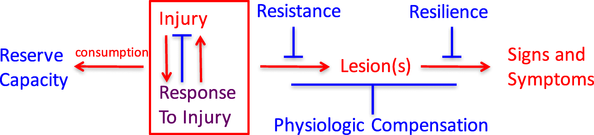
(Fig. 1).
While some may be examined individually and in exquisite molecular
detail in animal or in vitro models, most will require careful,
longitudinal validation in humans. From this perspective, it is not
surprising that so far we have had only limited success in identifying
risk factors and their underlying mechanisms to guide effective primary
and secondary preventive interventions.
Fig. 1
Relationships among
adverse (red), protective (blue), and mixed (purple) processes that
culminate in signs and symptoms of neurodegenerative diseases
Until
quite recently, “late onset Alzheimer’s disease” was widely viewed as a
specific disease entity responsible for the vast majority of late-life
dementia. However, longitudinal epidemiologic studies of brain aging and
cognitive decline with brain autopsy have consistently demonstrated a
central role for multiple co-morbidities as the dominant determinants of
late-life dementia. It is important to recognize that current intra
vitam measures of these several common diseases of the aging brain are
limited, and consequently, despite limitations, brain histopathologic
evaluation remains the only means to assess comprehensively the impact
of co-morbid diseases on cognitive performance during life.
In
combination with functional assessments obtained during life,
histopathologic features (lesions) determined with brain autopsy define
the presence of specific clinico-pathologic entities, which may or may
not reliably correspond to specific mechanism(s) of disease. As a result
of the highly consistent findings from longitudinal epidemiologic
studies with brain autopsy from across the globe, the view of cognitive
decline and dementia in older adults is shifting from being the result
of a single disease to a conspiracy of multiple, common age-related
disease processes that combine idiosyncratically in each individual. The
most common is Alzheimer’s disease, defined by amyloid beta
accumulation and neurofibrillary degeneration in certain regions of the
brain. Four other commonly recognized pathophysiologic processes that
can contribute to cognitive decline and dementia in late life include
Lewy body disease, vascular brain injury (especially from small vessel
disease), hippocampal sclerosis, and generalized atrophy beyond what can
be explained by these other diseases. While the brain lesions of AD are
more prevalent at autopsy than any of the other lesions, the combined
frequencies of the non-AD abnormalities are usually greater. Indeed, in
both the Nun Study and the Honolulu Asia Aging Study, > 90% of
participants with severe cognitive impairment can be fully attributed to
the collective or individual influences of these five abnormalities [1].
It is critically important, but infrequently appreciated, that the
exponential influence of co-morbid disease is reflected in the
multiplication of individual relative risks (or odds ratios) for each
disease related to cognitive impairment or dementia (Table 1).
Table 1
Point estimates of
odds ratios (OR) from ordinal logistic regression of the impact of the
coprevalence of five brain lesions on cognitive performance within
2 years of death
Lesion co-morbidity index
OR for the Nun Study (n = 334)
OR for the Honolulu Asia Aging Study (n = 774)
0
1.0 (reference)
1.0 (reference)
0.4–0.8
2.8
2.4
1.0–1.8
5.0
4.6
2.0–2.4
23.1
16.3
2.6–4.4
99.1
37.6
Severity of each of
the five brain lesions (Braak stage for neurofibrillary degeneration,
cerebral cortical Lewy body disease, cerebral cortical microinfarcts,
hippocampal sclerosis, low brain weight) was scored as none/mild (0),
moderate (0.4), or severe (1.0) by established criteria, and the lesion
co-morbidity index was calculated as the sum of scores for each of the
five lesions [1]
To
frame a discussion of resistance, resilience, reserve, and
compensation, we conventionally consider the diseases that cause
late-life cognitive impairment and dementia to derive from injury and
response to injury that begin before there are signs or symptoms, but
that the resulting damage, distortion, disruption, and/or degeneration
ultimately becomes overwhelmingly evident as impairments of cognitive
and behavioral function.
The
recognition of risk factors linked to measures of different types and
amount of brain lesions may illuminate fundamental mechanisms and
primary instigating exposures. A systematic search to identify specific
protective factors and the mechanisms that underlie them has been
conducted relatively infrequently. We propose the following operational
definitions as a step toward systematically investigating each of these
processes in individuals:
Resistance
is inferred from an observed absence or lower level of
dementia-associated brain injury, relative to an expected greater
frequency or severity based on age, genetic factors, or other
characteristics of the individual. This state of unexpectedly low or
absent brain injury theoretically may be intrinsic, meaning in someone with greater defenses to forces that usually lead to brain lesions, or environmental,
meaning in someone with usual defenses but who avoided exposure to
these forces. While resistance now can be assessed comprehensively only
with neuropathologic evaluation, specific facets (e.g., beta amyloid,
pathologic tau burden, neuron damage) can be estimated during life with
biomarkers and imaging.
Resilience
is inferred from an observed level of cognitive functioning higher than
expected in the face of demonstrated brain injury. Resilience only can
be recognized or measured when injury exists and can be related to
(near) coincident assessment of function. We prefer to consider two
forms of resilience: apparent and essential. Apparent resilience
refers to a specific lesion type without consideration of common
co-morbidities. Consider two individuals who both are positive by PET
imaging for fibrillar amyloid and pathologic tau; one is cognitively
normal and the other has dementia. The first person has apparent
resilience to AD neuropathologic change. Imagine further a future state
when there also is a PET ligand for pathologic alpha-synuclein. Now, we
learn that the first person lacks Lewy body disease and the second has
co-morbid neocortical Lewy body disease. Is the difference between these
two individuals explained by resilience to AD neuropathologic change or
by resistance to Lewy body in the first person? Once comprehensive
assessment of brain lesions associated with dementia is achieved, then essential resilience
can be evaluated. Currently, this is accomplished best with
neuropathologic assessment, but even this approach is limited. Our brain
autopsy data suggest that much, and perhaps most, of what is referred
to currently as (apparent) resilience actually is resistance to
co-morbid disease.
Consumption or retention of reserve
can be measured or inferred either as brain structural and/or
physiological pre-morbid capacity. Examples might be greater than usual
synaptic density (analogous to computational “hardware”) or enhanced
cognitive effectiveness or redundancy because of learned language,
educational richness, or occupational complexity (analogous to
computational “software”) prior to the onset of disease. The salutary
influence of such resources may be apparent in cognitive test
performance well before the onset of cognitive decline. This definition
requires that measures of reserve capacity must have been estimated or
inferred prior to the development of brain injury. Mechanisms underlying
physiologic compensation
also may be changes in “hardware” or “software,” but in distinction to
pre-existing reserve capacity, physiologic compensation occurs following
injury rather than developing prior to injury/response to injury. An
example of physiologic compensation might be recruitment of additional
regions of the brain to subserve memory function following damage to the
hippocampus or in recovery of language functioning after an infarction
or brain injury.
With
operational definitions of resistance, resilience, and reserve, a
focused analytic search for their predictors and correlates can be
undertaken. This will require distinguishing and measuring each
independently, and then employing those measures as distinct endpoints
to identify their individual determinants. This approach, essentially a
search to identify protective risk factors and their mechanisms,
represents a relatively unexplored pathway toward the identification of
candidate preventive interventions.


 “There
is a tendency to equate aging with a decline in mental function,” said
Molly Wagster, Ph.D., chief of the Behavioral and Systems Neuroscience
Branch in NIA’s Division of Neuroscience. “We want to study these
cognitive super agers, who clearly break the expectation of cognitive
decline, and unravel their secrets.”
“There
is a tendency to equate aging with a decline in mental function,” said
Molly Wagster, Ph.D., chief of the Behavioral and Systems Neuroscience
Branch in NIA’s Division of Neuroscience. “We want to study these
cognitive super agers, who clearly break the expectation of cognitive
decline, and unravel their secrets.” In
a separate study, the Northwestern researchers found that super agers’
brains contained a much higher density of a particular type of cell
called von Economo neurons, which are linked to social intelligence and
awareness. Their brains had more of these neurons even than the brains
of younger adults.
In
a separate study, the Northwestern researchers found that super agers’
brains contained a much higher density of a particular type of cell
called von Economo neurons, which are linked to social intelligence and
awareness. Their brains had more of these neurons even than the brains
of younger adults. Another
new initiative that stemmed from the summit and that also is underway —
Collaboratory on Research Definitions for Cognitive Reserve and
Resilience — brings together investigators from around the globe to
develop a consensus on uniform definitions for terms such as “cognitive
reserve,” “brain maintenance,” “resilience,” and “compensation” so that
the research community will have a common language and understanding for
reporting of results. Use of uniform definitions offers many benefits,
including the pooling of data across studies to allow for adequately
powered analyses and better interpretation of data.
Another
new initiative that stemmed from the summit and that also is underway —
Collaboratory on Research Definitions for Cognitive Reserve and
Resilience — brings together investigators from around the globe to
develop a consensus on uniform definitions for terms such as “cognitive
reserve,” “brain maintenance,” “resilience,” and “compensation” so that
the research community will have a common language and understanding for
reporting of results. Use of uniform definitions offers many benefits,
including the pooling of data across studies to allow for adequately
powered analyses and better interpretation of data.