Use the labels in the right column to find what you want. Or you can go thru them one by one, there are only 34,032 posts. Searching is done in the search box in upper left corner. I blog on anything to do with stroke. DO NOT DO ANYTHING SUGGESTED HERE AS I AM NOT MEDICALLY TRAINED, YOUR DOCTOR IS, LISTEN TO THEM. BUT I BET THEY DON'T KNOW HOW TO GET YOU 100% RECOVERED. I DON'T EITHER BUT HAVE PLENTY OF QUESTIONS FOR YOUR DOCTOR TO ANSWER.
Changing stroke rehab and research worldwide now.Time is Brain!trillions and trillions of neuronsthatDIEeach day because there areNOeffective hyperacute therapies besides tPA(only 12% effective). I have 523 posts on hyperacute therapy, enough for researchers to spend decades proving them out. These are my personal ideas and blog on stroke rehabilitation and stroke research. Do not attempt any of these without checking with your medical provider. Unless you join me in agitating, when you need these therapies they won't be there.
What this blog is for:
My blog is not to help survivors recover, it is to have the 10 million yearly stroke survivors light fires underneath their doctors, stroke hospitals and stroke researchers to get stroke solved. 100% recovery. The stroke medical world is completely failing at that goal, they don't even have it as a goal. Shortly after getting out of the hospital and getting NO information on the process or protocols of stroke rehabilitation and recovery I started searching on the internet and found that no other survivor received useful information. This is an attempt to cover all stroke rehabilitation information that should be readily available to survivors so they can talk with informed knowledge to their medical staff. It lays out what needs to be done to get stroke survivors closer to 100% recovery. It's quite disgusting that this information is not available from every stroke association and doctors group.
Showing posts with label don't have a functioning stroke hospital. Show all posts
Showing posts with label don't have a functioning stroke hospital. Show all posts
You didn't specify what protocols they should be; SO FUCKING USELESS! My god the stupidity out there is stupendous! We still have no protocol on when stroke rehab should start with appropriate objective damage diagnosis starting points. You're screwed along with your children and grandchildren when they have strokes. We need stroke leadership and we need it now.
Conditions Published Aug 31, 2026 3:10 PM EDT By Joseph James Rehabilitation after a stroke should begin in the hospital as soon as a patient is medically stable, ideally within 48 hours, and it should be built around cognition, communication, and mental health rather than physical function alone. That is the central instruction in the updated adult stroke rehabilitation and recovery guideline, published by the American Stroke Association in the journal Stroke and replacing guidance that had stood since 2016. The change lands on a very large group of families. Nearly 800,000 people in the United States have a stroke each year, and about 2.7 million people in the country are caring for a stroke survivor. By the American Heart Association's latest statistics, stroke is now the fourth leading cause of death nationally and remains a leading cause of serious long-term disability. or a household, the practical meaning is that the questions asked in the first days matter more than many families realize. Whether a therapist has evaluated the patient, whether a depression screen has been done, and whether a caregiver has been included in planning are now things a guideline says should happen, not favors to request.
The Timing Rule and Its Deliberate Exception
Studies have shown that starting rehabilitation early leads to better outcomes, which is why the writing group set the 48-hour target. The guideline pairs that with a limit which is easy to miss: moderate to high-intensity exercise and task practice should not be performed in the first 24 hours after a stroke. (Why? You don't understand that the neuronal cascade of deathis killing off neurons in the first days and you incorrectly ascribed that damage to early exercise. Doesn't anyone in stroke know how to think?)That is not a contradiction. Early rehabilitation on day one means assessment, positioning, safe mobility, and preventing the complications of immobility. Intensity comes after the first 24 hours. Beyond that point, the guideline calls for enough task practice to reduce impairment and teach compensatory strategies, and it advises transferring patients with moderate to severe stroke to an inpatient rehabilitation facility when that is feasible. Lorie Gage Richards, the occupational therapist and University of Utah associate professor who chaired the volunteer writing group, framed the difficulty in the association's announcement. "Stroke rehabilitation is complicated. A stroke can affect many of the skills people rely on for daily living, and even the most routine activities involve multiple physical, cognitive and communication abilities working together," she said.
Recovery Redefined to Include Mood, Memory and Speech
The most consequential shift is what now counts as rehabilitation. The guideline calls for people who have had a stroke to be screened for depression, anxiety and other mental health concerns early, rescreened later, and treated when needed. Post-stroke depression is described as common and treatable, and as something that can make physical recovery harder. Sara Kutzle, a stroke survivor who served on the writing group, had a hemorrhagic stroke in 2021 at age 39 while working as a makeup artist in San Francisco. She spent a week in intensive care and a month in the hospital, then relearned walking, writing, and how to function as a right-handed artist. "At first, I was focused on getting my body to recover, but I quickly realized healing involved so much more than that," Kutzle said. "My confidence, my mental health, my relationships and my sense of independence were all affected." The guideline also addresses returning to work, parenting young children, sleep and pain problems, and changes in sexual function, and it notes that strokes among people under 50 are increasing. Recreational therapists are named as part of the team for helping survivors reconnect with community life.
Caregivers Enter the Care Plan by Name
The document treats the roughly 2.7 million caregivers of stroke survivors as people who need support rather than as an unpaid extension of the care team. Many experience stress, depression or anxiety of their own, and the guideline says caregivers should be included in rehabilitation planning, education and support throughout the process. That reframing has household consequences. A caregiver trained in transfers and swallowing precautions is less likely to be injured and less likely to see a preventable readmission. A caregiver whose own depression is recognized is more likely to sustain the work over months. Where rehabilitation happens depends on stroke severity. Some survivors go home and receive outpatient therapy with support from a relative. Others need a skilled nursing facility, an inpatient rehabilitation facility, or a long-term care hospital. The guideline identifies telehealth as a growing part of recovery, particularly for people who cannot travel to a clinic, and credits it with improving both access and cost-effectiveness.
Scientists have for the first time mapped in exquisite, three-dimensional detail six major conformations of a membrane in the brain related to learning, memory and fear-related behavior.
Researchers used state-of-the-art cryo-electron microscopy housed in OHSU’s South Waterfront Campus to capture the most detailed view yet of a specific type of membrane protein: an acid-sensing ion channel known as ASIC1a.
The findings could form the blueprint for drug development useful in treating stroke, said senior author Isabelle Baconguis, Ph.D., assistant professor in the OHSU Vollum Institute.
“Previous studies show that when you block this channel, it can be
neuroprotective
(Meaning stopping the the neuronal cascade of death in the first week! Neuroprotection is such a bland word, doesn't impart immediacy at all!)
,” Baconguis said. “If you’re able to design a drug that infuses an inhibitor to this channel, it could lengthen the survival of the tissue in cases of stroke.”
Already, scientists in Australia are using a molecule derived from spider venom that explicitly targets ASIC1a to improve outcomes in heart attacks and stroke.
OHSU researchers used recombinant DNA technology to express the human gene to generate human proteins they imaged using cryo-EM. Because acid-sensing ion channels respond to variations of extracellular pH in the central and peripheral nervous systems, researchers were able to capture six distinct conformations by varying their exposure to acidity.
“In our bodies, there are locations where cells undergo different pH conditions, especially in the brain,” Baconguis said. “In neuronal injuries such as stroke, where brain tissue undergoes a drop in pH, these channels can be activated causing tissue damage.”
The images provide a blueprint for designing new drugs capable of inhibiting this one specific acid-sensing ion channel in cases of stroke.
“The sooner you can protect brain tissue from damage, the less severe disability stroke survivors will have,” Baconguis said. “Time is of the essence when it comes to stroke.”
In addition to Baconguis, co-authors include James Cahill, Kimberly A. Hartfield, Ph.D., Craig Yoshioka, Ph.D., of OHSU; Stephan Alexander Pless, Ph.D., Nadine Ritter, Ph.D., and Mette Homann Poulsen, Ph.D., of the University of Copenhagen; and Stephanie Andrea Heusser, Ph.D., of the University of Copenhagen in Denmark and Linköping University in Sweden.
The research was supported by the National Institutes of Health, grant award R24GM154185, RO1GM138862 from the National Institute of General Medical Sciences (NIGMS); and the Lundbeck Foundation, grant award R313-2019-571. Electron microscopy was performed at the Multiscale Microscopy Core, part of OHSU’s university-shared resource cores. The content is solely the responsibility of the authors and does not necessarily represent the official views of the NIH.
What further research will your competent?
doctor initiate to figure out how to successfully prevent cognitive
decline? Oh sorry; YOUR DOCTOR PLANS ON DOING NOTHING, RIGHT!
You are finding out now that you don't have a functioning stroke doctor/hospital even after unsuccessfully getting you 100% recovered! THAT IS DOCTOR FAILURE!
High-dose DHA successfully
reached the brains of older adults at increased risk of Alzheimer's
disease, but the two-year clinical trial found no improvements in memory
or brain structure, challenging assumptions that greater omega-3
delivery alone can slow cognitive decline.
A clinical trial published in eBioMedicine
found that high-dose docosahexaenoic acid (DHA) supplementation
successfully increased brain DHA levels in older adults at risk of
dementia, including those carrying the APOE ε4 Alzheimer's risk variant.
However, despite reaching the brain, the supplement did not improve
cognitive performance or brain structure over two years, raising new
questions about how DHA is used within the brain.
Why APOE ε4 alters brain DHA metabolism
DHA is a fatty acid that is part of the nerve cell membrane, playing a
key role in synaptic function and modulating neuroinflammation. Its
levels tend to be lower in the presence of dementia-linked changes like
amyloid deposition and cognitive decline, and in patients with
late-onset Alzheimer's disease (AD).
The APOE ε4 gene variant is the strongest genetic risk factor for AD.
Previous research suggests it is associated with accelerated DHA
catabolism and lower plasma and cerebrospinal fluid DHA levels in people
with AD dementia compared with non-carriers.
Observational studies have suggested modest associations between
higher omega-3 intake and lower risk of cognitive decline, but
randomized trials have produced inconsistent results. Of 24 randomized
trials in people without dementia, only five reported positive cognitive
effects following DHA supplementation. Conversely, no improvement was
seen in patients with AD.
Thus, two important questions remain unanswered: is early
intervention necessary in patients with low omega-3 levels before
dementia sets in, and are higher doses required to ensure adequate brain
uptake? Previous imaging studies suggest that younger cognitively
healthy carriers have increased brain DHA incorporation, which may
reflect greater DHA demand, compared to non-carriers. This has not been
studied in older adults prior to the onset of dementia.
In the current study, researchers investigated whether high-dose DHA
supplementation could effectively raise brain DHA levels and potentially
support cognitive and structural brain health in older adults with low
dietary omega-3 intake before dementia develops.
Testing high-dose DHA before dementia develops
The investigators conducted a randomized, double-blind,
placebo-controlled trial that enrolled 365 adults without dementia, aged
55–80 years, with low DHA intake and at least one dementia risk factor
at baseline. Participants received either 2 g/day of DHA or a placebo
for 24 months.
Pittcon Highlights: Pharmaceutical & Biologic eBookCheck out the highlights from Pittcon in the Pharmaceutical & Biologic industriesDownload the latest edition
The
mean participant age was 66 years, with 58% being female. Approximately
47% of the participants were APOE ε4 carriers, and 39% were Hispanic.
The participants were first classified by willingness to undergo a
lumbar puncture (LP) to obtain cerebrospinal fluid (CSF) for analysis.
The two groups were assessed for the CSF DHA: arachidonic acid (AA)
ratio after six months, which reflects the extent to which DHA is
delivered to the brain. Various brain volumes were also assessed.
Increased DHA delivery
The 365 participants were divided into two arms: 181 in the LP
arm and 184 in the non-LP arm. In both arms, DHA supplementation
significantly increased the CSF DHA/AA ratio at six months compared with
placebo, indicating successful delivery of DHA to the brain. There was
also a 17% increase in CSF DHA. The red cell omega-3 index also
increased from 4.9% to 11%.
The increases in DHA delivery to the brain and in the red cells were
independent of APOE ε4 status. This suggests that the gene variant did
not influence this process.
However, APOE ε4 non-carriers showed greater improvement in cognitive
scores than carriers, with a mean improvement of 3.8 and 1.6 in the two
groups, respectively, regardless of treatment group. Importantly, the
study demonstrates that inadequate brain delivery is unlikely to explain
the disappointing results of previous DHA supplementation trials,
because high-dose supplementation successfully increased CNS DHA levels.
The authors hypothesize that simply improving DHA delivery to the
brain may not be sufficient to enhance cognitive function, given the
enzymatic catabolism of DHA within synaptic membranes, which are crucial
for cognitive processing.
There was no difference in brain volumes or in cognitive performance
over the whole study period between the intervention and control groups.
Adverse events were comparable between groups, and the treatment was
generally safe and well-tolerated.
Strengths and limitations
The sample included White and Hispanic participants with a high
proportion of APOE ε4 carriers. The low baseline omega-3 intake, CSF DHA
measurement, and multiple outcome assessments, coupled with a stringent
trial design, were among the study’s strengths.
However, it had several limitations. The participants were relatively
young, well-educated, and at an early stage of disease, which might
have limited their ability to detect treatment effects over just 24
months.
The study showed a relatively high dropout rate at 38%. Most of this
was related to the coronavirus disease 2019 (COVID-19) pandemic. The
consequent reduction in sample size might have affected its ability to
detect smaller effects on cognitive function or brain structure. Those
who dropped out of the study were more likely to be Hispanic, to have
lower education levels and baseline cognitive scores, and to have lower
plasma DHA concentrations than those who completed the study, which
might have affected the generalizability of the findings.
The study used a single supplement, but the authors point out that
this could be insufficient in the face of multiple disease processes
affecting neuronal health and DHA metabolism in the brain. This is even
more true when the participants have vascular risk factors like
hypertension and physical inactivity, all of which need to be addressed
simultaneously.
The current study included only cognitively healthy individuals, but
future studies may benefit from testing supplementation in individuals
who already have biochemical signs of early neurodegeneration, such as
elevated biomarkers (phosphorylated tau in blood, advanced imaging
markers) or more granular neuropsychological testing to detect small
changes in executive function. This would improve the detection of
treatment changes. A longer follow-up may also be necessary.
Conclusion
The findings show that high-dose DHA supplementation can
substantially increase brain DHA levels within six months in older
adults at risk of dementia, regardless of APOE ε4 status. Conversely,
this did not translate into observable improvements in cognition or
brain structure over 24 months.
These results suggest that high DHA intake alone may not be
sufficient to improve cognitive outcomes or preserve brain structure in
relatively healthy older adults over a 2-year period, despite adequate
brain delivery. They also imply that APOE ε4 carriers experience normal
DHA delivery to the brain before dementia, despite the dysregulation in
established dementia reported in prior research.
Future research should focus on examining DHA metabolism in the brain
rather than on additional supplementation trials. Because brain DHA
delivery was successfully achieved without improving cognition, future
work should focus on how DHA is processed and used within brain cells
rather than simply increasing DHA intake.
Yassine, H. N., Pour, S. G., Juarez, M., et al. (2026). CNS target
engagement of high-dose DHA supplementation in older adults at risk for
dementia: a randomised, double-blind, placebo-controlled trial. eBioMedicine. DOI: https://doi.org/10.1016/j.ebiom.2026.106316. https://www.thelancet.com/journals/EBIOM/article/PIIS2352-3964(26)00198-2/fulltext
And your competent? doctor has had protocols for all these for years already, right? OH NO; NOT! So, you DON'T have a functioning stroke doctor or hospital? And you haven't had everyone in that stroke hospital fired and blackballed from stroke forever? Because this is massively incompetent not creating protocols on all this!
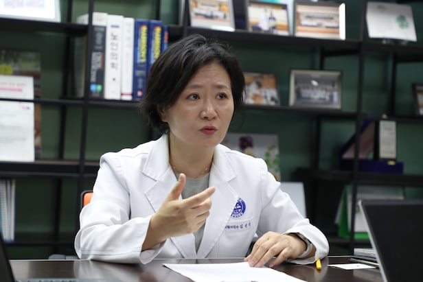
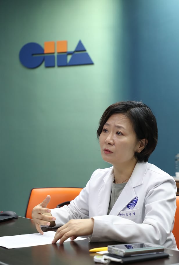
ha Bundang professor Kim Min-young blends stem cells, VR, and magnetic stimulation to restore brain function and curb inflammationAt Bundang CHA Hospital in Seongnam, Gyeonggi Province, Kim Min-young, a professor of rehabilitation medicine, says, "Rehabilitation is now the process of restoring lost function by using the brain's plasticity," adding, "The combination of cell therapy and digital technology will be the answer."/Courtesy of Jeon Gi-byeong
By Lee Young-wan Published 2026.04.15. 06:03 Updated 2026.04.15. 09:58
In his 1632 book "Dialogue Concerning the Two Chief World Systems," Galileo Galilei rejected the then-mainstream geocentric model and argued that Earth orbits the sun. Like Galileo's "Dialogo (dialogue)," we introduce people who, through in-depth interviews, are changing the world's paradigms.
Cerebral palsy is a disorder in which damage to an immature brain before birth, during delivery, or shortly after birth causes abnormalities in motor function. Muscles weaken and the body cannot be controlled. For a long time, people believed the brain could not be restored once damaged. Kim Min-young (59), a professor of rehabilitation medicine at CHA University Bundang CHA Hospital, in 2013 became the first in the world to successfully treat cerebral palsy by injecting stem cells from cord blood (umbilical cord blood).
Stem cells are a kind of primitive cell that can grow into various cells in the human body. If stem cells are injected into tissue damaged by disease, they may be replaced by healthy cells. In particular, Kim treated patients with cord blood stem cells from other people. This opened the way to treat patients who did not store their own cord blood at birth, as long as immune compatibility was confirmed.
So far, Kim has provided cord blood treatment to more than 600 patients with cerebral palsy solely as clinical research for medical study, not as an approved therapy. It is by far the highest treatment count in the world. Babies who could not even hold themselves up received cord blood injections and crawled and sat up on their own. As motor and sensory neurons in the brain regenerated, not only motor skills but also cognitive abilities improved.
Recently, following cerebral palsy, treatment results were achieved for stroke, autism spectrum disorder (ASD), and dementia. Kim tried various methods to regain brain function. In addition to cell therapy such as cord blood, the team mobilized transcranial magnetic stimulation, which stimulates brain nerves with magnetic fields, and Virtual Reality (VR) digital therapy. Kim said, "If we can treat patients, the boundaries of technology are meaningless," adding, "Meeting patients every day and thinking about how to treat them led us to find various methods."
Professor Kim Min-young says, "We have seen treatment benefits by injecting umbilical cord blood stem cells for patients with cerebral palsy, stroke, and autism," adding, "It is the result of neural regeneration and suppressed inflammation."/Courtesy of Jeon Gi-byeong
◇ "A damaged brain can change"
– People think rehabilitation means building muscle, but you protected and activated neurons with cord blood.
"It was believed that once the brain was damaged, it could never be restored, but in the late 1990s the theory of brain plasticity emerged, saying neural circuits can change. Now rehabilitation is not simply about building muscle; it is a process of regaining lost function by using the brain's plasticity."
– What is the principle of plasticity that restores brain function?
"First, repeated stimulation or training increases the connections (synapses) of neurons. There are also changes in the brain's network. A patient with poor memory may not answer properly if only asked verbally about a specific event, but if you show the music heard at the time, the people present, or the scenery seen together, the memory can be recalled more easily. The third is the neural function recovery effect of stem cells."
– What is the most effective means to restore brain function?
"In patients with cerebral palsy, combination therapy with cord blood cells and erythropoietin showed better results, but treatment effects were also achieved with cord blood cells alone. Continued research showed that improvement in motor function from cord blood therapy was due to an increase in innate immune responses and anti-inflammatory effects. The principle of treatment is precisely to suppress inflammation."
– We heard you are also focusing on controlling inflammation in treating other brain diseases.
"The more we studied, the more we found a common cause in cerebral palsy, stroke, dementia, and autism: inflammation in the brain. When brain nerves are damaged, chronic inflammation arises around them and hinders neural regeneration. If this is suppressed, other brain diseases can also be treated. The prevalence of cerebral palsy is only 0.2% to 0.3%, but autism spectrum disorder is reported up to 3%. Some of the children receiving rehabilitation turned out to have autism, which led us to conduct clinical research."
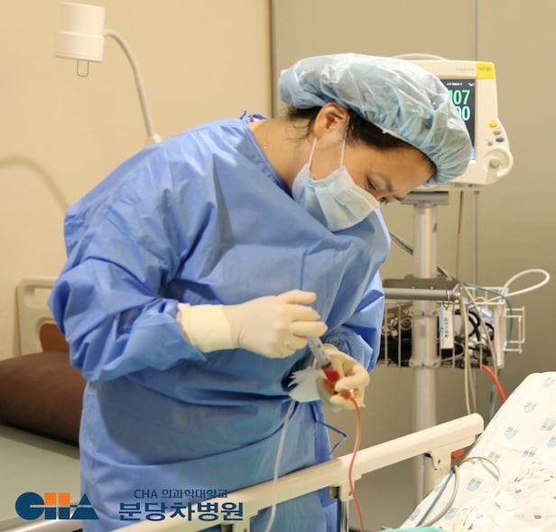
Kim Min-young, a professor of rehabilitation medicine at Bundang CHA Hospital of CHA University, administers an intravenous infusion of allogeneic (donor) cord blood to a child with cerebral palsy./Courtesy of CHA Hospital
◇ A medical student who went to the Electronics and Telecommunications Research Institute
– You are a medical school professor, yet your three years at ETRI (Electronics and Telecommunications Research Institute) stand out. That is very unusual for a physician.
"From 2000 to 2003, I worked at ETRI as an invited researcher on the VR interface research team. At the time, women were rare among medical school professors. I chose to become a researcher rather than a professor, learned programming languages with help from Professor Kim Jae-hee of Yonsei University's Department of Electronic Engineering during my master's, and developed VR therapy technology for patients with hemiparesis at ETRI."
– That means you started what we now call digital therapeutics (DTx) early on.
"Only the name has changed; the essence is the same. Although digital therapeutics began in earnest in 2019, the concept of software as a medical device (SaMD) existed before that. VR is one form of it. Since attempting treatment for patients with hemiparesis back then, we have applied it to treating various patients with brain injuries to this day."
– Which patient is most memorable?
"A person with apraxia. As the name suggests, the patient could not carry out intended actions. The command system from the brain to the muscles was tangled, leaving the patient unsure how to use the hands. After a handshake, the hand could not be released and the patient had to use the other hand to straighten each finger. When we put VR goggles on this patient and asked them to catch fish in a virtual space, the hands moved naturally. It was a case proving that a 3D (stereoscopic) virtual world, which offers a sense of space more than a flat monitor, activates the brain's entire neural network far more powerfully."
"We thought older adults were not familiar with digital technology such as VR, but in reality they were not. We had them choose clothing according to the weather in a virtual world and scored them, and they liked it more than expected. We confirmed that older adults who used VR had better attention and memory than the control group who were simply shown a movie."
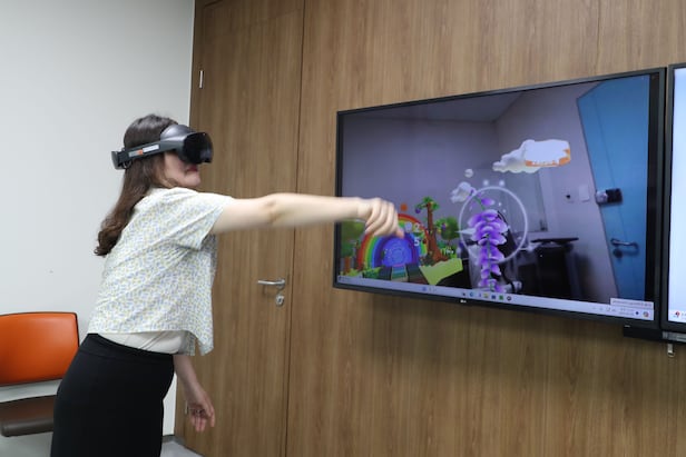
A Bundang CHA Hospital researcher uses a VR therapy device developed by Professor Kim Min-young of rehabilitation medicine. A patient who cannot properly raise an arm trains to catch butterflies in a virtual world and naturally gains rehabilitative benefits./Courtesy of Jeon Gi-byeong
◇ Mobilizing transcranial magnetic stimulation and soft robots
– VR likely offers high immersion for patients. Why did you also try the lower-immersion AR (Augmented Reality)?
"AR overlays virtual images on real objects. For the general public, VR is better for immersion, but for patients undergoing rehabilitation or for older adults, real visual information is obscured, so they may fall or get hurt in VR. Patients with risk factors must be able to feel the virtual world while also seeing reality to ensure safety."
– We heard you also tried treatment that stimulates the brain with magnetic fields.
"In patients with vascular dementia, transcranial magnetic stimulation, which stimulates specific brain nerves with magnetic fields, reduced inflammatory gene expression in peripheral blood, and this led to improved cognitive function. The correlation between improved cognitive scores and reduced inflammation was as high as 0.9. We are also pursuing studies that use VR and transcranial stimulation simultaneously."
– Are there any other innovative technologies useful for rehabilitation?
"I am paying attention to wearable soft robots that are not made of metal but are worn like clothing to build muscle strength. Conventional robotic rehabilitation devices are too large and heavy, but if thin, lightweight fibers themselves contract like muscles to assist force, patients could wear them like clothes and undergo rehabilitation 24 hours a day. The technological prototypes already exist. The problem is the speed of commercialization, and it is unfortunate that engineers develop separately without working together with physicians."
Professor Kim Min-young notes, "Regulations often fail to keep pace with medical technology and only hold it back," adding, "Recently, Korea has also opened the way for stem cell therapy, but we are far behind Japan."/Courtesy of Jeon Gi-byeong
◇ "There are no answers if you research without seeing patients"
Since 2011, Kim has published 86 papers in international journals, averaging six papers a year. Of those, 58 were as lead author. This was the result of reading and researching papers two to three hours every day amid a busy clinical schedule. Wouldn't there be more成果 if Kim did only research? Kim asserted, "If you leave the clinical field, you cannot conduct proper research."
– Physician-scientists who focus more on research than on clinical practice are drawing attention these days. But you stress that true physician-scientists must have clinical experience.
"As a medical student, I learned to hold children with autism tightly when they have a seizure. The seizures in children with autism result from proprioception, which recognizes the body's position and the existence of the limbs, working excessively. In hindsight, holding them was a stimulus to control the sensory problem. Without such clinical experience, physicians who do not know the limits patients face can do research for papers but cannot produce solutions for patients. I advise juniors to complete the residency and then pursue research or startups."
– Is that why you use various treatment methods to restore brain function?
"While advising on government-affiliated committees that review research and development projects, I often saw developers who did not know how technology is used in clinical settings and developed based only on their own assumptions and guesses. It was the same for both medical devices and advanced regenerative therapies. If a technology is to be used for patients, collaborating with physicians who have rich clinical experience is the shortcut to success. There is no royal road to treatment. Whether it is cell therapy or VR, everything is needed depending on the situation. Insisting only on what one knows harms patients."
– There must be many institutional shortcomings as well.
"With the enforcement of the Act on Advanced Regenerative Medicine and Advanced Biopharmaceuticals, the door to stem cell therapy has opened, but often the pace of scientific progress is still not matched by laws and regulations. In addition, there is a conservative culture in which specialists in related diseases do not actively adopt new treatments. Despite meaningful clinical research results for cord blood in cerebral palsy, pediatric patients still are not receiving treatment."
☞ Professor Kim Min-young
Kim graduated from Yonsei University College of Medicine and received master's and doctoral degrees from the same graduate school. From 2000 to 2003, Kim worked as an invited researcher on the VR interface research team at the Electronics and Telecommunications Research Institute (ETRI), pursuing convergence of medicine and engineering. Kim is currently a professor in the Department of Rehabilitation Medicine at CHA University Bundang CHA Hospital and head of the Center for Digital Innovative Medicine at CHA Advanced Research Institute. A world-renowned authority in cord blood cell therapy for cerebral palsy, Kim received the prime minister's commendation on Health Day in 2024 and the Seokjeon Shin Jeong-sun Academic Award in 2025. Kim is a full member of the National Academy of Medicine of Korea, served as policy director of the Korean Medical Association and chair of the Korean Society of Pediatric Rehabilitation and Developmental Medicine, and is now a director at the Korean Society for Stem Cell Research and the Korean Society of Digital Therapeutics.
※ This article has been translated by AI. Share your feedback here.
Will you competent? doctor and hospital IMMEDIATELY CREATE A PROTOCOL TO IMPLEMENT THESE VACCINES UPON ENTERING THE HOSPITAL POST STROKE?
NO? So, you don't have a functioning stroke doctor or hospital? They don't read or implement research, do they? And your board of directors is so incompetent they can't recognize incompetence in their hospital!
Influenza vaccination could significantly reduce the risk of influenza-associated heart attack and stroke even among people who get infected after getting the vaccine, according new research.
Catching influenza increases the short-term risk of cardiovascular conditions, and existing evidence has shown that the vaccine reduces the risk of heart attack and stroke by preventing infection in the first place.
The study, which included 1,221 adults aged 40 or older in Denmark, looked into how much the risk increased after infection and if the vaccine could reduce this added risk even if people were infected anyway.
“If confirmed by additional studies in other settings, this would strengthen the case for prioritising influenza vaccination among people at risk of heart disease or stroke and would support refining recommendations across Europe,” the researchers wrote.
The study used Danish health registry data from 2014 to 2025 and included individuals aged 40 and above with a first-ever hospital admission for a heart attack or stroke within about a year after an influenza virus infection.
It included all laboratory-confirmed influenza virus infections that occurred during nine consecutive influenza seasons with an observation window of 365 days before and after a positive test for influenza.
Testing, hospitalisation, vaccination and mortality records were matched through Denmark’s unique personal identifiers.
The study population comprised 660 males and 561 females aged 40 years and above, with a median age of 75.
Most patients were hospitalised with a stroke (65 per cent), while 35 per cent had a heart attack. Of 1,231 influenza virus infections confirmed by PCR testing, about half were happening after patients had been vaccinated.
Researchers used conditional Poisson regression to estimate incidence rate ratios (IRRS) and 95 per cent confidence intervals using the self-controlled case series design, a study design that compares event timing within the same person, thereby controlling for other individual factors such as comorbidities, genetic predisposition, and socioeconomic status.
To minimise reverse causality, i.e. where cardiovascular symptoms may have prompted testing, a pre-exposure period of two weeks before testing was excluded.
The risk for a first-time hospitalisation for heart attack and stroke during the first week after testing positive for influenza was found to be significantly higher than for any other period before or after; it increased threefold for a stroke and fivefold for a heart attack.
This increased risk was reduced by half for people who were infected but had been vaccinated against influenza for that influenza season.
The study did not account for differences in effectiveness between influenza vaccines, which can vary depending on how well the vaccine formulation matches the viral strains circulating in that season.
It could also not assess whether vaccination timing or gender affected outcomes.
Results might also not apply directly to populations or settings with different influenza epidemiology, healthcare systems, or vaccination strategies.
Didn't your competent? doctor put together a protocol on this for your recovery already? NO? So, you DON'T have a functioning stroke doctor, hospital or board of directors? Good to know you're in fucking incompetent hands!
An elevated neutrophil-to-lymphocyte ratio was significantly associated with an increased prevalence of depressive disorder and a higher risk for suicide.
HealthDay News — An elevated neutrophil-to-lymphocyte ratio (NLR) is associated with a higher prevalence of depressive disorder (DD) and with increased suicide risk among those with DD, according to a review published in the November/December issue of the Harvard Review of Psychiatry.
Yuanyuan Zhou, from the Institute of Acupuncture and Moxibustion in Jinan, China, and colleagues conducted a systematic literature review to identify studies assessing the relationship between NLR and DD. The primary outcome was the prevalence of DD and suicide risk in individuals with DD.
The review included 37 studies, with 88,019 participants. The researchers found that high NLR was significantly associated with DD presence (odds ratio, 1.57). Individuals diagnosed with DD had elevated NLR levels in continuous analyses (standardized mean difference, 0.73). In addition, heightened suicide risk was seen among those with DD in association with increased NLR (categorical outcome odds ratio, 1.56; continuous outcome standardized mean difference, 0.42).
“These findings underscore the clinical relevance of systemic inflammation in DD pathophysiology and highlight NLR as a cost-effective, accessible tool to inform risk stratification and tailored treatment strategies in psychiatric care,” the authors write.
There is a wide diversity in studies investigating the effects of stretching on spasticity,
and the available evidence on its clinical benefit is overall inconclusive. We recognize
the need for consensus on a paradigm for stretching and for good-quality studies.
Future research should address this issue and should investigate the clinical importance
of the short- and long-term effects.
Hand spasticity is one of the most frustrating challenges many stroke survivors face. Muscles in the hand and fingers tighten involuntarily, making it hard to open the palm, pick up objects, or even rest comfortably.
The good news is that stretching can make a meaningful difference. By gently lengthening stiff muscles, you can reduce tension, improve range of motion, and help your hand feel more relaxed. While progress takes time, consistent stretching is one of the most effective ways to support recovery.
In this guide, we’ll cover eight stretching exercises specifically designed for hand spasticity after stroke. Each one targets common problem areas, from clenched fists to tight fingers. In addition, we will also cover some practical tips for how to gradually progress from wherever you are starting out!
Understanding Hand Spasticity After Stroke and Why Stretching Is Important
A stroke disrupts communication between the brain and muscles. When the signals are unclear or blocked, certain muscles can involuntarily contract and don’t release properly. In the hand, this often causes the fingers to curl inward or the thumb to pull tightly across the palm.
Over time, spasticity can lead to:
Reduced flexibility and stiffness
Difficulty grasping or releasing objects
Risk of contractures (permanent shortening of muscles)
Pain or discomfort in the wrist and fingers
Stretching helps combat these issues by gradually loosening the muscles, improving circulation, and maintaining joint mobility. It’s not a quick fix, but when practiced daily, it can improve both comfort and function.
A Few Tips for Stretching Before You Begin
Before trying the stretches, keep these safety tips in mind:
Start slow. Move into each stretch gradually. Forcing your hand open can cause pain or trigger spasms.
Hold gently. Aim to hold each stretch for 15–60 seconds, depending on your comfort level. Ease further into the stretch throughout this time if possible.
Breathe steadily. Deep breathing can help your body relax and reduce resistance.
Stay consistent. Daily practice is more effective than occasional effort.
Use assistance if needed. A caregiver, therapist, or soft object like a rolled-up towel can help with positioning.
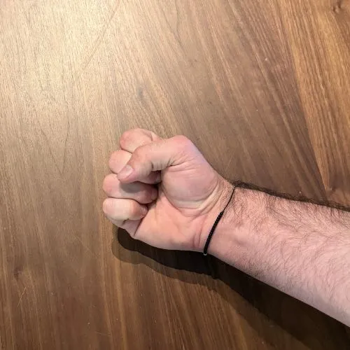
1. Palm Opening Stretch
Why it helps
One of the most common effects of hand spasticity is a clenched fist. This stretch helps gently open the palm and extend the fingers.
How to do it
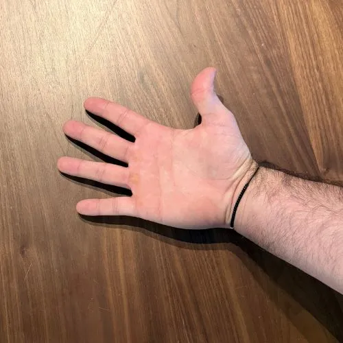
Rest your affected hand on a flat surface, palm facing upward.
With your other hand, gently press the fingers and thumb outward, encouraging the palm to open.
Hold for 20–40 seconds, then relax.
Repeat 2–3 times.
Tips
If your hand resists, start with small movements.
Don’t worry if you can’t fully open the hand—progress happens gradually.
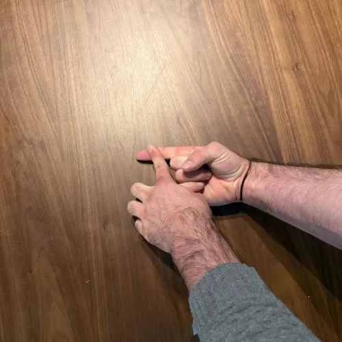
2. Finger Extension Stretch
Why it helps
Tightness in the fingers makes it hard to straighten them. This stretch targets each finger individually to improve flexibility.
How to do it
Place your hand palm-up on a table.
Use your other hand to gently lift one finger at a time, extending it outward.
Hold for 15–30 seconds before moving to the next finger.
Tips
Focus on one finger at a time for better control.
Skip any finger that causes sharp pain and return later when the muscles are more relaxed.
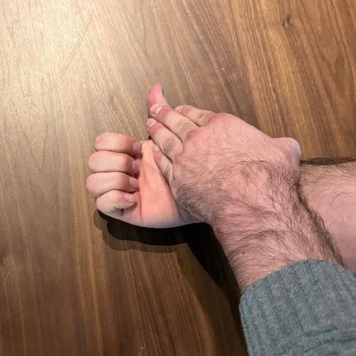
3. Thumb Stretch
Why it helps
Spasticity often pulls the thumb tightly across the palm, making grasping objects very difficult. This stretch increases thumb mobility.
How to do it
Hold your affected hand palm-up.
With your other hand, gently move the thumb away from the palm.
Hold for 20–30 seconds.
Repeat 2–3 times.
Tips
If your thumb is very tight, use a soft ball or rolled-up washcloth to keep the thumb slightly open throughout the day.
4. Wrist Flexor Stretch
Why it helps
The muscles in the forearm that connect to the hand often tighten during spasticity. Stretching them supports hand relaxation.
How to do it
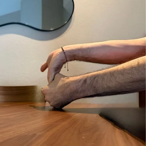
Rest your forearm on a table with your palm facing upward.
Use your other hand to gently bend the wrist backward, extending the palm.
Hold for 20–40 seconds.
Tips
You should feel the stretch in your forearm, not just the hand.
Move slowly to avoid triggering spasms.
5. Wrist Extensor Stretch
Why it helps
Balancing the muscles that open and close the wrist is important. This stretch targets the opposite group of muscles.
How to do it
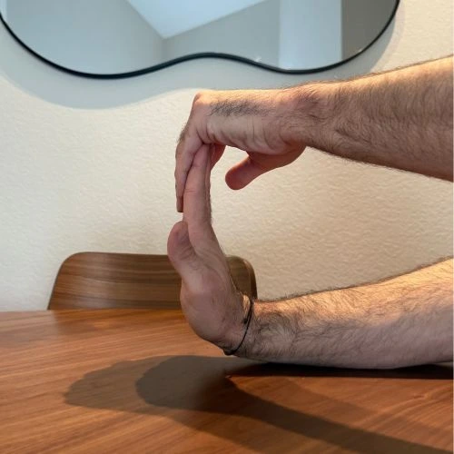
Rest your forearm on a table with your palm facing downward.
Use your other hand to gently press the back of your hand downward, bending the wrist.
Hold for 20–30 seconds.
Tips
Keep the movement gentle and steady.
This stretch pairs well with the wrist flexor stretch for balanced relief.
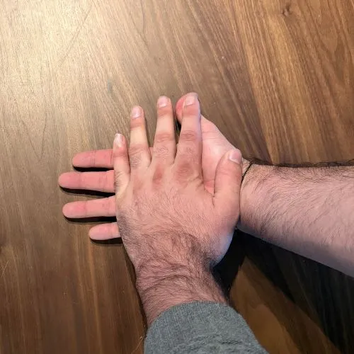
6. Tabletop Finger Spread
Why it helps
Hand spasticity often draws the fingers inward. This exercise encourages the opposite movement by spreading them apart.
How to do it
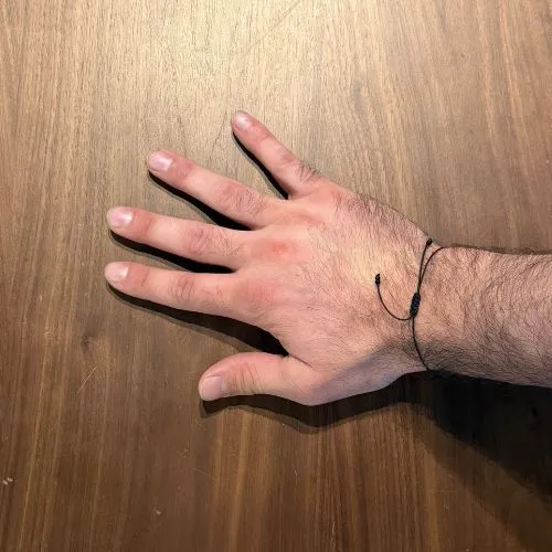
Place your hand flat on a table, palm down.
Use your other hand to gently guide the fingers apart.
Hold for 20–30 seconds, then relax.
Tips
If you struggle to spread your fingers, start with very small movements.
A therapy putty exercise afterward can help reinforce this stretch.
Try a similar stretch by gently interlocking the fingers together with the palms facing toward each other.
7. Hand Rotation Stretch (Supination to Pronation)
Why it helps
Supination (turning the palm upward) and pronation (turning it downward) are often limited after stroke. Improving rotation helps with daily activities like eating or holding objects.
How to do it
Rest your elbow on a table with your hand raised slightly.
Use your other hand to gently guide your palm upward, then downward.
Hold each position for 10–15 seconds.
Tips
Keep your elbow stable to focus on wrist and hand rotation.
Only rotate as far as feels comfortable.
8. Passive Stretch with Object Support
Why it helps
Sometimes the hand is too tight to stretch manually. Using an object can provide gentle, prolonged stretching without extra effort.
How to do it
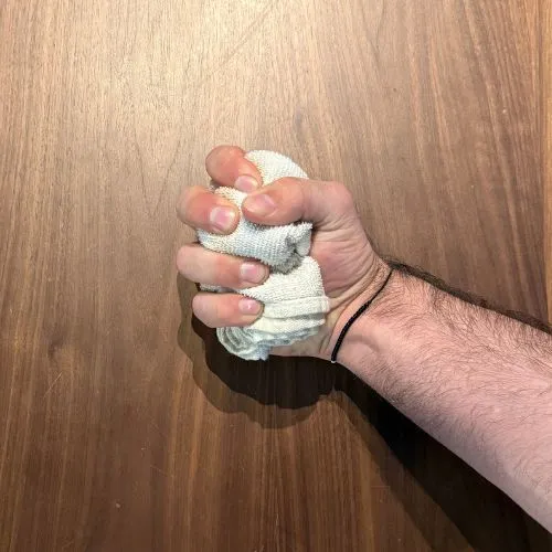
Place a rolled-up washcloth or soft ball in your palm.
Wrap your fingers and thumb around it gently, letting the object keep the hand slightly open.
Hold for several minutes as tolerated.
Tips
This is especially helpful for severe spasticity or during rest periods.
Rotate between different-sized objects for variety.
Building a Routine for Hand Stretching and Improving Spasticity
Stretching once in a while won’t bring lasting changes. To make progress, aim to include hand stretches into your daily routine. Here are some practical strategies:
Morning routine: Stretch after waking to reduce stiffness from the night.
Activity breaks: Stretch after tasks that increase tightness, such as typing or gripping objects.
Evening wind-down: Stretch before bed to help muscles relax for better sleep.
Try to commit to at least 10–20 minutes of stretching each day. Breaking it into smaller sessions can make it easier to stay consistent. Some find stretching after a hot shower to be particularly effective, as heat can help the muscles to relax.
Seeking Help from a Professional for Hand Spasticity After Stroke
If stretching feels too difficult on your own, don’t get discouraged. Physical and occupational therapists can guide you through the safest techniques and provide tools like splints or braces to support your progress.
You should contact your care team if you notice:
Severe pain during stretches
Increasing stiffness despite regular practice
Inability to safely perform stretches independently
Development of skin breakdown from prolonged tightness
Therapists can also suggest advanced techniques, such as functional electrical stimulation, to complement your stretching routine.
Final Thoughts on Hand Stretches For Spasticity After Stroke
Hand spasticity after stroke can interfere with independence and quality of life, but daily stretching is a powerful way to regain control. The eight exercises above ranging from simple palm opening to thumb and finger stretches can help reduce stiffness, improve mobility, and keep your hand more functional.
Recovery takes patience, but every small improvement matters. Whether you’re able to open your palm a little wider, rotate your wrist more smoothly, or hold objects with less effort, these changes add up over time.
By staying consistent and working within your comfort level, you’ll give your hand the best chance to improve.
Flint Rehab is leading the way in neuro-rehabilitation with products that
are backed by research and clinically proven to help you recover more
effectively from stroke, TBI, and SCI. Trusted by over 300+ rehab facilities


 Professor Kim Min-young notes, "Regulations often fail to keep pace with medical technology and only hold it back," adding, "Recently, Korea has also opened the way for stem cell therapy, but we are far behind Japan."/Courtesy of Jeon Gi-byeong