Changing stroke rehab and research worldwide now.Time is Brain! trillions and trillions of neurons that DIE each day because there are NO effective hyperacute therapies besides tPA(only 12% effective). I have 523 posts on hyperacute therapy, enough for researchers to spend decades proving them out. These are my personal ideas and blog on stroke rehabilitation and stroke research. Do not attempt any of these without checking with your medical provider. Unless you join me in agitating, when you need these therapies they won't be there.

What this blog is for:

My blog is not to help survivors recover, it is to have the 10 million yearly stroke survivors light fires underneath their doctors, stroke hospitals and stroke researchers to get stroke solved. 100% recovery. The stroke medical world is completely failing at that goal, they don't even have it as a goal. Shortly after getting out of the hospital and getting NO information on the process or protocols of stroke rehabilitation and recovery I started searching on the internet and found that no other survivor received useful information. This is an attempt to cover all stroke rehabilitation information that should be readily available to survivors so they can talk with informed knowledge to their medical staff. It lays out what needs to be done to get stroke survivors closer to 100% recovery. It's quite disgusting that this information is not available from every stroke association and doctors group.

Showing posts with label growth hormone. Show all posts

Sunday, November 23, 2025

What does growth hormone do?

 Does your competent? doctor have a protocol for its' use?

Do you prefer your doctor, hospital and board of director's incompetence NOT KNOWING? OR NOT DOING?

What does growth hormone do?

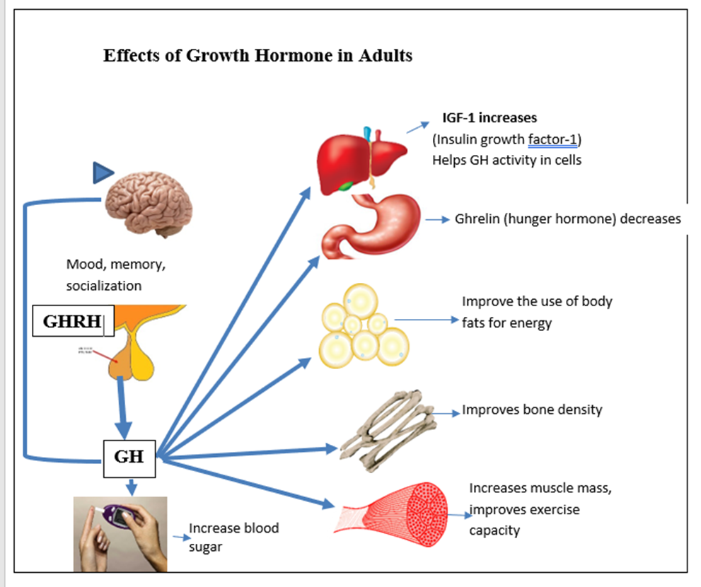
In childhood, GH is needed for growth of muscles, bones. It distributes and controls body fat and helps with glucose control.

In adulthood GH helps to decrease total body fat, improve muscle function, exercise capacity, energy, and decreases the risk of heart attacks and strokes. GH improves bone density and decreases the risk of bone fractures. Psychologic improvements in mood, interest in social activities and overall quality of life have been reported.(All these seem incredibly important for stroke recovery!)

Note:

The total amount of growth hormone produced is different for men and women.  The highest rates of growth hormone secretion occur during puberty, which is responsible for the adolescent growth spurt. Growth hormone level becomes lower as we grow older.

All pituitary functions are usually evaluated if growth hormone is suspected of being abnormal to help determine if other levels are affected.

Wednesday, April 24, 2024

Growth hormone promotes myelin repair after chronic hypoxia via triggering pericyte-dependent angiogenesis

 Ask you competent? doctor if you need myelin repair post stroke, and don't accept not knowing as an answer. YOUR FUCKING DOCTOR IS SUPPOSED TO BE COMPETENT! Only over a decade for your doctor to become competent in myelin!

In here is both myelin repair topics and whether stroke causes myelin damage.

  • myelin (73 posts to April 2011)

Growth hormone promotes myelin repair after chronic hypoxia via triggering pericyte-dependent angiogenesis


Highlights

  • GH treatment promotes myelin repair and functional recovery after hypoxia
  • GHR is selectively expressed by a subpopulation of pericytes
  • GHR-positive pericyte-tip cells lead blood vessel bridging and branching
  • GHR-positive pericytes modulate angiogenesis and govern myelination indirectly

Summary

White matter injury (WMI) causes oligodendrocyte precursor cell (OPC) differentiation arrest and functional deficits, with no effective therapies to date. Here, we report increased expression of growth hormone (GH) in the hypoxic neonatal mouse brain, a model of WMI. GH treatment during or post hypoxic exposure rescues hypoxia-induced hypomyelination and promotes functional recovery in adolescent mice. Single-cell sequencing reveals that Ghr mRNA expression is highly enriched in vascular cells. Cell-lineage labeling and tracing identify the GHR-expressing vascular cells as a subpopulation of pericytes. These cells display tip-cell-like morphology with kinetic polarized filopodia revealed by two-photon live imaging and seemingly direct blood vessel branching and bridging. Gain-of-function and loss-of-function experiments indicate that GHR signaling in pericytes is sufficient to modulate angiogenesis in neonatal brains, which enhances OPC differentiation and myelination indirectly. These findings demonstrate that targeting GHR and/or downstream effectors may represent a promising therapeutic strategy for WMI.

Graphical abstract

Thursday, April 11, 2024

Combination therapy of Epidermal Growth Factor and Growth Hormone-Releasing Hexapeptide in acute ischemic stroke: a phase I/II non-blinded, randomized clinical trial

 So with further research suggested will your competent? doctor and hospital ensure it gets done? Oh, you don't have a functioning stroke doctor or hospital that even knows about this, much less get more research done?

Combination therapy of Epidermal Growth Factor and Growth Hormone-Releasing Hexapeptide in acute ischemic stroke: a phase I/II non-blinded, randomized clinical trial

\r\nFrancisco Hernndez-Bernal,&#x;&#x;Francisco Hernández-Bernal1,2Donner Estenoz-García&#x;Donner Estenoz-García3Juan H. Gutirrez-Ronquillo&#x;Juan H. Gutiérrez-Ronquillo4Yenima Martín-Bauta&#x;Yenima Martín-Bauta1Karen Catasús-lvarez&#x;Karen Catasús-Álvarez1Mario Gutirrez-Castillo&#x;Mario Gutiérrez-Castillo4Marbelys Guevara-Rodríguez&#x;Marbelys Guevara-Rodríguez4Aliuska Castro-Jerz&#x;Aliuska Castro-Jeréz5Yoandra Fuentes-Gonzlez&#x;Yoandra Fuentes-González5Yulemis Pinto-Cruz&#x;Yulemis Pinto-Cruz3Carmen Valenzuela-Silva&#x;Carmen Valenzuela-Silva6Verena L. Muzio-Gonzlez&#x;Verena L. Muzio-González1Hctor Prez-Saad&#x;Héctor Pérez-Saad7Nelvys Subirs-Martínez&#x;Nelvys Subirós-Martínez7Gerardo E. Guilln-Nieto,&#x;Gerardo E. Guillén-Nieto8,9Diana Garcia-del-Barco-Herrera,,
&#x;&#x;Diana Garcia-del-Barco-Herrera7,9,* on behalf of the COURAGE (COmbined therapeUtic appRoAch durinG acute strokE) Research Group
  • 1Clinical Trial Direction, Center for Genetic Engineering and Biotechnology, Havana, Cuba
  • 2Department of Comprehensive General Medicine, Latin American School of Medicine (ELAM), Havana, Cuba
  • 3Neurology Department, “Antonio Luaces” Hospital, Ciego de Ávila, Cuba
  • 4Neurology Department, “Arnaldo Milián” Hospital, Santa Clara, Villa Clara, Cuba
  • 5Neurology Department, “Celia Sánchez” Hospital, Manzanillo, Gramma, Cuba
  • 6Institute of Cybernetics, Mathematics, and Physics, Havana, Cuba
  • 7Neuroprotection Project, Biomedical Research Direction, Center for Genetic Engineering and Biotechnology, Havana, Cuba
  • 8Biomedical Research Direction, Center for Genetic Engineering and Biotechnology, Havana, Cuba
  • 9Department of Physiology, Latin American School of Medicine (ELAM), Havana, Cuba

Objective: This study tested the hypothesis that a neuroprotective combined therapy based on epidermal growth factor (EGF) and growth hormone-releasing hexapeptide (GHRP6) could be safe for acute ischemic stroke patients, admitting up to 30% of serious adverse events (SAE) with proven causality.

Methods: A multi-centric, randomized, open-label, controlled, phase I-II clinical trial with parallel groups was conducted (July 2017 to January 2018). Patients aged 18–80 years with a computed tomography-confirmed ischemic stroke and less than 12 h from the onset of symptoms were randomly assigned to the study groups I (75 μg rEGF + 3.5 mg GHRP6 i.v., n=10), II (75 μg rEGF + 5 mg GHRP6 i.v., n=10), or III (standard care control, n=16). Combined therapy was given BID for 7 days. The primary endpoint was safety over 6 months. Secondary endpoints included neurological (NIHSS) and functional [Barthel index and modified Rankin scale (mRS)] outcomes.

Results: The study population had a mean age of 66 ± 11 years, with 21 men (58.3%), a baseline median NIHSS score of 9 (95% CI: 8–11), and a mean time to treatment of 7.3 ± 2.8 h. Analyses were conducted on an intention-to-treat basis. SAEs were reported in 9 of 16 (56.2%) patients in the control group, 3 of 10 (30%) patients in Group I (odds ratio (OR): 0.33; 95% CI: 0.06–1.78), and 2 of 10 (20%) patients in Group II (OR: 0.19; 95% CI: 0.03–1.22); only two events in one patient in Group I were attributed to the intervention treatment. Compliance with the study hypothesis was greater than 0.90 in each group. Patients treated with EGF + GHRP6 had a favorable neurological and functional evolution at both 90 and 180 days, as evidenced by the inferential analysis of NIHSS, Barthel, and mRS and by their moderate to strong effect size. At 6 months, proportion analysis evidenced a higher survival rate for patients treated with the combined therapy. Ancillary analysis including merged treated groups and utility-weighted mRS also showed a benefit of this combined therapy.

Conclusion: EGF + GHRP6 therapy was safe. The functional benefits of treatment in this study supported a Phase III study.

Clinical Trial Registration: RPCEC00000214 of the Cuban Public Registry of Clinical Trials, Unique identifier: IG/CIGB-845I/IC/1601.

1 Introduction

Ischemic stroke remains an important target for novel preventive and therapeutic strategies. It is estimated that 15 million people worldwide are affected by stroke every year, 5 million of them die and another 5 million of them suffer from long-term disability (1). The global population aged 65 years and over is growing faster than all other age groups, with a concomitant increase in stroke incidence (2). Furthermore, stroke among COVID-19 patients is associated with a significant risk of early mortality (3).

Currently, reperfusion therapy with thrombolytic drugs or endovascular thrombectomy represents the only approved therapeutic approach for acute stroke (4, 5). However, these approaches are associated with a narrow therapeutic window, increased risk of hemorrhagic transformation, and the high cost required to deliver these treatments (6), which limit their suitability for patients (~3%−6%), particularly in low-income countries (7). Moreover, despite treatment, recovery may be incomplete in a significant proportion of patients (8), as the vascular dynamics following recanalization do not invariably reduce tissue injury or reverse functional deficits (9, 10), and there remains scope for additional pharmacological-based neuroprotective interventions in addition to recanalization in acute ischemic stroke.

Considering the strong interdependence of elements in the neurovascular unit (11) and the limitations of neuron-protective strategies in clinical trials (12, 13), the scientific community has moved to combination strategies that seek to enhance endogenous mechanisms of neuroprotection (14). Combined therapies for stroke are likely to be more effective as they simultaneously target multiple levels of the ischemic pathophysiological cascade. However, few clinical trials have been published to date (2).

Molecules that trigger cytoprotective effects can be considered candidates for combined therapies (8). Particularly, a combined therapy based on epidermal growth factor (EGF) and growth hormone-releasing hexapeptide (GHRP6) has demonstrated benefit in preclinical contexts by activating pleiotropic endogenous mechanisms of survival and brain protection (1519). Both molecules cross the blood-brain barrier (2023), and their receptors are widely distributed in brain tissues (24, 25). EGF and GHRP6 share common properties such as anti-apoptotic (26, 27) and anti-excitotoxic effects (28, 29). In addition, both molecules have independent biological effects. Specifically, EGF promotes neurogenesis and remyelination (30), while GHRP6 induces endogenous neuroprotective factors such as growth hormone and insulin, like growth factor 1 (31).

Previous results of our group demonstrated the therapeutic benefits of this combination in animal models of multiple sclerosis (15), proximal axonopathy mimicking ALS, and focal and global ischemic stroke (16). Later, EGF+GHRP6 combined therapy improved both clinical and pathological aspects as it reduced neurological symptoms and brain infarct volume, preserving neuronal density (17). Additionally, EGF+GHRP6 combined therapy achieved similar results in a preclinical context when compared to therapeutic hypothermia (18). Moreover, both active ingredients exhibited a high safety profile in preclinical and clinical trials (32, 33).

Supported by these data, a phase I/II randomized clinical trial was designed to test the hypothesis that administration of a combined therapy based on rEGF and GHRP6 at two dose levels is safe for acute ischemic stroke (AIS) patients, admitting up to 30% of serious adverse events (SAE) in relation to proven causality. The therapeutic effect was assessed as a secondary endpoint. The current report has been written in compliance with the Consolidated Standards of Reporting Trials (CONSORT) guidelines for randomized controlled trials (34, 35).

More at link

Monday, January 3, 2022

One more good reason to exercise

 It is your doctor's responsibility to get you to be able to exercise enough in 35 days to generate GH(growth hormone), or I suppose second best would be to administer it to get the benefits. What is your doctor's solution? No solution, fire them.

One more good reason to exercise

As we start the New Year with a fervent hope that it’s better than the last two, many people are making a resolution to get more exercise. A new study suggests that might not just benefit the body, it could also help the brain. At least if you are a mouse.

Researchers at the University of Queensland Brain Institute found that 35 days of exercise could improve brain function and memory.

Futurity, Dan Blackmore, one of the lead researchers on the study, says they not only showed the benefits of exercise, but also an explanation for why it helps.

“We tested the cognitive ability of elderly mice following defined periods of exercise and found an optimal period or ‘sweet spot’ that greatly improved their spatial learning. We found that growth hormone (GH) levels peaked during this time, and we’ve been able to demonstrate that artificially raising GH in sedentary mice also was also effective in improving their cognitive skills. We discovered GH stimulates the production of new neurons in the hippocampus—the region of the brain critically important to learning and memory.

The study was published in the journal iScience.

Obviously, this is great for mice, but they hope that future research could show similar benefits for people. But don't wait for that study to come out, there's already plenty of evidence that exercising has terrific benefits for the body. Here's just seven ways it can give you a boost.


Tuesday, October 19, 2021

An exercise ‘sweet spot’ reverses cognitive deficits of ageing by growth hormone induced neurogenesis

You can have your doctor read the 45 pages at the link.

An exercise ‘sweet spot’ reverses cognitive deficits of ageing by growth hormone induced neurogenesis

 Daniel G. Blackmore, Frederik J. Steyn, Alison Carlisle, Imogen O’Keeffe, King-Year Vien, Xiaoqing Zhou, Odette Leiter, Dhanisha Jhaveri, Jana Vukovic, Michael J. Waters, Perry F. Bartlett PII: S2589-0042(21)01244-X DOI: https://doi.org/10.1016/j.isci.2021.103275 Reference: ISCI 103275 To appear in: ISCIENCE Received Date: 21 July 2021 Revised Date: 9 September 2021 Accepted Date: 12 October 2021 Please cite this article as: Blackmore, D.G., Steyn, F.J., Carlisle, A., O’Keeffe, I., Vien, K.-Y., Zhou, X., Leiter, O., Jhaveri, D., Vukovic, J., Waters, M.J., Bartlett, P.F., An exercise ‘sweet spot’ reverses cognitive deficits of ageing by growth hormone-induced neurogenesis, ISCIENCE (2021), doi: https:// doi.org/10.1016/j.isci.2021.103275. This is a PDF file of an article that has undergone enhancements after acceptance, such as the addition of a cover page and metadata, and formatting for readability, but it is not yet the definitive version of record. This version will undergo additional copyediting, typesetting and review before it is published in its final form, but we are providing this version to give early visibility of the article. Please note that, during the production process, errors may be discovered which could affect the content, and all legal disclaimers that apply to the journal pertain.

Monday, September 20, 2021

Growth Hormone (GH) Enhances Endogenous Mechanisms of Neuroprotection and Neuroplasticity after Oxygen and Glucose Deprivation Injury (OGD) and Reoxygenation (OGD/R) in Chicken Hippocampal Cell Cultures

 Many earlier pieces of research on this that obviously your doctors and hospital did no followup on to create stroke protocols for this. Our stroke associations also did nothing with this, they need to be destroyed and run by survivors.

Growth Hormone (GH) Enhances Endogenous Mechanisms of Neuroprotection and Neuroplasticity after Oxygen and Glucose Deprivation Injury (OGD) and Reoxygenation (OGD/R) in Chicken Hippocampal Cell Cultures

Academic Editor: Clive Bramham
Received25 Mar 2021
Accepted14 Aug 2021
Published16 Sep 2021

Abstract

As a classical growth promoter and metabolic regulator, growth hormone (GH) is involved in development of the central nervous system (CNS). This hormone might also act as a neurotrophin, since GH is able to induce neuroprotection, neurite growth, and synaptogenesis during the repair process that occurs in response to neural injury. After an ischemic insult, the neural tissue activates endogenous neuroprotective mechanisms regulated by local neurotrophins that promote tissue recovery. In this work, we investigated the neuroprotective effects of GH in cultured hippocampal neurons exposed to hypoxia-ischemia injury and further reoxygenation. Hippocampal cell cultures obtained from chick embryos were incubated under oxygen-glucose deprivation (OGD, <5% O2, 1 g/L glucose) conditions for 24 h and simultaneously treated with GH. Then, cells were either collected for analysis or submitted to reoxygenation and normal glucose incubation conditions (OGD/R) for another 24 h, in the presence of GH. Results showed that OGD injury significantly reduced cell survival, the number of cells, dendritic length, and number of neurites, whereas OGD/R stage restored most of those adverse effects. Also, OGD/R increased the mRNA expression of several synaptogenic markers (i.e., NRXN1, NRXN3, NLG1, and GAP43), as well as the growth hormone receptor (GHR). The expression of BDNF, IGF-1, and BMP4 mRNAs was augmented in response to OGD injury, and exposure to OGD/R returned it to normoxic control levels, while the expression of NT-3 increased in both conditions. The addition of GH (10 nM) to hippocampal cultures during OGD reduced apoptosis and induced a significant increase in cell survival, number of cells, and doublecortin immunoreactivity (DCX-IR), above that observed in the OGD/R stage. GH treatment also protected dendrites and neurites during OGD, inducing plastic changes reflected in an increase and complexity of their outgrowths during OGD/R. Furthermore, GH increased the expression of NRXN1, NRXN3, NLG1, and GAP43 after OGD injury. GH also increased the BDNF expression after OGD, but reduced it after OGD/R. Conversely, BMP4 was upregulated by GH after OGD/R. Overall, these results indicate that GH protective actions in the neural tissue may be explained by a synergic combination between its own effect and that of other local neurotrophins regulated by autocrine/paracrine mechanisms, which together accelerate the recovery of tissue damaged by hypoxia-ischemia.

1. Introduction

Ischemic stroke is a serious cerebrovascular event caused by a blockage of blood supply and oxygen to the brain, leading to damage or death of brain cells, which produces a severe neurological impairment, or even decease [1]. It is well established that cerebral ischemia induces a pathophysiological response in the neural tissue that leads to apoptotic and necrotic cell death [2], neural structural damage, and synaptic loss, which then contribute to the drastic deficiency of neurological functions [3].

In addition to its classical actions on growth and metabolism, growth hormone (GH) has been reported to play a relevant role, as a neurotrophic factor, on brain repair after traumatic brain injury (TBI) and stroke [46]. The neurotrophic actions of GH in the central nervous system (CNS) include prosurvival effects during embryonic development [7, 8], neurogenesis in the adult brain [9], structural plasticity [10, 11], and synaptogenesis [12], among others. These effects could be associated with the cognitive and motor improvement observed in TBI patients, with or without growth hormone deficiency (GHD), who received GH therapy [6, 1315].

It has been reported that after neural injury, there is an activation of local mechanisms that induce neuroprotection and neuroplasticity which, in some cases, also promote proliferation of newly born neurons and migration of neural precursor cells into the lesioned peri-infarct region [16, 17]. The cellular and molecular mechanisms behind the brain capacity to repair an infarcted region are still largely undetermined; although, the expression and release of endogenous neurotrophic factors have been shown to be significantly increased during ischemic events [1821]. Interestingly, GH is also synthesized by cells surrounding the peri-infarcted area suggesting that local autocrine/paracrine mechanisms are triggered after a neural injury [22]. Moreover, it has been shown that the expression of growth hormone receptor (GHR) is increased in the injured tissue, facilitating the neuroprotective action of this hormone [23].

Neuroprotective actions of GH treatments on either brain ischemia in vivo or oxygen-glucose deprivation (OGD) injury in vitro have been previously documented [2426]. In the hippocampus, GH significantly reduced apoptotic cell death rate after an experimental stroke [25], decreased loss of neural tissue, and increased the expression of neurotrophic factors, synaptogenesis, and myelination biomarkers, as well as the formation of new blood vessels within the peri-infarct area, and provoked an improvement in cognitive function in experimentally stroked mice [26]. Recent studies showed that the administration of GH after experimental stroke promoted neurogenesis and stimulated synaptic plasticity and angiogenesis within the peri-infarct region, which were associated with an improvement in the motor function [27]. Furthermore, GH treatment promoted remote hippocampal plasticity and enhanced cognitive recovery after cortical injury [28]. Relevantly, GH addition also increased the expression of neurotrophic factors, such as BDNF and IGF-1, which in turn could participate in the endogenous neuroprotective response that occurs after ischemic injury [4, 25, 29].

Given the increasing number of reports regarding the beneficial effects of GH treatment in patients with brain injury and stroke [4, 5, 14, 3032], as well as its therapeutic potential to treat neurodegenerative diseases [33, 34], it is pertinent to further investigate the interactions between the administration of GH and the expression of endogenous neurotrophic factors that may be involved in local neuroprotection mechanisms. Thus, the aim of the present study was to evaluate the neuroprotective role of GH in cultured hippocampal neurons that were injured by exposition to OGD and then submitted to an additional reoxygenation (OGD/R) period.

This work shows that OGD injury (24 h) significantly affects cell survival, reduces neurite outgrowth, and alters the expression of several synaptogenic markers, such as neurexins (NRXN1, NRXN3), neuroligins (NLG1), and growth associated protein 43 (GAP43), in hippocampal neurons. Interestingly, exposure of the harmed cultures to reoxygenation and normal glucose incubation conditions (OGD/R), for another 24 h, reverses most of the adverse effects of OGD. Additionally, it is shown that the expression of several neurotrophic factors (i.e., BDNF, NT-3, IGF-1, and BMP4) is significantly increased after OGD, whereas the GHR expression was upregulated only in the OGD/R condition. Furthermore, it was found that administration of GH treatments, both under OGD and OGD/R conditions, clearly stimulated significant plastic changes by promoting cell survival, enabling an increase of neurite outgrowth, and inducing a rise of synapse formation markers, in levels above those observed in the OGD/R condition. These protective neurotrophic actions of GH are probably mediated through a synergistic mechanism between GH and other endogenous neurotrophins.
 

Wednesday, January 22, 2020

Growth Hormone Promotes Motor Function after Experimental Stroke and Enhances Recovery-Promoting Mechanisms within the Peri-Infarct Area

I see nothing here that suggests that a protocol was written and distributed to all stroke hospitals in the world.  So because we have no great stroke association with a database of all research and  protocols and the means to distribute stroke research to all stroke medical professionals and every one of the 10 million yearly stroke survivors, this will fall by the wayside. 


Growth Hormone Promotes Motor Function after Experimental Stroke and Enhances Recovery-Promoting Mechanisms within the Peri-Infarct Area

Sonia Sanchez-Bezanilla 1,2 , N. David Åberg 3,4, Patricia Crock 2,5, Frederick R. Walker 1,2,6,7, Michael Nilsson 1,2,6,7,8, Jörgen Isgaard 1,3,4,*,† and Lin Kooi Ong 1,2,6,9,*,† 1 School of Biomedical Sciences and Pharmacy and the Priority Research Centre for Stroke and Brain Injury, the University of Newcastle, University Dr, Callaghan, NSW 2308, Australia; sonia.sanchezbezanilla@uon.edu.au (S.S.-B.); rohan.walker@newcastle.edu.au (F.R.W.); michael.nilsson@newcastle.edu.au (M.N.) 2 Brain and Mental Health, Hunter Medical Research Institute, Lot 1, Kookaburra Cct, New Lambton Heights, NSW 2305, Australia; patricia.crock@newcastle.edu.au 3 Department of Internal Medicine, University of Gothenburg, 405 30 Gothenburg, Sweden; david.aberg@medic.gu.se 4 Department of Internal Medicine, Region Västra Götaland, Sahlgrenska University Hospital, Blå stråket 5, 413 45 Gothenburg, Sweden 5 Department of Paediatric Endocrinology and Diabetes, John Hunter Children’s Hospital, Kookaburra Cct, New Lambton Heights, NSW 2305, Australia 6 NHMRC Centre of Research Excellence Stroke Rehabilitation and Brain Recovery, 245 Burgundy Street, Heidelberg, VIC 3084, Australia 7 Centre for Rehab Innovations, Lot 1, Kookaburra Cct, New Lambton Heights, NSW 2305, Australia 8 LKC School of Medicine, Nanyang Technological University, 50 Nanyang Ave, Singapore 639798, Singapore 9 School of Pharmacy, Monash University Malaysia, Bandar Sunway, Subang Jaya 47500, Selangor, Malaysia * Correspondence: jorgen.isgaard@medic.gu.se (J.I.); ong.linkooi@monash.edu (L.K.O.) † These authors contributed equally to this work.
Received: 20 December 2019; Accepted: 15 January 2020; Published: 17 January 2020

Abstract: Motor impairment is the most common and widely recognised clinical outcome after stroke. Current clinical practice in stroke rehabilitation focuses mainly on physical therapy, with no pharmacological intervention approved to facilitate functional recovery. Several studies have documented positive effects of growth hormone (GH) on cognitive function after stroke, but surprisingly, the effects on motor function remain unclear. In this study, photothrombotic occlusion targeting the motor and sensory cortex was induced in adult male mice. Two days post-stroke, mice were administered with recombinant human GH or saline, continuing for 28 days, followed by evaluation of motor function. Three days after initiation of the treatment, bromodeoxyuridine was administered for subsequent assessment of cell proliferation. Known neurorestorative processes within the periinfarct area were evaluated by histological and biochemical analyses at 30 days post-stroke. This study demonstrated that GH treatment improves motor function after stroke by 50%–60%, as assessed using the cylinder and grid walk tests. Furthermore, the observed functional improvements occurred in parallel with a reduction in brain tissue loss, as well as increased cell proliferation, neurogenesis, increased synaptic plasticity and angiogenesis within the periinfarct area. These findings provide new evidence about the potential therapeutic effects of GH in stroke recovery

Tuesday, September 18, 2018

Growth Hormone Deficiency Is Frequent After Recent Stroke

And what is your doctor doing about it? It is useful in our recovery.


Growth Hormone Deficiency Is Frequent After Recent Stroke

Thomas Lillicrap1, Carlos Garcia-Esperon1, Frederick Rohan Walker2, Lin Kooi Ong2, Michael Nilsson2, Neil Spratt1,2, Christopher R. Levi1,2, Mark Parsons1,2, Jörgen Isgaard2,3 and Andrew Bivard1,2*
  • 1Department of Neurology, John Hunter Hospital, University of Newcastle, Newcastle, NSW, Australia
  • 2Hunter Medical Research Institute, University of Newcastle, Newcastle, NSW, Australia
  • 3Department of Internal Medicine, Sahlgrenska Academy at the University of Gothenburg, Gothenburg, Sweden
Introduction: The incidence of pituitary dysfunction after severe ischemic stroke is unknown, however given the increasing attention to pituitary dysfunction after neurological injuries such as traumatic brain injury, this may represent a novel area of research in stroke.
Methods: We perform an arginine and human growth hormone releasing hormone challenge on ischemic stroke patients within a week of symptom onset.
Results: Over the study period, 13 patients were successfully tested within a week of stroke (baseline NIHSS 10, range 7–16). Overall, 9(69%) patients had a poor response, with 7(54%) of these patients meeting the criteria for had human growth hormone deficiency. Other measures of pituitary function were within normal ranges.
Conclusion: After major ischemic stroke, low GH levels are common and may play a role in stroke recovery.

Introduction

Enhancement of recovery after ischemic stroke has been a challenging area, with no intervention being widely adopted to promote recovery in clinical practice. In the search for innovative treatment targets post stroke, we decided to investigate pituitary function after stroke with a focus on human growth hormone (hGH). Other acute brain insults such as traumatic brain injury (TBI) and subarachnoid hemorrhage have demonstrated hypopituitarism in around one quarter of patients shortly after injury (1). Although TBI has previously been considered as a rare cause of hypopituitarism, an increased prevalence of neuroendocrine dysfunction in patients with TBI has been reported over the last 15 years as endocrinological testing has become more common in this patient group. The symptoms of hypopituitarism depend on which hormone is deficient, but for hGH, they include impaired attention and memory, reduced energy, cognitive dysfunction, a decrease in muscle mass and strength, reduced bone mass and density and cardiac dysfunction, all of which are common in stroke survivors (2). We hypothesized that patients with a severe ischemic stroke resulting in significant residual disability would experience hypopituitarism, particularly affecting hGH secretion. The aim of this study was to investigate the potential incidence of hGH deficiency from 7 days after stroke onwards.

Methods

Consecutive patients presenting to the John Hunter Hospital Emergency Department with first ever clinical diagnosis of an ischemic stroke were assessed for this study. The acute clinical diagnosis was confirmed by multi modal computed tomography (CT) which included CT angiography, CT perfusion and a noncontrast CT. Patients presenting with symptoms typical of transient ischemic attack or stroke mimics such as migraine with aura, seizures, acute confusional states, hypoglycaemia, diabetes, or patients with symptoms related to known prior stroke, or patients with a known history of conversion disorder were excluded from the study.
Baseline clinical data recorded included demographics (age, sex), vascular risk factors (hypertension, diabetes mellitus, dyslipidemia, atrial fibrillation, smoking history, prior stroke/TIA, and ischemic heart disease), clinical features such as the baseline and 24 h National Institutes of Health Stroke Scale (NIHSS), and premorbid modified Rankin Score (mRS), mRS at discharge and 90 days post stroke.
If the patient consented to the study, researchers would organize a fasting blood assessment for the next morning to be taken between 8 and 10 a.m. All enrolled patients underwent growth hormone (GH) provocative test with a GH-releasing-hormone (GHRH) and arginine infusion (3). GH reserve was assessed by GHRH (1 μg/kg bolus) plus arginine (ARG; 0.5 g/kg iv up to maximum of 30 g in 60 ml saline, infused over a 30 min period) testing. A GH response peak < 11 μg/liter in patients with BMI < 25 kg/m2, < 8 μg/liter in patients with BMI between 25 and 30 kg/m2, and < 4.2 μg/liter in those with BMI 30 kg/m2 or greater was considered diagnostic for GHD (3). From the initial 10 mL sample we will also test the levels of serum insulin-like growth factor 1 (IGF-1), thyroid function with thyroid specific hormone (TSH) and T1 and T2 and cortisol. In addition to the GH test, participants were tested for levels of TSH, T3, T4, ACTH, LH, testosterone, FSH, and cortisol. These tests were processed and analyzed by the local pathology department on the same day as the GH test.
If the patient was able, study specific assessments were also performed at the time of blood testing and included the Montreal Cognitive Assessment (MoCA) and the NIHSS.

Results

From July 2016 to July 2017, 13 patients were enrolled in this prospective study. Median time of testing was 7 days post stroke (range 4–10, Table 1) and median age was 71 (range 54–78). Median hospital admission National Institutes of Health Stroke Scale (NIHSS) was 10 (range 7–16). Seven patients had middle cerebral artery stroke, two had lacunar infarction, two patients had posterior circulation stroke, and two had brain stem stroke. The mean 24 h post stroke MRI DWI lesion was 55 mL (range 8–92 mL). The median 90 day modified Rankin Score median was 2 (range 1–4). Of the 13 patients, 7 (54%) fulfill criteria for hGH deficiency when the GH peak level was related to BMI and two patients had a borderline response (mean abnormal GH peak 4.4, Table 2). In the 8 included men, 6 showed low levels of testosterone below the normal reference range (mean low testosterone result was 3.2 nmol/L), and two showed normal levels (mean normal testosterone result was 12.8 nmol/L). Lastly, only one of the studies 5 females showed low levels of luteinising hormone (LH). FSH was elevated in 8 patients (mean abnormal FSH level 46.3, reference range 1–12) as can be expected in elderly subjects. Insulin-like growth factor 1 (IGF-1) was mostly within the normal range in the tested patients, however two patients had low IGF-1 concomitant with GH deficiency. TSH, T3, T4, and cortisol were all within normal reference ranges.