Wrong, wrong, wrong. You should be using imaging to diagnose all the damage
(dead and dying neurons), then map protocols to recover from such damage. This is so fucking easy to understand, why isn't it being done? No protocols is not a valid answer. Lots of words here but nothing useful for recovery.
https://www.news-medical.net/whitepaper/20180626/Using-Imaging-Techniques-to-Assess-the-Effects-of-a-Stroke.aspx
Cerebral ischaemia
Cerebral ischaemia is a type of stroke causing high rates of disability and mortality
1.
It occurs when blood vessels serving the brain become blocked or burst.
This prevents the affected area of the brain from receiving sufficient
oxygen and nutrients to meet its metabolic demands.
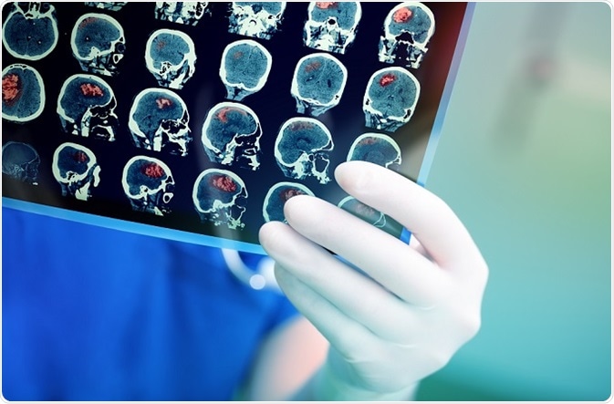 © sfam_photo/Shutterstock.com
Consequently, cells in the affected area cannot work properly and the
bodily functions for which they are responsible are impaired. Cerebral
ischaemia can affect a specific localised area of the brain or be more
widespread.
Symptoms of cerebral ischaemia may be fleeting if the interruption of
blood supply to the brain is restored quickly, but if oxygen
deprivation is prolonged the affected brain tissue will die causing
irreversible brain damage. Cerebral ischaemia symptoms include sight
disturbances, dizziness, loss of co-ordination, muscle weakness or
paralysis, and difficulty speaking.
Changes in the brain after cerebral ischaemia
Cerebral ischaemia results in changes in energy usage, disruption to
the metabolism of neurotransmitters and lipids and alterations to
protein synthesis. In addition, intracellular cytosolic calcium
concentration increases, as does the level of damaging free radicals. It
is these metabolic, biochemical and ionic perturbations that ultimately
cause neuronal death. Understanding the precise nature of these changes
may therefore help in treatments to minimise the debilitating
consequences of a stroke. However, since such a wide range of
metabolites is affected, leading to disruption of several complex
pathways, unravelling the effects of cerebral ischaemia is not
straightforward.
Furthermore, it appears that the pathological changes of stroke may
be even more far-reaching than initially thought. Research indicates
that metabolic changes occur not only in the area of the brain that has
become ischaemic but may also induce secondary effects in other areas of
the brain. Such functional impairment of the brain at a location
removed from the injury site is known as diaschisis, which appears to be
a form of shock response.
Defining and understanding the metabolic disturbances caused by
oxygen and nutrient deprivation have this been identified as key to the
development of treatments to minimise morbidity in patients suffering
cerebral ischaemia
2. With the possibility of diaschisis, the
physiological effects of cerebral ischaemia must be evaluated across the
brain and not just in the ischaemic area.
Magnetic resonance imaging and positron emission tomography have
successfully measured remote metabolic changes after cerebral ischaemia.
Such techniques have provided insight into the more far-reaching
effects of stroke in areas of the brain far-removed from the ischaemic
area. However, although cerebellar diaschisis has been well studied
3,
there are few data relating to the less common interhemispheric
diaschisis. It remains unclear whether ischaemic damage in one
hemisphere can lead to diaschisis in another hemisphere.
Measuring metabolic changes
Metabolomics is the study of biochemical pathways through the
measurement of the metabolites. By determining the relative changes in
concentration of substrate and product metabolites, it can be inferred
whether specific pathways have been up or down-regulated. This
methodology has been widely used across a range of disciplines to
investigate the effects of drugs or contaminants.
Nuclear magnetic resonance spectroscopy detects molecules based on
their chemical shift on application of a magnetic field. Since different
types of molecules give different spectral peaks, it can measure the
many different components in a complex mixture. It has been widely used
to investigate the pathological mechanisms of cerebral ischaemia, but
has not to study the effects on biochemical processes within the brain.
Investigating inter-hemisphere effects of cerebral ischemia
A recent study investigated metabolite changes after cerebral artery
occlusion in both the left and right cerebral hemispheres of rats
4. In order to simultaneously measure the quantities of numerous metabolites,
1H nuclear magnetic resonance (
1H
NMR) spectroscopy was employed for the analysis using a Bruker AVANCE
III 600 MHz NMR spectrometer. Characteristic changes in metabolites
indicated that biochemical changes had occurred in both the ischaemic
and contralateral cerebral hemispheres.
In the ischaemic cerebral hemisphere, the changes in metabolites
after cerebral ischaemia indicated an increase in anaerobic glycolysis, a
perturbation of choline metabolism, neuronal cell damage and
neurotransmitter imbalance. In the contralateral hemisphere, an increase
in anaerobic glycolysis was also observed, along with changes in energy
metabolism and alteration in the balance of neurotransmitters. Since
the two cerebral hemispheres are connected by a large mass of neural
fibres called the corpus callosum, it is likely that this is the means
by which ischaemic damage in one hemisphere can initiate metabolic
changes in the other hemisphere.
These findings indicate that interhemispheric diaschisis can indeed
occur after cerebral ischaemia. Furthermore, they support cerebral
metabolic analysis as valuable tool for understanding the biochemical
mechanisms of cerebral ischaemia and its effects on distant areas of the
brain.
References
- Anuncibay-Soto B, et al. Neuroprotection by salubrinal treatment in
global cerebral ischemia. Neural Regen Res 2016;11:1744‑1745.
- Yang M, et al. NMR analysis of the rat neurochemical changes
induced by middle cerebral artery occlusion. Talanta 2012;88:136‑144.
- Madai VI, et al. Crossed cerebellar diaschisis after stroke: can
perfusion-weighted MRI show functional inactivation? J Cereb Blood Flow
Metab 2011;31:1493‑1500.
- Ruan L, et al. Metabolite changes in the ipsilateral and
contralateral cerebral hemispheres in rats with middle cerebral artery
occlusion. Neural Regen Res 2017;12(6):931-937.


 Francisco Páscoa dos Santos
 Paul F. M. J. Verschure