Changing stroke rehab and research worldwide now.Time is Brain! trillions and trillions of neurons that DIE each day because there are NO effective hyperacute therapies besides tPA(only 12% effective). I have 523 posts on hyperacute therapy, enough for researchers to spend decades proving them out. These are my personal ideas and blog on stroke rehabilitation and stroke research. Do not attempt any of these without checking with your medical provider. Unless you join me in agitating, when you need these therapies they won't be there.

What this blog is for:

My blog is not to help survivors recover, it is to have the 10 million yearly stroke survivors light fires underneath their doctors, stroke hospitals and stroke researchers to get stroke solved. 100% recovery. The stroke medical world is completely failing at that goal, they don't even have it as a goal. Shortly after getting out of the hospital and getting NO information on the process or protocols of stroke rehabilitation and recovery I started searching on the internet and found that no other survivor received useful information. This is an attempt to cover all stroke rehabilitation information that should be readily available to survivors so they can talk with informed knowledge to their medical staff. It lays out what needs to be done to get stroke survivors closer to 100% recovery. It's quite disgusting that this information is not available from every stroke association and doctors group.

Showing posts with label Dr. Michael Tymianski. Show all posts

Friday, February 6, 2026

Neuroprotective Agent Improved Outcomes After Acute Ischemic Stroke

 WOW! Incompetent for well over a decade and still no protocol on this! (We will never get any urgency to solve this as long as we still call it neuroprotection rather than the neuronal cascade of death. Which term suggests urgency to lay people?)

  • PSD95 (8 posts to February 2012)
  • Neuroprotective Agent Improved Outcomes After Acute Ischemic Stroke

    The PSD-95 inhibitor also had a favorable safety profile in phase III trial by Nicole Lou, Senior Staff Writer, MedPage Today

    NEW ORLEANS -- Loberamisal improved functional outcomes after acute ischemic stroke in a placebo-controlled phase III trial, raising its prospects as an investigational neuroprotective therapy.

    In patients with relatively moderate strokes who went without recanalization therapies for the most part, 10-day treatment with loberamisal led to excellent functional outcomes, defined as modified Rankin Scale (mRS) scores of 0-1 at 90 days (69.7% vs 56.3% with placebo; risk ratio 1.24, 95% CI 1.12-1.36), reported Shuya Li, MD, PhD, of Beijing Tiantan Hospital in China, at the International Stroke Conference.

    What's more, the postsynaptic density (PSD)-95-targeting, dual antidepressant-anxiolytic also had a favorable safety profile based on the 1,000-person LAIS trial, Li reported.

    "The results of the study are intriguing," commented Michael Tymianski, MD, PhD, of the University of Toronto. "This treatment is aimed at stroke patients who do not fit the acute ischemic stroke treatment protocol in that they do not receive recanalization therapies."

    "The drug as used in this study seems to be administered over a 10-day period after the stroke onset in the absence of reperfusion. Thus the mechanism of action is less likely to be neuroprotection, but perhaps through some form of recovery enhancement," Tymianski told MedPage Today.

    Loberamisal marks a notable development in the long search for a good neuroprotectant with clinical efficacy for reducing secondary neuronal damage and promoting neurological recovery. The LAIS trial arrives after years of mixed or outright failed neuroprotection trials in acute ischemic stroke.

    In the ESCAPE-NA1 trial, for instance, the PSD-95 inhibitor nerinetide failed to improve stroke patients' outcomes when given in conjunction with endovascular therapy (EVT), though those not given alteplase did appear to benefit.

    More recently, the novel sublingual combination of edaravone (Radicava) with dexborneol was shown to improve functional outcomes in the phase III TASTE-SL trial from China -- though its relatively low power and nonsignificant secondary results promptly invited skepticism.

    Loberamisal is a novel dual-target, small-molecule inhibitor of the PSD-95-neuronal nitric oxide synthase interaction that selectively potentiates alpha-2 GABA A receptors. The 40-mg dose was selected for phase III study following a dose-finding study, Li reported.

    LAIS was conducted from 2024 to 2025 in 32 Chinese centers and included patients with acute ischemic stroke within 48 hours of symptom onset who had NIH Stroke Scale scores of 7-20. Patients were excluded if they were planned for EVT, were on antidepressant or anxiolytic medications, or had used other neuroprotective agents (e.g., edaravone or edaravone dexborneol) before enrollment.

    Patients were randomized to loberamisal (n=502; 40 mg daily for 10 days) or placebo (n=495).

    The randomized cohort had an average age of 63 years, and roughly 65% were men. About one in three had a history of stroke, and median baseline NIH Stroke Scale score was 8.0. The mean time from stroke onset to treatment was just over 25 hours. About 17% received IV thrombolysis in the trial.

    As for secondary efficacy outcomes, the loberamisal group was no more likely to have significant improvements in NIH Stroke Scale scores; the two study groups also had similarly few limitations in activities of daily living per the Barthel Index.

    The incidence of serious adverse events was also similar between groups (8.6% with loberamisal vs 10.7% with placebo, P=0.283), as were all-cause mortality rates (1.2% vs 2.0%, P=0.300) and discontinuation of treatment due to adverse events (1.2% for both).

    Tymianski highlighted the oddities in these findings: the efficacy outcomes outside of the mRS scores would be expected to have been positive -- "since they are other measures of clinical benefit that should co-correlate with improved functional outcome" -- and the serious adverse event rate was so low in this population, as opposed to rates approaching 50% in his own experience.

    "Future trials would need to clarify whether these results, both safety and efficacy, are applicable to populations outside of China, whether they are useful for stroke patients who also receive reperfusion therapies, whether there is potential for these agents to improve outcomes for patients with more severe strokes, and whether there is potential for this therapy to improve on functional independence (mRS 0-2), which may be of relevance to patients with moderate and severe strokes," he said.

    In any case, he added, the LAIS group is to be congratulated "for having accessed a small molecule that purportedly targets PSD-95."

    "To our knowledge, this has been elusive, and no small molecule that we have tested to date has actually bound to this target," he noted.

    Saturday, February 10, 2024

    Role of Neuroprotective Approaches in the Recanalization Era

    This is the Michael Tymianski that years ago stated this.

    Dr. Michael Tymianski, of the Toronto Western Hospital Research Institute in Canada states;  over the last half-century, there have been more than 1,000 drugs (So what are they?)aimed at preventing brain damage that have failed to work in people, even though they worked well in mice or rats. If you called it by the correct name, neuronal cascade of death, it sounds like it needs solving immediately rather than the milquetoast term 'neuroprotection'.

    NOW WE JUST NEED STROKE LEADERSHIP to solve the neuronal cascade of death.

    It's that fucking simple, solve that and you'll save hundreds of millions to billions of neurons from dying for each patient. I bet a lot of patients could easily recover if they didn't lose so many neurons due to the neuronal cascade of death.

    Role of Neuroprotective Approaches in the Recanalization Era

    Originally publishedhttps://doi.org/10.1161/STROKEAHA.123.044229Stroke. 2024;0
    First page image

    Tuesday, March 29, 2022

    Systematic Review - Combining Neuroprotection With Reperfusion in Acute Ischemic Stroke

     We've known for years that neuroprotection studies have failed. 1000+ according to Dr. Michael Tymianski, of the Toronto Western Hospital Research Institute in Canada states;  over the last half-century, there have been more than 1,000 drugs (So what are they?)aimed at preventing brain damage that have failed to work in people, even though they worked well in mice or rats. If you called it by the correct name, neuronal cascade of death, it sounds like it needs solving immediately rather than the milquetoast term 'neuroprotection'.

    Systematic Review - Combining Neuroprotection With Reperfusion in Acute Ischemic Stroke

    E. M. Vos1*, V. J. Geraedts1,2, A. van der Lugt3, D. W. J. Dippel4, M. J. H. Wermer2, J. Hofmeijer5,6, A. C. G. M. van Es7,8, Y. B. W. E. M. Roos9, C. M. P. C. D. Peeters-Scholte2 and I. R. van den Wijngaard1,2
    • 1Department of Neurology, The Hague Medical Center, The Hague, Netherlands
    • 2Department of Neurology, Leiden University Medical Center, Leiden, Netherlands
    • 3Department of Radiology and Nuclear Medicine, Erasmus University Medical Center, Rotterdam, Netherlands
    • 4Department of Neurology, Erasmus University Medical Center, Rotterdam, Netherlands
    • 5Department of Neurology, Rijnstate Hospital, Arnhem, Netherlands
    • 6Department of Clinical Neurophysiology, Technical Medical Centre, University of Twente, Enschede, Netherlands
    • 7Department of Radiology, Leiden University Medical Center, Leiden, Netherlands
    • 8Department of Radiology, The Hague Medical Center, The Hague, Netherlands
    • 9Department of Neurology, Amsterdam University Medical Center, Amsterdam, Netherlands

    Background: Clinical trials of neuroprotection in acute ischemic stroke (AIS) have provided disappointing results. Reperfusion may be a necessary condition for positive effects of neuroprotective treatments. This systematic review provides an overview of efficacy of neuroprotective agents in combination with reperfusion therapy in AIS.

    Methods: A literature search was performed on the following databases, namely PubMed, Embase, Web of Science, Cochrane Library, Emcare. All databases were searched up to September 23rd 2021. All randomized controlled trials in which patients were treated with neuroprotective strategies within 12 h of stroke onset in combination with intravenous thrombolysis (IVT), endovascular therapy (EVT), or both were included.

    Results: We screened 1,764 titles/abstracts and included 30 full reports of unique studies with a total of 16,160 patients. In 15 studies neuroprotectants were tested for clinical efficacy, where all patients had to receive reperfusion therapies, either IVT and/or EVT. Heterogeneity in reported outcome measures was observed. Treatment was associated with improved clinical outcome for: 1) uric acid in patients treated with EVT and IVT, 2) nerinetide in patients who underwent EVT without IVT, 3) imatinib in stroke patients treated with IVT with or without EVT, 4) remote ischemic perconditioning and IVT, and 5) high-flow normobaric oxygen treatment after EVT, with or without IVT.

    Conclusion: Studies specifically testing effects of neuroprotective agents in addition to IVT and/or EVT are scarce. Future neuroprotection studies should report standardized functional outcome measures and combine neuroprotective agents with reperfusion therapies in AIS or aim to include prespecified subgroup analyses for treatment with IVT and/or EVT.

    Introduction

    Intravenous thrombolytic therapy (IVT) has become standard care for acute ischemic stroke (AIS), but only a small minority (12%) of patients is eligible for IVT because of the limited time window and contra-indications (1). The absolute benefit of treatment with IVT is limited and is estimated to be 4–10% (2). In the last decade, endovascular therapy (EVT) to mechanically reopen the occluded cerebral artery has led to an improvement of functional outcome in patients with AIS caused by large vessel occlusion (LVO) (3). However, despite high recanalization rates (70–90%) chances of good functional outcome after EVT remain relatively low (30–60%) (3, 4). Currently only 10% of patients after EVT are without stroke symptoms at 3 months follow-up with a modified Rankin Scale (mRS) score of 0 (3, 5). This implies the need for additional treatment and systems-based interventions to further improve recovery of patients with AIS. A wide range of neuroprotective agents has been investigated in the past to reduce brain injury and thereby improve patient recovery. Despite promising results from animal studies, none of the tested neuroprotective strategies appeared effective in clinical trials (6). Earlier trials may have failed due to a lack of recanalization in treating patients with AIS. As ischemic tissue will eventually become infarcted if blood flow is not restored, adequate reperfusion is probably a necessary condition for recovery with or without additional neuroprotective treatments (4, 7–9). The four primary treatment targets are reduction of excitotoxicity, oxidative stress, inflammation, and cellular apoptosis (10). In patients with adequate recanalization, another targeted mechanism is reducing reperfusion injury (7, 11). With the introduction of IVT and EVT, drugs with neuroprotective properties can now be investigated in combination with reperfusion therapy. This systematic review provides an overview of randomized controlled trials (RCTs) of neuroprotective agents in AIS as an adjunct to IVT and/or EVT.

    More at link.


    Sunday, March 20, 2022

    Oxidative Stress in Ischemia/Reperfusion Injuries following Acute Ischemic Stroke

    These are all causes of the neuronal cascade of death. SOLVE THEM!  I don't care that there have been thousands of failures.

    These failures of clinical trials have been known for years. WHOM  is cataloging and solving them to make the next ones better?

    This is not going to be easy as Dr. Michael Tymianski, of the Toronto Western Hospital Research Institute in Canada states;  over the last half-century, there have been more than 1,000 drugs (So what are they?)aimed at preventing brain damage that have failed to work in people, even though they worked well in mice or rats.

    The latest here:

    Oxidative Stress in Ischemia/Reperfusion Injuries followingAcute Ischemic Stroke

    Anamaria Jurcau 1,2,* and Adriana Ioana Ardelean 3,4 1 Department of Psycho-Neurosciences and Rehabilitation, Faculty of Medicine and Pharmacy, University of Oradea, 410087 Oradea, Romania 2 Department of Neurology, Clinical Municipal Hospital Oradea, Louis Pasteur Street nr 26, 410054 Oradea, Romania 3 Department of Preclinical Sciences, Faculty of Medicine and Pharmacy, University of Oradea, Universitatii Street nr 1, 410087 Oradea, Romania; adriana_toadere@yahoo.com 4 Department of Cardiology, Clinical Emergency County Hospital Oradea, Gh. Doja Street nr 65, 410169 Oradea, Romania * Correspondence: anamaria.jurcau@gmail.com; Tel.: +40-744-600-833 

    Abstract: 

    Recanalization therapy is increasingly used in the treatment of acute ischemic stroke. However, in about one third of these patients, recanalization is followed by ischemia/reperfusion injuries, and clinically to worsening of the neurological status. Much research has focused on unraveling the involved mechanisms in order to prevent or efficiently treat these injuries. What we know so far is that oxidative stress and mitochondrial dysfunction are significantly involved in the pathogenesis of ischemia/reperfusion injury. However, despite promising results obtained in experimental research, clinical studies trying to interfere with the oxidative pathways have mostly failed. The current article discusses the main mechanisms leading to ischemia/reperfusion injuries, such as mitochondrial dysfunction, excitotoxicity, and oxidative stress, and reviews the clinical trials with antioxidant molecules highlighting recent developments and future strategies. 
    Keywords: 
    ischemic stroke; reactive oxygen species; mitochondria; oxidative stress; antioxidants; nanoparticles; stem cells 1. Introduction Although the proper management of vascular risk factors and increasing use of prophylactic measures between the 1970s and early 2000s resulted in an annual 1–1.5% decrease in stroke incidence in high income countries [1], stroke is still the main cause of disability in adults and the second leading cause of death worldwide. Moreover, the increasing prevalence of diabetes mellitus [2] and obesity [3], together with aging of the population, will probably increase the incidence of stroke [4]. The treatment of ischemic stroke entered a new era in 1995, with the release of the results of the National Institute of Neurological Disorders and Stroke trial with recombinant tissue plasminogen activator (r-tPA) [5], which showed that reestablishing blood flow in the first 3 h after stroke onset is able to salvage much of the hypoperfused cerebral tissue and improve patient outcome. Subsequent trials refined the recanalization methods by extending the time window to 4.5 h in certain subsets of patients [6], using intra-arterial thrombolysis [7], ultrasound-enhanced thrombolysis [8], or various devices for mechanical clot extraction [9] within 24 h from stroke onset [10]. However, still only 2–20% of acute ischemic stroke patients are eligible for recanalization treatments [11]. In addition, successful recanalization rates vary around 46% for intravenous thrombolysis, 63% for intra-arterial thrombolysis, or 83% for mechanical thrombectomy, and recanalization does not always translate into efficient reperfusion of the tissue at risk, leading to neurological worsening of the patient through cerebral edema, hemorrhagic transformation, or ischemia/reperfusion injuries (I/R injuries) [12]. Oxidative stress and neuroinflammation have been shown to significantly contribute to these complications. Thus, understanding the mechanisms of I/R injuries and finding ways to prevent them would significantly improve the outcome of ischemic stroke patients [13]. In the following sections we will review the literature on the pathophysiology of these injuries, focusing on oxidative stress, sources of reactive oxygen species (ROS), and neurotoxic oxidative and neuroprotective antioxidative pathways in the central nervous system (CNS). The second part reviews the studies done so far with antioxidants in ischemic stroke and discusses promising novel antioxidant approaches. 2. Oxidative Stress in the Pathophysiology of Ischemia/Reperfusion Injuries after Acute Ischemic Stroke Oxidative stress is an imbalance between the rate of generation of ROS and the biological system’s ability to clear these highly reactive molecules [14]. The cerebral tissue is particularly sensitive to oxidative stress due to a series of features, such as [15–18]: - It has the highest metabolic activity per unit weight compared to other organs; - It has low levels of antioxidant enzymes, such as superoxide dismutase, catalase, glutathione peroxidase, heme oxygenase-1; - Upon release, neurotransmitters contribute to cellular calcium overload and, through their metabolism, generate ROS; - Brain cells have a higher membrane surface/cytoplasmic volume ratio, and the plasmalemma is rich in cholesterol, is arranged in lipid rafts, has polyunsaturated fatty acids, and is very susceptible to oxidative damage; - The brain has lower levels of cytochrome c oxidase, leading to increased superoxide generation during adenosine triphosphate (ATP) generation; - Iron, released from damaged cerebral tissue, can catalyze the generation of free radicals. Restoration of blood supply to ischemic tissue, although necessary for restoration of aerobic metabolism, will also result in ROS production, which overwhelms the ability of cerebral tissue to neutralize these ROS and leads to increased oxidative stress. Research has shown that cerebral ischemia is accompanied by increased serum concentrations of markers of oxidative stress [19–21]. The main ROS are superoxide anions, (O2 −), hydroxyl radicals (OH−), and hydrogen peroxide (H2O2) [22], stemming from the activity of mitochondria, cyclooxygenases, lipoxygenases, nitric oxide synthases (NOSs), NADPH oxidase (NOX), and xanthine oxidase [23]. Once generated, ROS interact with various biological molecules: - ROS oxidize, degrade, or cleave proteins, leading to protein aggregation, modifications in ion channel activities, and enzyme inactivation [24]. - By attacking the carbon–carbon bonds of polyunsaturated fatty acids, ROS initiate lipid peroxidation, a self-propagating chain of events leading to the generation of unstable lipid radicals which further react with oxygen to form lipid peroxyl radicals [25]. Peroxidation of membrane lipids alters the bi-layer thickness, membrane fluidity, and membrane permeability. - ROS can directly damage deoxyribonucleic acids (DNA) by causing double strand breaks, structural changes, DNA mutations, or protein-DNA cross-links [26]. - They also regulate several apoptosis and necrosis signaling cascades. ROS can activate p53, a key molecule in ROS-induced cell death [27], which, in turn, upregulates PUMA (p53 upregulated modulator of apoptosis). ROS can open the mitochondrial permeability transition pore (MPTP), leading to mitochondrial swelling and cytochrome c release, thereby initiating apoptosis [28]. The MAPK (mitogen activated protein kinase) pathway, also triggered by ROS, has 3 main members: c-Jun NH2-terminal kinase (JNK), extracellular signal-regulated kinase 1/2 (ERK 1/2), and p38 MAPK. While ERK 1/2 has a controversial role in cell death and appears to be rather neuroprotective against ischemia/reperfusion injuries [15], JNK and p38 MAPK, activated by ROS through ASK1 (apoptosis signal-regulating kinase 1), significantly contribute to apoptosis during reperfusion after an ischemic insult [29,30]. 2.1. Mitochondria as a Source of ROS and Their Implication in Cerebral Ischemia/Reperfusion Injuries Mitochondria, the powerhouse of the cell, generate over 90% of the ATP in the brain through beta-oxidation of fatty acids, the Krebs cycle, and oxidative phosphorylation (OxPhos) [31]. They also use pyruvate from cytosolic glycolysis to reduce flavin adenine dinucleotide and nicotinamide adenine dinucleotide, which serves in energy transfer to the electron transport chain (ETC) [32]. The mitochondrial electron transport chain (ETC) consists of a series of protein complexes situated in the inner mitochondrial membrane which use the electrons removed by reduced nicotinamide adenine dinucleotide (NADH) and flavin adenine dinucleotide (FADH2) from the Krebs cycle to pump protons from the matrix into the intermembrane space, thereby generating a potential gradient across the inner mitochondrial membrane, which will be used in the final step of OxPhos to synthesize ATP [33]. NADH binds to NADH dehydrogenase (complex I), located on the inner mitochondrial membrane, and donates two electrons which will be passed down to ubiquinone to produce ubiquinol, a process coupled with the translocation of four protons from the matrix through the inner mitochondrial membrane [34]. Complex II, or succinate dehydrogenase, also participates in the Krebs cycle and contains FAD as a prosthetic group. It oxidizes succinate to fumarate and reduces ubiquinone [35]. Ubiquinol diffuses through the inner mitochondrial membrane and donates its electrons to cytochrome c reductase (complex III), which passes these electrons onto two molecules of cytochrome c while translocating two protons from the mitochondrial matrix and depositing an additional two protons in the intermembrane space [36]. At complex IV (cytochrome c oxidase), four cytochrome c molecules donate each one electron which will serve to form two H2O molecules from one O2 molecule, a process coupled with pumping of four protons from the matrix into the intermembrane space [37]. The final step is the synthesis of ATP from ADP and phosphate, achieved by ATP synthase (complex V), which uses the energy of the proton electrochemical gradient in a complex process, the elucidation of which led Boyer and Walker to achieve the 1997 Nobel Prize in Chemistry [38]. The transfer of protons from the mitochondrial matrix to the intermembrane space by the reactions of complexes I, III, and IV establishes a negative potential difference (∆Ψm) of 150–180 mV (with respect to the cytosol) across the inner mitochondrial membrane, which, together with the pH difference, drives complex V to generate ATP and cytosolic calcium ions to accumulate via the mitochondrial calcium uniporter in the matrix [32,39], where calcium stimulates the activity of dehydrogenases in the Krebs cycle and modulates the function of complexes IV and V [40]. As such, the balance between phosphorylation and dephosphorylation of the OxPhos complexes as well as intramitochondrial calcium concentrations maintain the cellular respiration rate and the ∆Ψm [41] by interfering with their electron transfer kinetics and allosteric regulation by ATP and ADP (adenosine diphosphate) [42]. Under normal conditions, more than 90% of oxygen is reduced to water, while about 2% of electrons may leak from complexes I and III and react with oxygen, generating superoxide anions [41]. During ischemia, the intramitochondrial calcium levels increase [42], activating mitochondrial phosphatases and leading to dephosphorylation of the OxPhos complexes, especially of cytochrome c and of cytochrome c oxidase [43], and ultimately to loss of allosteric inhibition by ATP [41]. Because oxygen as the final electron acceptor is lacking, OxPhos is maximally activated in a feed-forward mechanism. Upon restoration of oxygen supply, increased OxPhos activity restores ∆Ψm within 1 min and cellular levels of ATP within 15 min [44], after which it hyperpolarizes the mitochondrial membrane potential with dramatic effects on ROS production. Research has shown that a 10 mV increase in the ∆Ψm above 140 mV leads to a 70–90% increase in the generation of ROS [45].
     
    More at link.

    Tuesday, January 25, 2022

    The nutraceutical potential of omega-3 alpha-linolenic acid in reducing the consequences of stroke

     You can't do anything with this until maybe 50 years from now when your doctor finally gets around to reading this research and creates protocols from it. Hope you aren't dead yet.

    These failures of clinical trials have been known for years. WHOM  is cataloging and solving them to make the next ones better?

    This is not going to be easy as Dr. Michael Tymianski, of the Toronto Western Hospital Research Institute in Canada states;  over the last half-century, there have been more than 1,000 drugs (So what are they?)aimed at preventing brain damage that have failed to work in people, even though they worked well in mice or rats.

    The nutraceutical potential of omega-3 alpha-linolenic acid in reducing the consequences of stroke

    Affiliations

    Abstract

    Stroke is a worldwide major cause of mortality and morbidity. Preclinical studies have identified over 1000 molecules with brain-protective properties. More than 200 clinical trials have evaluated neuroprotective candidates for ischemic stroke yet, to date almost all failed, leading to a re-analysis of treatment strategies against stroke. An emerging view is to seek combinatory therapy, or discovering molecules able to stimulate multiple protective and regenerative mechanisms. A pertinent experimental approach to identify such candidates is the study of brain preconditioning, which refers to how the brain protects itself against ischemia and others stress-inducing stimuli. The recent discovery that nutrients like alpha-linolenic acid (ALA is an essential omega-3 polyunsaturated fatty acid required as part of our daily diet), may be an efficient brain preconditionner against stroke fosters the novel concept of brain preconditioning by nutraceuticals. This review stresses the underestimated role of nutrition in preventing and combating stroke. Although there is a consensus that increased consumption of salt, fatty foods and alcoholic beverages may promote pathologies like hypertension, obesity and alcoholism - all of which are well known risk factors of stroke - few risk factors are attributed to a deficiency in an essential nutrient in the diet. The ALA deficiency observed in the Western modern diets may itself constitute a risk factor. This review outlines how ALA supplementation by modification of the daily diet prevented mortality and cerebral damage in a rodent model of ischemic stroke. It also describes the pleiotropic ability of ALA to trigger responses that are multicellular, mechanistically diverse, resulting in neuronal protection, stimulation of neuroplasticity, and brain artery vasodilation. Overall, this review proposes a promising therapeutic opportunity by integrating a nutritional-based approach focusing on enriching the daily diet in ALA to prevent the devastating damage caused by stroke.

    Keywords: Brain preconditioning; Functional food; Ischemia; Neurogenesis; Neuroprotection; Synaptogenesis.


    Sunday, July 25, 2021

    Cerebroprotection for Acute Ischemic Stroke: Looking Ahead

     If we had ANY LEADERSHIP AT ALL IN STROKE, we would have assigned researchers to solve this way back when years ago Dr. Michael Tymianski of the Toronto Western Hospital Research Institute in Canada referenced 1000+ failed neuroprotective clinical trials. Of course nobody knows of them and what knowledge they provided, but your doctor should know every one of those failed trials. 

    This tells us nothing we didn't know years ago, if we didn't try solving it then we won't solve it now since we still have fucking crapola for stroke leadership. 

    Oops, I'm not playing by the polite rules of Dale Carnegie,  'How to Win Friends and Influence People'. 

    Telling stroke medical persons they know nothing about stroke is a no-no even if it is true. 

    Politeness will never solve anything in stroke. Yes, I'm a bomb thrower and proud of it. Someday a stroke 'leader' will try to ream me out for making them look bad by being truthful , I look forward to that day.


    Cerebroprotection for Acute Ischemic Stroke: Looking Ahead

    Originally publishedhttps://doi.org/10.1161/STROKEAHA.121.032241Stroke. ;0:STROKEAHA.121.032241

    We search for ischemic stroke treatment knowing we have failed—intensely and often—to translate mechanistic knowledge into treatments that alleviate our patients’ functional impairments. Lessons can be derived from our shared failures that may point to new directions and new strategies. First, the principle criticisms of both preclinical and clinical assessments are summarized. Next, previous efforts to develop single-mechanism treatments are reviewed. Finally, new definitions, novel approaches, and different directions are presented. In previous development efforts, the basic science and preclinical assessment of candidate treatments often lacked rigor and sufficiency; the clinical trials may have lacked power, rigor, or rectitude; or most likely both preclinical and clinical investigations were flawed. Single-target agents directed against specific molecular mechanisms proved unsuccessful. The term neuroprotection should be replaced as it has become ambiguous: protection of the entire neurovascular unit may be called cerebral cytoprotection or cerebroprotection. Success in developing cerebroprotection—either as an adjunct to recanalization or as stand-alone treatment—will require new definitions that recognize the importance of differential vulnerability in the neurovascular unit. Recent focus on pleiotropic multi-target agents that act via multiple mechanisms of action to interrupt ischemia at multiple steps may be more fruitful. Examples of pleiotropic treatments include therapeutic hypothermia and 3K3A-APC (activated protein C). Alternatively, the single-target drug NA-1 triggers multiple downstream signaling events. Renewed commitment to scientific rigor is essential, and funding agencies and journals may enforce quality principles of rigor in preclinical science. Appropriate animal models should be selected that are suited to the purpose of the investigation. Before clinical trials, preclinical assessment could include subjects that are aged, of both sexes, and harbor comorbid conditions such as diabetes or hypertension. With these new definitions, novel approaches, and renewed attention to rigor, the prospect for successful cerebroprotective therapy should improve.


    Sunday, February 21, 2021

    The Next Step in the Treatment of Stroke

    So if they have solved the failures of 1000+ neuroprotective trials, then every single stroke hospital needs the protocols.

    Well, years ago Dr. Michael Tymianski of the Toronto Western Hospital Research Institute in Canada referenced 1000+ failed neuroprotective clinical trials. Of course nobody knows of them and what knowledge they provided, but your doctor should know every one of those failed trials.


    The Next Step in the Treatment of Stroke

    Nathanael Matei 1
    , Justin Camara2 and John H. Zhang2,3,4
    *
    1 Department of Ophthalmology, University of Southern California, Los Angeles, CA, United States, 2 Department of
    Physiology and Pharmacology, Loma Linda University, Loma Linda, CA, United States, 3 Department of Anesthesiology,
    Loma Linda University, Loma Linda, CA, United States, 4 Department of Neurosurgery, Loma Linda University, Loma Linda,
    CA, United States
    Although many patients do not receive reperfusion therapy because of delayed
    presentation and/or severity and location of infarct, new reperfusion approaches are
    expanding the window of intervention. Novel application of neuroprotective agents
    in combination with the latest methods of reperfusion provide a path to improved
    stroke intervention outcomes. We examine why neuroprotective agents have failed to
    translate to the clinic and provide suggestions for new approaches.
    New developments
    in recanalization therapy in combination with therapeutics evaluated in parallel animal
    models of disease will allow for novel, intra-arterial deployment of therapeutic agents over
    a vastly expanded therapeutic time window and with greater likelihood success. Although
    the field of neuronal, endothelial, and glial protective therapies has seen numerous large
    trials, the application of therapies in the context of newly developed reperfusion strategies
    is still in its infancy. Given modern imaging developments, evaluation of the penumbra
    will likely play a larger role in the evolving management of stroke. Increasingly more
    patients will be screened with neuroimaging to identify patients with adequate collateral
    blood supply allowing for delayed rescue of the penumbra. These patients will be ideal
    candidates for therapies such as reperfusion dependent therapeutic agents that pair
    optimally with cutting-edge reperfusion techniques

    Tuesday, November 26, 2019

    Here's Why Drugs That Work So Well in Mouse Brains Often Fail Miserably in Humans

    So our stroke leadership should identify which research needs to be redone. But since we have NO STROKE LEADERSHIP, nothing will occur. 

    Did your stroke leadership do anything with this from last year?

    Drugs That Work In Mice Often Fail When Tried In People Sept. 2018

     These are probably the candidates needing rework, your doctor should be able to instantly identify them all if they are up-to-date in stroke research.  

    Well, years ago Dr. Michael Tymianski of the Toronto Western Hospital Research Institute in Canada referenced 1000+ failed neuroprotective clinical trials. Of course I don't know what they are, but your doctor should know every one of those failed trials.

    Here's Why Drugs That Work So Well in Mouse Brains Often Fail Miserably in Humans


    Neuroscientists face a major obstacle in developing drugs to treat brain disorders — if the drugs work really well on mice, they often fall short when humans are treated. Now, a new study suggests a potential reason why:  Brain cells in mice turn on genes that are very different from the ones in human brain cells.
    Mice and humans have evolutionarily conserved brains, meaning they have very similar brain architectures made up of similar types of brain cells. In theory, that makes mice ideal test subjects for neuroscientists, who don't typically have the ability to peer into living human brains.


    Yet for mysterious reasons, treatments that worked beautifully in the mouse brain often don't pan out when tested in humans. 
    To figure out why that may be, a group of scientists from the Allen Institute for Brain Science in Seattle analyzed brains donated from deceased people and brain tissue donated by epilepsy patients after brain surgery. They specifically looked at a part of the brain called the medial temporal gyrus, which is involved in language processing and deductive reasoning.
    Researchers sorted through nearly 16,000 cells from this brain region and identified 75 different cell types. When they compared the human cells with a data set of mouse cells, they found that mice had counterparts that were similar to almost all of those human brain cells.
    But when they looked at which genes were switched on or off inside those cells, they found stark differences between the mouse and human cells.
    For example, serotonin is a neurotransmitter — or brain chemical — that regulates appetite, mood, memory and sleep. It does so by binding to brain cells via a receptor on the cell surface, which acts like a glove that is made to catch a baseball.
    But a mouse's serotonin receptors are not found on the same cells that they're found in humans, the researchers discovered. So a drug that increases serotonin levels in the brain, such as those used to treat depression, might deliver it to vastly different cells in mice than in humans.
    They also found differences in the expression of genes that help build connections between neurons. In essence, the cellular roadmap in our brains may look very different from what it looks like in a mouse.
    "The bottom line is there are great similarities and differences between our brain and that of the mouse," co-senior author Christof Koch, the chief scientist and president of the Allen Institute for Brain Science, said in a statement. "One of these tells us that there is great evolutionary continuity, and the other tells us that we are unique."
    "If you want to cure human brain diseases, you have to understand the uniqueness of the human brain," he added. The findings were published yesterday (Aug. 21) in the journal Nature.
    Originally published on Live Science.

    Wednesday, August 28, 2019

    Here's Why Drugs That Work So Well in Mouse Brains Often Fail Miserably in Humans

    Well, years ago Dr. Michael Tymianski of the Toronto Western Hospital Research Institute in Canada referenced 1000+ failed neuroprotective clinical trials. Of course I don't know what they are, but your doctor should know every one of those failed trials. 

    Everyone of those failed trials should be looked at based on this new data and rerun. But that assumes we have a great stroke association and leader we can talk to. Well instead we have fucking failures of stroke associations doing nothing to get to 100% recovery.  

    Here's Why Drugs That Work So Well in Mouse Brains Often Fail Miserably in Humans

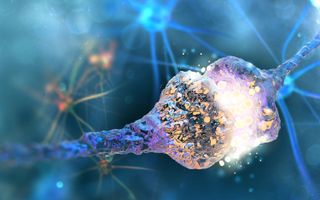
    A synapse where a signal travels from one neuron to the next.
    (Image: © Shutterstock)
    Neuroscientists face a major obstacle in developing drugs to treat brain disorders — if the drugs work really well on mice, they often fall short when humans are treated. Now, a new study suggests a potential reason why:  Brain cells in mice turn on genes that are very different from the ones in human brain cells.
    Mice and humans have evolutionarily conserved brains, meaning they have very similar brain architectures made up of similar types of brain cells. In theory, that makes mice ideal test subjects for neuroscientists, who don't typically have the ability to peer into living human brains.
    Yet for mysterious reasons, treatments that worked beautifully in the mouse brain often don't pan out when tested in humans.
    Related: 7 Ways to Trick Your Brain
    CLOSE
    To figure out why that may be, a group of scientists from the Allen Institute for Brain Science in Seattle analyzed brains donated from deceased people and brain tissue donated by epilepsy patients after brain surgery. They specifically looked at a part of the brain called the medial temporal gyrus, which is involved in language processing and deductive reasoning.
    Researchers sorted through nearly 16,000 cells from this brain region and identified 75 different cell types. When they compared the human cells with a data set of mouse cells, they found that mice had counterparts that were similar to almost all of those human brain cells.
    But when they looked at which genes were switched on or off inside those cells, they found stark differences between the mouse and human cells.
    For example, serotonin is a neurotransmitter — or brain chemical — that regulates appetite, mood, memory and sleep. It does so by binding to brain cells via a receptor on the cell surface, which acts like a glove that is made to catch a baseball.
    But a mouse's serotonin receptors are not found on the same cells that they're found in humans, the researchers discovered. So a drug that increases serotonin levels in the brain, such as those used to treat depression, might deliver it to vastly different cells in mice than in humans.
    They also found differences in the expression of genes that help build connections between neurons. In essence, the cellular roadmap in our brains may look very different from what it looks like in a mouse.
    "The bottom line is there are great similarities and differences between our brain and that of the mouse," co-senior author Christof Koch, the chief scientist and president of the Allen Institute for Brain Science, said in a statement. "One of these tells us that there is great evolutionary continuity, and the other tells us that we are unique."
    "If you want to cure human brain diseases, you have to understand the uniqueness of the human brain," he added. The findings were published yesterday (Aug. 21) in the journal Nature.
    Originally published on Live Science.

    Sunday, August 25, 2019

    Neuroscientists publish a ‘parts list’ for the brain, detailing differences between mice and humans

    Based on this ask your doctor to explain EXACTLY why these referenced trials failed and what is being done to fix them. I most certainly am not doing a damn thing with this, I'm not employed in the area and not medically trained for any of this.  I'm even less than a laboratory rat as Amy Farber puts it. This just proves once again what Amy Farber has to say.
    For the past five years Farber has been battling not only her own disease but also the wall of resistance erected by those who believe that a patient can make about as much of a meaningful contribution to the process of scientific discovery as a laboratory rat.

    Well, years ago Dr. Michael Tymianski of the Toronto Western Hospital Research Institute in Canada referenced 1000+ failed neuroprotective clinical trials. Of course I don't know what they are, but your doctor should know every one of those failed trials.

    Alan Boyle,GeekWire Wed, Aug 21 1:00 PM EDT

    Wednesday, April 3, 2019

    The future of stroke patients may depend on the part-time job of a Canadian surgeon

    This points out the complete lack of stroke leadership. Leaders would take these promising ideas and run them to completion.  My god, do we have incompetency in stroke, 6 years and nothing seems to have happened.

    What happened to this?

    Safety and efficacy of NA-1 in patients with iatrogenic stroke after endovascular aneurysm repair (ENACT): a phase 2, randomised, double-blind, placebo-controlled trial  October 2012 

    Is this enough to push out to all stroke hospitals?
    Who is going to do that? I want a name.

    The future of stroke patients may depend on the part-time job of a Canadian surgeon

    By Oliver Staley
    There are roughly 100 billion neurons in the human brain.(Wrong, 80 billion) These microscopic cells transmit and process information we receive from the outside world and turn our thoughts into action. They are responsible for how we talk, how we move, and how we think. Neurons are, in many ways, what make us us.
    Strokes kill neurons. By starving them of the blood that carries glucose and oxygen, strokes trigger a biochemical cascade that destroys neurons in vast numbers. Ischemic strokes—the most common form, caused by a blocked blood vessel—kill an average of 1.9 million neurons for every minute the patient is untreated. Those dead neurons add up, and in 10 hours, stroke patients can lose as many neurons as they would in 36 years of normal aging.
    Roughly 15 million men, women, and children suffer strokes every year, and about half of them are fatal.(1/3 are fatal) Stroke is the second-leading killer globally, after its close cousin, heart disease, and far more deadly than cancer and the most life-threatening communicable diseases like AIDS and malaria.
    Global Rank Cause Deaths (millions) % of total deaths
    1 Ischemic heart disease 9.43 16.6
    2 Stroke 5.78 10.2
    3 Chronic obstructive pulmonary disease 3.04 5.3
    4 Lower respiratory infections 2.96 5.2
    5 Alzheimer’s disease and other dementias 1.99 3.5
    6 Trachea, bronchus, and lung cancers 1.71 3.0
    7 Diabetes 1.60 2.8
    8 Road injury 1.40 2.5
    9 Diarrheal diseases 1.38 2.4
    10 Tuberculosis 1.29 2.3
    Yet despite the enormous toll of stroke, the pharmaceutical industry has been virtually powerless to treat it. After decades spent pouring billions of dollars into the pursuit of drugs to protect neurons after strokes without success, most drug companies abandoned the field by the mid-2000s.
    Today, fewer than 5% of all stroke victims worldwide receive any treatment beyond basic palliative care, and the lack of effective stroke drugs remains one of the most glaring unmet needs in medicine.
    Stroke is a maddeningly complex problem. The intricacy of the brain, the need for immediate action, and the variability of both strokes and the people who have them make designing and testing drugs an enormous challenge. But the medical establishment has failed stroke patients not just because the research is hard, but because of misaligned incentives, the financial pressures of an industrial drug-development model, and sloppy science.

    Stroke nihilism

    “Time is brain” is a longtime cliche among stroke professionals, but it’s largely true.
    Death follows when a stroke causes the brain to swell, starving it of oxygen, or because the stroke destroys the body’s ability to regulate breathing or blood flow. Others die from complications like pneumonia, which can affect up to one-third of all stroke patients. Stroke weakens the immune system, making it harder for the body to fight lung infections that can occur when stroke victims, who can no longer swallow properly, wind up with food, water, or saliva in their lungs.
    For most of history, health workers had no way to help stroke victims. Once a stroke was identified, the patient was made comfortable and family members were given the bad news. Stroke was viewed as a dead end—for patients, for researchers, and for neurologists looking for solutions—and stroke nihilism still permeates the medical establishment.
    The bulk of progress in reducing stroke deaths has come from prevention, particularly the introduction of medicines to lower high blood pressure, a leading cause of stroke.
    The first—and to date only—medical breakthrough for stroke treatment came when a drug called tissue plasminogen activator (tPA, sold globally under the brand names Activase and Actilyse) was approved by the US Food and Drug Administration (FDA) in 1996.
    The goal of tPA is “reperfusion,” the act of returning blood flow to the injured part of the brain. While neurons in the immediate vicinity of the stroke can’t be saved, there’s a larger zone, called the ischemic penumbra, that can be rescued if blood flow can be restored. The longer the penumbra is deprived of blood, the less brain there is to save.
    With tPA, emergency-room doctors at last had a way to treat patients. But, as with most things in the world of stroke, there were complications.
    In this case, the issue was that there are two kinds of stroke. While the majority (about 85% in the US) are ischemic and caused by a blockage that can potentially be treated with tPA, the rest are hemorrhagic, caused by a ruptured blood vessel, and tPA can be be fatal in these strokes because it prevents the blood from clotting. (For that reason, the drug is also not given to patients on blood thinners or who have other complications. As many as 65% ischemic stroke patients are not eligible for tPA).
    Doctors can’t administer tPA without determining the nature of the stroke and that can only be done by examining the brain with a CT scan or some other advanced brain-imaging device. There are less than 4.5 hours after the onset of stroke for doctors to  administer the drug—in many cases, not nearly enough time for a patient to get scanned. For patients over 80 or those who had a previous stroke, the window is only three hours.

    Read more: With the pharma industry’s repeated failures to develop stroke treatments, stroke doctors have turned to mechanical devices that can clear blocked blood vessels in the brain.

    Further, tPA is expensive. The drug, developed by Genentech, has no generic competition, and a 100 milligram vial used in a typical treatment can cost more than $8,300. It also needs to be refrigerated, a challenge for clinics in some parts of the world. As a result, the use of tPA is limited to affluent nations with sophisticated healthcare systems, and even then it is only rarely administered. Since its introduction, tPA has also been plagued by doubts about its safety, stemming from long-standing criticisms of its initial clinical trials. As a result, some doctors won’t prescribe it, even in eligible patients. Fewer than 5% of patients diagnosed with ischemic stroke in the US received the drug, according to a 2014 study. In poorer parts of the world, the number is closer to zero.
    Despite tPA’s limited reach, it’s enormously profitable, estimated to make $1.5 billion (pdf) in revenue this year for Roche, the Swiss pharma giant that owns Genentech.
    “The lesson they learned is that they should pursue something else.”
    Given tPA’s limitations—and the enormous potential market—researchers have focused on finding a drug that preserves neurons until the brain is reperfused. These drugs, called “neuroprotective agents,” could either save the brain cells in the penumbra until the brain heals, or extend the window of time to preserve neurons in stroke patients until a clot is dissolved by tPA or removed with a mechanical device. In theory, a neuroprotective drug that could be given safely to the 15 million victims of ischemic and hemorrhagic stroke each year—and that could be administered without scanning them first—could generate many times tPA’s revenues.
    Jeffrey Saver, a University of California-LA neurologist at the forefront of stroke research for decades, calls neuroprotection the “Holy Grail” of stroke treatment, and like that sacred relic, its pursuit has been an epic tale of frustration and failure.
    According to one landmark study, 1,026 potential neuroprotective drugs were tested between 1957 and 2003, in 8,516 separate experiments. Researchers experimented with aged-garlic extracts, uric acid, and compounds engineered from pigs’ brains. Their trials have alluring names, built out of complicated acronyms, that suggest important science is taking place: VENUS, ACTION, SAINT.
    None worked.
    Those failures cost billions of dollars and wasted the productive years of thousands of scientists. Worse, they salted the ground for future research, ushering in what one researcher called “the nuclear winter” for neuroprotection research. The pharma industry saw more lucrative opportunities elsewhere, and moved on.
    “Sadly, the lesson they learned is that they should pursue something else,” says Myron Ginsberg, a neurologist at the University of Miami who has studied the industry’s failures.
    But not all scientists accepted that conclusion. On the fringes of industrial medicine, one neurosurgeon has spent the last two decades doggedly developing a neuroprotective agent.

    The great white north of neuroscience

    The best hope for stroke patients may come not from the giant research labs of industrial pharma, or the biotech hotbeds of Boston or San Francisco, but from the relative backwater of Toronto, Ontario.
    Michael Tymianski, now 55, has been working on his drug, called NA-1, since the late 1990s, when researchers were still infused with optimism about developing a stroke treatment. A tall, balding man with a furious work ethic, Tymianski poured himself into developing NA-1 while holding down his day job as a neurosurgeon at a Toronto hospital. His plan was always to develop the drug to the point where it could be tested in humans, then sell it to a pharma company. But no buyers materialized, and eventually Tymianski stopped looking.

    Thursday, February 21, 2019

    Brain protein critical to recovery from stroke identified

    WHOM  is the person responsible for shepherding this through human clinical studies to a translational protocol?  With no one identified it will fall thru the cracks like the thousands of other research studies that showed promise in animals. Well, years ago Dr. Michael Tymianski of the Toronto Western Hospital Research Institute in Canada referenced 1000+ failed neuroprotective clinical trials. Of course nobody knows of them and what knowledge they provided, but your doctor should know every one of those failed trials. 

    The answers are out there if someone with a few functioning brain cells would put together a strategy to follow up promising research. We have to direct the research being done for stroke, we can't let researchers flail about.

    Brain protein critical to recovery from stroke identified

    Every 40 seconds, someone in the United States suffers a stroke and available therapies, such as clot busting drugs or clot removal devices, are focused on limiting the extent of brain damage. Now, research from the University of Pittsburgh School of Medicine and the VA Pittsburgh Healthcare System shows that a brain protein called UCHL1 may be critical to how nerve cells repair themselves after stroke damage. The research, conducted in animal models, could aid in the development of therapies that enhance stroke recovery by improving the underlying biological repair process.
    "Even though traditional stroke therapies are very effective when available, the treatment must be started in the first hours after a stroke and most patients are not able to get these treatments. So there is a clear need for new approaches that can improve recovery days after a patient experiences a stroke," said co-senior author Steven Graham, M.D., Ph.D., professor of neurology at Pitt's School of Medicine, and associate chief of staff for research at VA Pittsburgh. "We think we have identified a protein that is at the root of how the brain recovers from stroke, making it an attractive target for developing drugs that help improve recovery."
    UCHL1 is an enzyme that is highly active in the brain and plays a role in clearing away abnormal proteins. Mutations in the gene coding for UCHL1 have been thought to cause motor function deficits in humans. Previous research from Graham's lab had provided some hints as to UCHL1's function, showing that cyclopentenone prostaglandins (CyPgs) - fatty acid molecules - released in nerve cells after a stroke bind to UCHL1 and impair its function.
    Graham teamed up with Feng Zhang, Ph.D., an assistant professor of neurology at Pitt's School of Medicine and a co-senior author on the current study published in the Proceedings of the National Academy of Sciences, to tease out the exact role of UCHL1 in stroke and to determine if it could be a viable drug target.
    The researchers created a mouse model in which they inserted an altered version of the UCHL1 gene that was resistant to the effects of the CyPgs. They then surgically modelled the effect of a stroke in both genetically engineered and normal mice to compare how the nerve cells recovered.
    Preventing CyPgs from inhibiting UCHL1 decreased the amount of injury to the axons after stroke when compared to normal mice. Axons - the long cables projecting outward from the center of the nerve cell - are needed to carry electrical signals and connect to other neurons and make up the bulk of the 'white matter' in the brain.
    Further experiments showed that keeping UCHL1 active after a stroke helped preserve the function of neurons and brain tissue by activating cellular repair mechanisms that quickly cleaned up damaged proteins, preventing further nerve cell loss. The mice with the resistant form of UCHL1 also had improved recovery of waking, balance and other motor functions.
    "While most stroke therapies focus on preventing neuronal death, preserving axonal integrity and decreasing white matter injury could be equally important for improved recovery," said Graham, who also is a neurologist at the UPMC Stroke Institute. "UCHL1 is a central player in that process."
    Graham and his colleagues are now engaged in efforts to identify new drugs that could prevent CyPgs from binding to UCHL1 or to replace damaged UCHL1 proteins with a derivative that can be given intravenously.