Changing stroke rehab and research worldwide now.Time is Brain! trillions and trillions of neurons that DIE each day because there are NO effective hyperacute therapies besides tPA(only 12% effective). I have 523 posts on hyperacute therapy, enough for researchers to spend decades proving them out. These are my personal ideas and blog on stroke rehabilitation and stroke research. Do not attempt any of these without checking with your medical provider. Unless you join me in agitating, when you need these therapies they won't be there.

What this blog is for:

My blog is not to help survivors recover, it is to have the 10 million yearly stroke survivors light fires underneath their doctors, stroke hospitals and stroke researchers to get stroke solved. 100% recovery. The stroke medical world is completely failing at that goal, they don't even have it as a goal. Shortly after getting out of the hospital and getting NO information on the process or protocols of stroke rehabilitation and recovery I started searching on the internet and found that no other survivor received useful information. This is an attempt to cover all stroke rehabilitation information that should be readily available to survivors so they can talk with informed knowledge to their medical staff. It lays out what needs to be done to get stroke survivors closer to 100% recovery. It's quite disgusting that this information is not available from every stroke association and doctors group.

Showing posts with label further research. Show all posts

Wednesday, September 16, 2026

Blood Protein Linked to Lower Risk of Depression and PTSD After Traumatic Brain Injury

 Your incompetent? doctor and hospital won't get stroke testing going, will they? 

And all this earlier research should have created protocols;
  • NMDAR (8 posts to November 2012)

Blood Protein Linked to Lower Risk of Depression and PTSD After Traumatic Brain Injury

Summary: Individuals with high baseline circulating levels of naturally occurring anti-NMDAR1 antibodies had a 25% lower risk of developing post-deployment depressive symptoms and a 22% lower risk of PTSD symptoms following a lifetime history of TBI.

Mechanistic mouse models revealed that antibody isotype size drives functional divergence: while small IgG isotypes penetrate synaptic clefts and impair memory (as seen in anti-NMDAR encephalitis), large, naturally occurring IgM isotypes selectively block extrasynaptic NMDA receptors that mediate excitotoxic injury after TBI, providing long-lasting neuroprotection.

Key Facts:

  • Psychiatric Resilience Biomarker: Active-duty Marines in the highest quartile for circulating anti-NMDAR1 antibodies exhibited 25% lower depression scores and 22% lower PTSD symptom scores following TBI compared to those in the lowest quartile.
  • Symptom-Specific Protection: Higher antibody levels protected against moderate-to-severe depression and PTSD and correlated with lower post-deployment psychiatric medication use, but showed no association with generalized anxiety symptoms.
  • Long-Term Serum Stability: Anti-NMDAR1 antibodies remained detectable in serum for over 12 months, indicating that elevated expression functions as an enduring biological trait rather than a transient acute-phase reaction.
  • Isotype Size Determines Pathology vs. Protection: Preclinical experiments showed that small IgG antibodies enter the synaptic cleft, driving cognitive dysfunction similar to autoimmune encephalitis. In contrast, larger IgM isotypes cannot fit into the synapse and instead bind to outer, extrasynaptic NMDA receptors.
  • Inhibition of Excitotoxic Injury: Traumatic brain injury triggers pathological glutamate release that hyperactivates extrasynaptic NMDA receptors, causing secondary neuronal injury. By shielding these extrasynaptic sites, large IgM antibodies blunt excitotoxic cascades in a manner analogous to long-acting ketamine.

Source: UCSD

Every year, an estimated 20 million people worldwide sustain a concussion or other traumatic brain injury (TBI), which leads to a two- to four-fold increase in the risk of depression, anxiety and post-traumatic stress disorder (PTSD). Yet not everyone who experiences a TBI goes on to develop psychiatric symptoms.

Now, researchers at University of California San Diego School of Medicine and Veterans Affairs San Diego Healthcare System have identified an antibody (immune system protein) that could make people more resilient to these conditions.

They found that active-duty U.S. Marines with higher levels of the naturally-occurring anti-NMDAR1 antibody had a significantly lower risk of developing depression and PTSD symptoms following a TBI than those with lower levels of the antibody.

Anti-NMDAR1 targets NMDA receptors, key proteins responsible for storing new information and forming memories in the brain. The antibody is known to be involved in a rare brain disease called anti-NMDAR encephalitis, leading to memory deficits and neurological distress among other symptoms.

However, the study, published in Molecular Psychiatry, provides evidence that natural anti-NMDAR1 may act differently and serve a protective role in the brain.

The researchers analyzed blood samples and clinical psychiatric assessments collected from 1,025 Marines before and after a seven-month combat deployment to Afghanistan between 2011 and 2013. They found:

  • Among participants with a lifetime history of TBI, those in the top quarter for natural anti-NMDAR1 antibody levels had about 25% lower depression symptom scores and 22% lower PTSD symptom scores after deployment than those with lower levels.
  • These participants were also significantly less likely to report moderate-to-severe depression and they used fewer psychiatric medications after returning from deployment.
  • These antibodies were detectable in blood for more than one year, suggesting that having high levels of these antibodies could be a relatively long-term trait.
  • No association was found between anti-NMDAR1 levels and anxiety, suggesting these antibodies are associated with specific kinds of psychiatric symptoms.

The analysis was part of the Marine Resiliency Study II, a long-term research project investigating the factors that contribute to heightened risk for — or resilience to — PTSD.

“We were excited to find that a naturally-occurring immune marker could act almost like a built-in protective factor against some of the most disabling consequences of brain injury,” said co-senior author Victoria B. Risbrough, PhD, professor and vice chair of academic affairs in the Department of Psychiatry at UC San Diego School of Medicine and VA Research Career Scientist at VA San Diego Healthcare System. “If we can understand how these antibodies work, it may open a new path toward identifying who is most at risk after a TBI, and eventually, toward new ways to intervene.”

One hypothesis for the antibody’s protective effect is that natural anti-NMDAR1 antibodies found in Marines are of a type called IgM, which are too large to fit into synapses — the tiny gaps between brain cells where chemical signals are transmitted. Instead, this version may only latch on to receptors outside of that gap. Because these outer receptors are primary drivers of brain damage after a TBI, being blocked by IgM anti-NMDAR1 may shield the brain from further injury.

In a previous study, the research team tried to mimic psychiatric symptoms of human anti-NMDAR encephalitis in mice. They found that the mice carrying anti-NMDAR1 antibodies in their blood exhibited impaired cognitive function. However, their antibodies were of a much smaller “IgG” version than the naturally-occurring IgM antibodies found in Marines — small enough, in fact, to fit into the synaptic gaps. There is evidence that the smaller IgG version of anti-NMDAR1 may cause memory deficits in humans.

“We suspected that size difference determines whether an antibody reaches the receptors involved in brain injury versus the ones needed for everyday brain function,” said co-senior author Xianjin Zhou, associate professor of psychiatry at UC San Diego School of Medicine and faculty member at VA Mental Illness Research and Clinical Center. “When we tested that idea directly in mice, it held up: the smaller IgG antibodies impaired memory, while the larger IgM antibodies appeared to protect it.”

Ketamine, an FDA-approved treatment for depression and PTSD, is thought to work by blocking the same type of receptor, But unlike ketamine’s short-lived effects, naturally-occurring anti-NMDAR1 appears to persist in the blood for a year or longer.

However, the authors caution that these findings are correlational, and more research needs to be done to determine if natural anti-NMDAR1 antibodies truly play a protective role following TBI.

Additional co-authors on the study include: Melonie N. Vaughn, Jenna M. DeWit and Dewleen Baker at  UC San Diego; Dean T. Acheson, Susan B. Powell, Caroline M. Nievergelt at UC San Diego and VA San Diego Healthcare System; and Kate A. Yurgil at Loyola University. Disclosures: Zhou is the inventor on a provisional patent filing by the University of California San Diego.

Funding: The study was funded, in part, by the National Institutes of Health (grant R01NS135620) and the U.S. Department of Veterans Affairs.

Sunday, September 13, 2026

Study Shows This Supplement Helps People 45+ Keep Muscle & Lose Fat by mindbodygreen


Ask your competent? doctor EXACTLY what should be done with this! Hemming or hawing is grounds for firing! But if your doctor and hospital don't get research going in stroke survivors; THEY ARE COMPLETELY FUCKING INCOMPETENT!

creatine (23 posts to February 2015)

Study Shows This Supplement Helps People 45+ Keep Muscle & Lose Fat

Sunday, September 6, 2026

New injectable treatment helps the brain rebuild after stroke

 Have your competent? doctor and hospital ensure that human testing occurs and EXACT PROTOCOLS ARE CREATED!  

Not doing so IS PURE INCOMPETENCE!

New injectable treatment helps the brain rebuild after stroke

Date:
September 3, 2026
Source:
Duke University
Summary:
Duke researchers developed an injectable scaffold that helped stroke-damaged brains grow new blood vessels, support nerve regrowth, and recover movement in mice. The treatment appears to work partly by recruiting the body’s own immune cells, including neutrophils that may switch from damaging to helpful under the right conditions.

Counting twitches: automated mechanomyography of muscle fatigue in healthy adults

 Do your stroke medical 'professionals' have two functioning brain cells that will  be used to objectively determine your fatigue and then measure the recovery you get from their EXACT RECOVERY PROTOCOLS?

NO? So you have blithering idiots in charge! You'll never get recovered with them! GET THEM FIRED!

Counting twitches: automated mechanomyography of muscle fatigue in healthy adults

    We’re sharing this article early to provide faster access to peer-reviewed, accepted research. It is citable and carries a permanent DOI. This version is subject to further edits and will be replaced automatically by the final Version of Record. All legal disclaimers apply.

    Abstract

    Background

    Assessing skeletal muscle fatigue is essential for diagnosing neuromuscular impairment, but conventional methods rely on maximal or tetanic contractions that can be impractical or uncomfortable in clinical populations. Surface mechanomyography (MMG) provides a non-invasive alternative; however, current MMG-based fatigue protocols require time-consuming manual peak-to-peak analysis. This study evaluated an automated algorithm for extracting MMG-derived fatigue metrics from electrically evoked muscle twitches.

    Methods

    Eighteen healthy adults completed a standardized fatigue protocol on the wrist extensors and ankle dorsiflexors using electrical stimulation at 2, 4, and 6 Hz over 9 min. A triaxial accelerometer captured MMG signals from > 2,100 contractions per muscle (approximately 4,320 per participant across the two muscles). A custom algorithm automatically extracted peak-to-peak values and computed the endurance index; the mean contraction amplitude was derived from peak and trough points identified manually by the research team. Repeated-measures ANOVAs assessed differences across stimulation frequencies and muscle groups.

    Results

    Endurance index declined significantly across the fixed ascending 2-, 4-, and 6-Hz stimulation sequence (p < 0.001) and was lower in ankle dorsiflexors than wrist extensors (p = 0.010), averaging approximately 6% points lower across frequencies. Mean contraction amplitude was significantly lower in the dorsiflexors (p < 0.001) and greater at 6 Hz than at 2–4 Hz (p < 0.001).

    Conclusion

    This study demonstrates the feasibility of an automated peak-to-peak extraction algorithm for deriving the MMG-based endurance index, enabling rapid processing of > 2,100 contractions per muscle. The mean contraction amplitude reported here was measured manually, and extending automated extraction to that measure remains to be implemented. The combination of the endurance index and the mean contraction amplitude provides a dual-metric approach to characterizing muscle performance. As a feasibility study in healthy adults, it does not establish clinical validity. Future work should validate the algorithm against manual methods and test it in clinical populations(Like stroke), including patients with ICU-acquired weakness.

    Wednesday, September 2, 2026

    Some people carry Alzheimer’s damage yet stay sharp, and scientists think they know why


    Will your competent? doctor and hospital get further research going so you can be like Bernadette the nun?(The link has disappeared but any competent doctor will know exactly what Bernadette represents!)

    Some people carry Alzheimer’s damage yet stay sharp, and scientists think they know why

    Some people carry the biological hallmarks of Alzheimer’s disease in their brains yet stay mentally sharp, and scientists believe they are closing in on why. According to ScienceDaily, a study from the Netherlands Institute for Neuroscience suggests the answer may lie in how a rare group of brain cells responds to damage.

    The existence of people who resist Alzheimer’s despite having its physical signature has long puzzled researchers and hinted at a hidden protective mechanism. Understanding that resilience could be as valuable as understanding the disease itself, because it points toward what keeps a brain functioning even under attack.

    Resilience despite the damage

    Researchers have long puzzled over individuals whose autopsies reveal the plaques and tangles associated with Alzheimer’s but who showed little or no cognitive decline in life. The new work points to immature neurons — a small population of cells — and how they react when the brain is under assault, as a possible source of that resilience.

     Related video: Study identifies protein that could help protect memory as we age (WKYC-TV Cleveland) 

    These immature neurons, capable of adapting and maturing, may help the brain compensate for damage rather than succumb to it. If their response is what preserves memory in resilient individuals, then bolstering that same response could, in principle, help others withstand the disease’s physical toll — a very different goal from simply removing the toxic proteins.

    Why this angle is promising

    Much Alzheimer’s research has focused on removing the toxic proteins that accumulate in the disease. Studying resilience flips the question: instead of asking only what causes decline, it asks what protects against it. If the brain has natural defenses that keep some people sharp, understanding those defenses could point toward ways to strengthen them in others. Years of drug development aimed at clearing plaques have produced only modest results, which has pushed the field to look for complementary strategies. Learning how some brains stay sharp despite the damage offers one such path: rather than fighting the pathology directly, treatments might aim to enhance the brain’s own capacity to cope with it.

    A different route to treatment

    Identifying the cells and mechanisms behind cognitive resilience could eventually inform therapies aimed at preserving memory rather than only clearing plaque. That approach is early and far from a treatment, but it reflects a broader shift in the field toward understanding why some brains withstand Alzheimer’s pathology — a question whose answer could matter as much as understanding the damage itself. Turning insights about immature neurons into a therapy would require far more research, and animal or laboratory findings do not always translate to patients. But the resilience angle broadens the search for solutions beyond the plaque-clearing strategies that have dominated the field, and it holds out the possibility of protecting cognition even when the disease’s physical hallmarks are present. This article was researched with the help of AI, with human editors creating the final content.

    Nanoparticles regenerate neurons and improve cognition in Alzheimer’s mice

    Will your competent? doctor and hospital get followup research initiated that will create protocols that prevent Alzheimers or be used in recovering from a stroke?

    Do you prefer your doctor, hospital and board of director's incompetence NOT KNOWING? OR NOT DOING? Your choice; let them be incompetent or demand action!

    Your doctor is responsible for preventing this! Is s/he willing to prevent this?

    1. A documented 33% dementia chance post-stroke from an Australian study?   May 2012.

    2. Then this study came out and seems to have a range from 17-66%. December 2013.`    

    3. A 20% chance in this research.   July 2013.

    4. Dementia Risk Doubled in Patients Following Stroke September 2018 

    The latest here:

     Nanoparticles regenerate neurons and improve cognition in Alzheimer’s mice

    The adult human brain has limited capacity to repair or regenerate neurons lost to Alzheimer's disease, the most common type of dementia. Existing treatments can slow disease progression but do not reverse cognitive decline. In a study publishing in the Cell Press journal Cell Biomaterials on August 26, researchers show that engineered nanoparticles can not only regenerate neurons in human brain organoids but also restore neural circuits and improve cognition in mice. 

    The new neurons can become mature and survive. We also confirmed much higher neuron density in the brains of treated mice." 

    Peisheng Xu, corresponding author, professor of pharmaceutics, University of South Carolina

    Xu's team studied a polymer nanogel system called Nano-ERASER that uses antibodies to degrade targeted proteins. They used the system to permeate the blood-brain barrier and enter astrocytes, which are star-shaped support cells abundant in the central nervous system. Within the astrocytes, Nano-ERASER deployed antibodies to break down a protein called PTBP1, triggering the astrocytes to convert to neurons. 

    Compared to gene-editing tools like CRISPR, Nano-ERASER does not modify DNA and its cell reprogramming is reversible. 

    "We hope this can be more effective and also safer," Xu says. "We don't need to worry about the potential side effects caused on the genetic level." 

    First, the researchers applied Nano-ERASER to human astrocyte cultures, as well as human organoids designed to mimic brains with Alzheimer's disease. In both models, PTBP1 levels were reduced, prompting the conversion of astrocytes to neurons. Further testing revealed these new neurons were functional. 

    Next, the team treated mice with Alzheimer's disease. Over several weeks, their nesting skills recovered, and they completed a water maze more efficiently than before, suggesting improved learning and memory. In addition, the mouse brains showed increased neuron density and reduced neuroinflammation and amyloid-beta protein buildup, a hallmark of Alzheimer's disease. 

    "After just two injections, these mice became smarter," Xu says. "Even after one injection, we already saw these mice's behavior differ from that of the nontreated ones." 

    The findings mark a critical step in regenerative neuroscience, Xu says, in part because previous research has debated whether PTBP1 suppression alone could induce in vivo neuroregeneration. 

    Though this study does not prove that Nano-ERASER treats Alzheimer's disease in humans, Xu says it offers a roadmap for a potential cure. He and his colleagues plan to evaluate the platform for longer-term efficacy, test it in nonhuman primates, and one day begin human clinical trials. 

    "If we can advance it to the clinic, then we can have hope for patients with Alzheimer's disease," Xu says. 

    Source:
    Journal reference:

    Wang, M., et al. (2026). Reverse the progression of Alzheimer’s disease through Nano-ERASER-based adult neuroregeneration. Cell Biomaterials. DOI: 10.1016/j.celbio.2026.100575. https://www.cell.com/cell-biomaterials/fulltext/S3050-5623(26)00231-X

    Monday, August 31, 2026

    Moving beyond single targets in neurological repair

     There are massive amounts of research needing followup if we had ANY LEADERSHIP AT ALL IN STROKE!

    But since we have blithering idiots in stroke; NOTHING IS EVER ACCOMPLISHED! 

    Moving beyond single targets in neurological repair

    Neuro-Innovators is testing whether a combination of existing medicines can promote neuroplasticity and improve outcomes for people living with chronic stroke disability.

    Stroke is a leading cause of long-term disability worldwide, leaving many survivors with persistent impairments despite advances in acute treatment and rehabilitation. A key determinant of post-stroke recovery is the brain’s ability to reorganize its structure and function following injury, known as neuroplasticity. This process involves a complex network of interacting mechanisms, including changes in synaptic connections, neural circuitry, blood vessel formation, and inflammation. However, harnessing these separate mechanisms is extremely difficult.

    Neuro-Innovators is taking a multi-pathway approach to that challenge. Rather than searching for a single new molecule, the company is combining existing FDA-approved drugs in an effort to influence multiple biological pathways involved in brain recovery.

    The company’s lead program, NIV-001, combines telmisartan, metformin, and cilostazol — three drugs with established clinical histories but different biological activities — and is now being evaluated in an investigator-initiated clinical study at Mass General Brigham’s Spaulding Rehabilitation Hospital. The trial is testing whether the combination, when paired with intensive robot-assisted rehabilitation, can improve motor recovery in people living with chronic stroke-related disability.

    “When you look at neuro drugs, the paucity of successful new single molecules is really mind-numbing. Think about the heart, lungs, liver or kidneys — these organs have a fairly fixed operational set, so a narrow, well-targeted mechanism of action can have quite an impact,” Howison Schroeder, CEO of Neuro-Innovators, told DDN. “The brain is much more complex and highly adaptive. You need to come up with something that is going to manage the system rather than simply treat a symptom.”

    Moving beyond single-target approaches

    Neuro-Innovators evaluated approximately 2,000 compounds with potential neuroplastic effects, including nutraceuticals, psychedelics, and FDA-approved drugs, eventually identifying around 160 candidates.

    From this group, the team looked for combinations that could influence multiple aspects of recovery while maintaining an established safety profile. “We optimized both the variety of mechanisms of plasticity, the variety of mechanisms of action within each of those mechanisms of plasticity, and then safety,” Schroeder said.

    NIV-001 was designed to affect several biological processes associated with recovery, including inflammation, neurogenesis, angiogenesis, and bioenergetics. Each of the three drugs has shown effects in preclinical or clinical research that could be relevant to neurological recovery. For example, metformin, best known for treating type 2 diabetes, has been shown to promote neurogenesis, reduce neuroinflammation, and support angiogenesis in preclinical studies.

    Telmisartan, a well-established antihypertensive drug, has also attracted interest for its effects beyond blood pressure control. The drug can regulate inflammation, oxidative stress, and cellular metabolism within the brain. Additionally, preclinical studies have shown that it can inhibit astrocyte and microglia activation and promote a shift from pro-inflammatory M1 microglia toward the more reparative M2 state, mitigating neuroinflammation and neuronal damage.

    Cilostazol is already commonly used for stroke prevention in several Asia-Pacific countries, as an antiplatelet agent. However, preclinical research has also suggested that cilostazol may influence processes relevant to neural repair, including myelin maintenance, and communication between astrocytes and neurons.

    In combination, these drugs could provide a way to influence several processes implicated in recovery rather than addressing any single mechanism in isolation. Now, the company needs to test whether those complementary effects can translate into greater functional recovery when the drugs are combined.

    Pairing pharmacology with rehabilitation

    A key concept underpinning the design of the clinical study is that patients will receive both NIV-001 and intensive robot-assisted upper-extremity rehabilitation. “The heavy hitter is that neurons that fire together, wire together,” Schroeder said. “It is key that [the patients] be doing something with their brain while it’s in this particularly responsive neurobiological state.”

    Paolo Bonato, Director of the Motion Analysis Laboratory at Spaulding Rehabilitation Hospital, said the combination of pharmacotherapy and high-intensity rehabilitation was one of the reasons his team was interested in evaluating the approach.

    “In the chronic stage, we do see improvements in motor function in response to high-intensity, high-dose interventions, and robotics is a good way to deliver that type of intervention. But the gains are still modest,” Bonato said. “The hope is that by combining pharmacotherapy with high-intensity interventions, we would significantly increase the motor gains that we’re achieving right now.”

    The current study is designed as an exploratory investigation, enrolling up to 50 participants and using the Fugl-Meyer Assessment as its primary endpoint. Schroeder said that the team will also collect biological measurements from patients before, during, and after treatment to explore whether specific biomarkers correlate with response.

    “If we can calibrate changes in blood markers to positive or negative outcomes, we have an opportunity to identify phenotypes that are particularly responsive to the therapy,” he said.

    Building a broader platform

    Although stroke is the company’s first clinical focus, Neuro-Innovators views NIV-001 as an initial test of a broader drug-combination strategy.

    Stroke provides a relatively well-defined starting point because it is an injury with measurable functional outcomes. Other neurological conditions, including Alzheimer’s disease and multiple sclerosis, present additional challenges because disease progression and biological markers are more complex.

    “We’ll also be looking at this sort of indication-wise,” Schroeder said. “Stroke is an injury. Alzheimer’s or MS are neurodegenerative diseases. Can we come up with other ways to alter their outcomes?”

    The company is continuing to explore the approximately 160 compounds identified through its initial literature review, with the aim of finding combinations tailored to different neurological conditions.

    For now, however, the focus is on whether influencing multiple biological pathways simultaneously can make the brain more responsive to rehabilitation. The answer could determine whether NIV-001 becomes a one-off approach to stroke recovery or the first example of a broader strategy for using combinatorial approaches to promote neurological repair.

    Saturday, August 29, 2026

    This vitamin may cut dementia risk almost in half, research says

     With your extra risk of dementia post stroke, WHAT IS YOUR DOCTOR'S EXACT PROTOCOL TO PREVENT THAT? Oh sorry, INCOMPETENCE PREVAILED, delivered nothing, doesn't even consider it part of the job!

    Your risk of dementia, has your doctor told you of this?  Your doctor is responsible for preventing this! Is s/he willing to prevent this?

    1. A documented 33% dementia chance post-stroke from an Australian study?   May 2012.

    2. Then this study came out and seems to have a range from 17-66%. December 2013.`    

    3. A 20% chance in this research.   July 2013.

    4. Dementia Risk Doubled in Patients Following Stroke September 2018 

    Didn't your competent? doctor start prescribing B vitamins 7+ years ago? Oh, you don't have a functioning stroke doctor , do you? Your doctor could start proving competence by getting further research done.

    This vitamin may cut dementia risk almost in half, research says

    Key Points

    • Higher riboflavin intake may lower dementia risk by up to 49%, research says.
    • Vitamin B6 and folate also linked to reduced dementia risk; no clear connection with B12.
    • A diet with B vitamin–rich foods like dairy, greens and lean proteins supports brain health.

    You know that moment when you open the pantry and can’t remember what you came looking for? Most of us shrug off those little lapses, but they still spark the same quiet question: Is there anything I can do now to help my brain age well? Diet is one of the few levers we actually control, and one study suggests that some humble B vitamins, the kind you’ll find in everyday foods, may matter more than we think.

    Researchers in Japan followed adults for more than 15 years and reported that people who ate the most riboflavin (vitamin B2) had up to a 49% lower risk of developing disabling dementia than those who ate the least. Folate and vitamin B6 also tracked with lower risk, while vitamin B12 didn’t show a clear link. It’s an intriguing pattern that fits what we already know about B vitamins: they’re workhorses in energy metabolism and help regulate homocysteine, a compound tied to vascular and brain health.

    No single nutrient is a magic shield, and this study can’t prove cause and effect. But it does add a real-world, long-term data point to the idea that what’s on your plate can support your brain over the decades. Here’s what the researchers did, what they found and how to turn those insights into everyday meals.

    How Was This Study Conducted?

    Researchers analyzed data from the community-based Circulatory Risk in Communities Study (CIRCS), which enrolled 4,171 Japanese adults ages 40 to 69. Diet was assessed by trained dietitians using a standardized 24-hour dietary recall. 

    Participants were then followed for a median of 15.4 years. They tracked new cases of dementia serious enough to require daily help, using Japan’s national long-term care insurance records—a trusted source researchers rely on. They compared people who ate the least B vitamins to those who ate the most and calculated their chances of developing dementia, accounting for factors like age, sex, body size, smoking, drinking, medications and diet.

     Related video: How to avoid dementia: 8 easy habits that doctors say protect your memory (Woman's World) What Did the Study Find? 

    Riboflavin stood out: Compared with the group with the lowest intake, the highest-intake group had about a 49% lower risk of disabling dementia. People who got more vitamin B6 and folate also tended to have a lower chance of dementia (by about 20%), while B12 didn’t stand out. The riboflavin and B6 connections were stronger among participants who had never had a stroke, hinting that these vitamins could be relevant for nonvascular forms of dementia.

    Like all studies, this one had certain limitations. Diet was captured from a single 24-hour recall, B-vitamin supplements weren’t recorded and the researchers couldn’t distinguish Alzheimer’s disease from other dementia subtypes. There were also gaps in surveillance timing in some communities—though sensitivity checks suggested the main findings were robust. As an observational study, the results show association, not causation.

    How Does This Apply to Real Life?

    While no single nutrient can prevent dementia, this study supports a food-first approach that regularly supplies B vitamin–rich choices. Some of those foods may already be waiting for you in your fridge and pantry—here are just a few:

    • Riboflavin (B2) is found in dairy (milk, yogurt), eggs, lean meats and fish, mushrooms, almonds and spinach.
    • Vitamin B6 can be found in poultry, salmon, potatoes, bananas, chickpeas and squash.
    • Folate is a key nutrient in leafy greens (spinach, romaine), asparagus, avocado, citrus, beans and lentils and fortified grains.

    And there are plenty of practical ways to incorporate these foods and the beneficial nutrients they offer into your eating plan. For instance, you can build a grain bowl with lentils, sautéed greens and salmon and bring it along to work for lunch. Or try tossing chickpeas into a spinach salad or wrap.

    At breakfast, choose fortified whole-grain cereal with milk and sliced banana, or opt for eggs with greens. For snacks, try yogurt with crushed almonds, or hummus with veggies.

    Because the study didn’t assess supplements and more isn’t always better, check with your health care provider before starting a B-complex supplement. Focus on consistent, varied eating patterns rich in plants, whole grains and lean proteins. That pattern naturally delivers the B vitamins highlighted here along with fiber and other brain-supportive nutrients.

    Our Expert Take

    In a large Japanese cohort followed for more than 15 years, higher intakes of riboflavin—and to a lesser extent vitamin B6 and folate—were linked with a lower risk of disabling dementia, while vitamin B12 showed no significant association. It’s another nudge toward a balanced diet that routinely includes B vitamin–rich foods as part of a broader brain-healthy lifestyle.

    Read the original article on EatingWell


    The health benefits of rooibos tea in humans (aspalathus linearis)-a scoping review

     Do you really think your competent? doctor will get human testing going?

    The health benefits of rooibos tea in humans (aspalathus linearis)-a scoping review

    PMCID: PMC10774856  PMID: 38204815

    Abstract

    Natural remedies in the treatment of health conditions are an appealing option for many individuals. Previous studies reported that fermented and unfermented rooibos tea have considerable anti-inflammatory and antioxidative properties. Most of this knowledge, however, originates from animal and cell culture studies. The aims of this review are to evaluate the existing, but limited, body of knowledge regarding rooibos tea interventions in humans and to identify the gaps in the literature. The PRISMA extension for Scoping Reviews (PRISMA-ScR) guidelines were followed in the collation of this scoping review. Among the databases searched were Google Scholar, PubMed, Cochrane Library, Scopus, and Web of Science. This review comprised 18 publications, with half (50%) of the studies being conducted in South Africa. There were 488 participants in all, ranging in age from six to 83 years, in the investigations. Rooibos tea was either fermented, unfermented, or black in 62% of the studies. Doses ranging from 200 to 1,200 ml were employed. In both healthy and at-risk individuals, rooibos has been shown to enhance lipid profiles, boost antioxidant status, and lower blood glucose levels. The existing findings suggests that rooibos consumption demonstrated to improve lipid profiles, boost antioxidant status, and lower blood glucose levels in both apparently healthy, and individual at-risk individuals or diagnosed of chronic conditions. Thus, it can be presumed that rooibos tea provides some health benefits, yet these findings are based on a limited number of human intervention studies and a small total sample size. Additionally, a variety of rooibos dosages and types of tea in the experiments had inconsistent results that were probably impacted by the amount consumed. Future studies should include a dose-response study in humans, as well as large scaled clinical trials to evaluate the health effects of Rooibos.


    Thursday, August 27, 2026

    Disrupted brain-immune signaling may help drive neurodegeneration

     Will your competent? doctor and hospital get followup research initiated that will create protocols that prevent neurodegeneration?

    Do you prefer your doctor, hospital and board of director's incompetence NOT KNOWING? OR NOT DOING? Your choice; let them be incompetent or demand action!

    Disrupted brain-immune signaling may help drive neurodegeneration

    From gut-primed T cells to microglial signaling and persistent gene-regulatory states, researchers map an intricate immune network that connects the brain with the rest of the body.

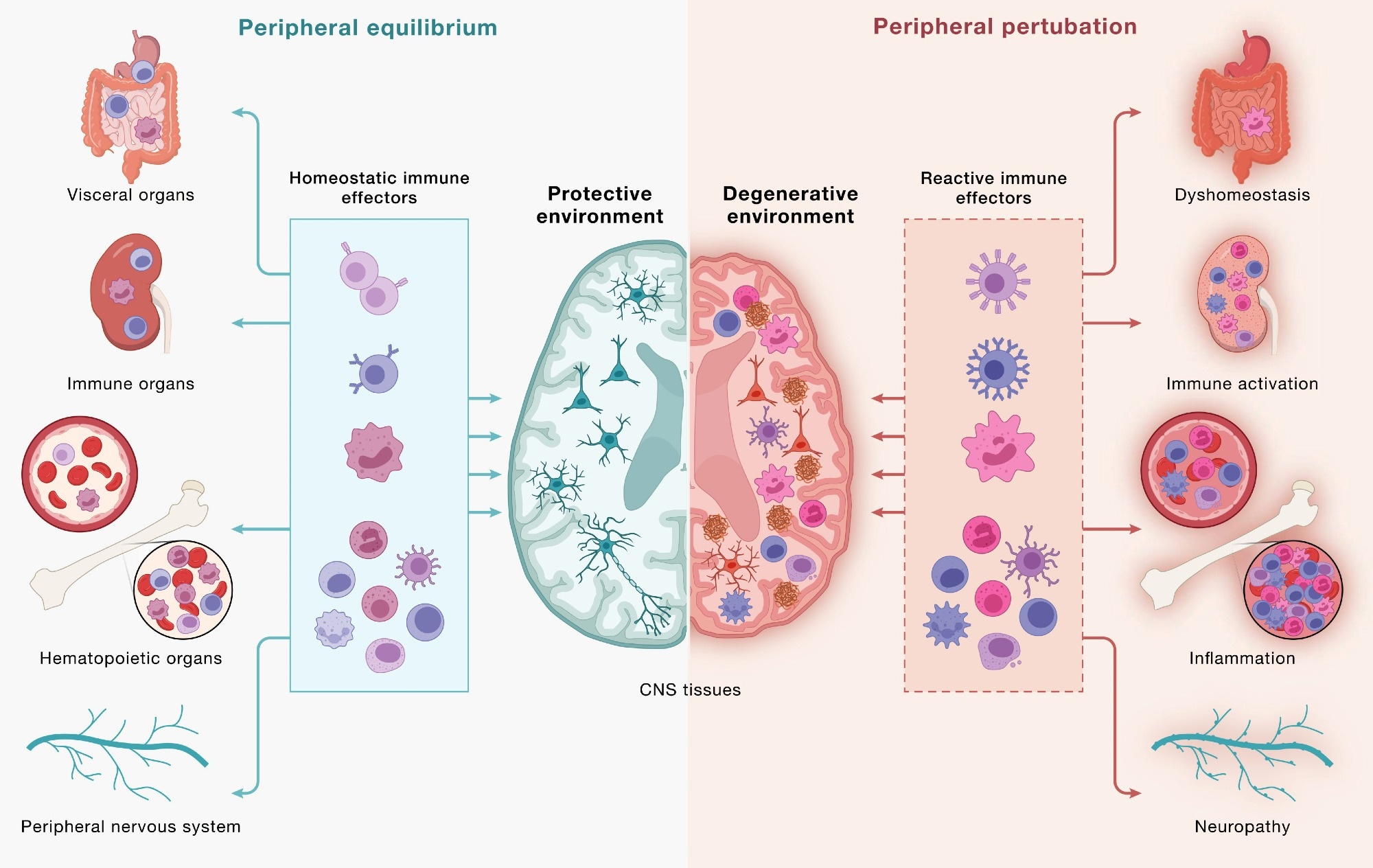
    Immune cells communicate across a continuous, bidirectional circuit between the CNS and periphery. Immune cells from across the body engage CNS tissues and, in turn, are shaped by signals returning from the brain. (Left) Under peripheral equilibrium, immune effectors mobilized from visceral organs, secondary lymphoid organs, hematopoietic tissues, and the peripheral nervous system (PNS) - including CD4+ and CD8+ T cells, B cells, monocytes (top 3 cell types), NK cells, dendritic cells, mast cells, neutrophils, and innate lymphoid cells (bottom cluster of cells) - engage the CNS in a homeostatic dialogue that supports neural cell health, plasticity, and repair, sustaining a protective environment. (Right) Age-associated dysfunction across the same peripheral compartments reshapes this circuit: altered immune effectors enter the CNS and contribute to shifting the local environment toward a degenerative state in which microglia adopt disease-associated programs, astrocytes become reactive, and neurons accumulate pathology. This model underscores how the same circuitry can yield protective or pathogenic outcomes and that neurodegeneration is downstream of dysfunctional brain-immune crosstalk. Study: Neurodegeneration as a dysregulation of neuroimmune crosstalk

    Immune cells communicate across a continuous, bidirectional circuit between the CNS and periphery. Immune cells from across the body engage CNS tissues and, in turn, are shaped by signals returning from the brain. (Left) Under peripheral equilibrium, immune effectors mobilized from visceral organs, secondary lymphoid organs, hematopoietic tissues, and the peripheral nervous system (PNS) - including CD4+ and CD8+ T cells, B cells, monocytes (top 3 cell types), NK cells, dendritic cells, mast cells, neutrophils, and innate lymphoid cells (bottom cluster of cells) - engage the CNS in a homeostatic dialogue that supports neural cell health, plasticity, and repair, sustaining a protective environment. (Right) Age-associated dysfunction across the same peripheral compartments reshapes this circuit: altered immune effectors enter the CNS and contribute to shifting the local environment toward a degenerative state in which microglia adopt disease-associated programs, astrocytes become reactive, and neurons accumulate pathology. This model underscores how the same circuitry can yield protective or pathogenic outcomes and that neurodegeneration is downstream of dysfunctional brain-immune crosstalk. Study: Neurodegeneration as a dysregulation of neuroimmune crosstalk

    A recent perspective published in the journal Cell synthesizes scientific evidence suggesting that neurodegeneration involves intricate crosstalk between neurons and immune cells, linking the brain to peripheral immunity through bidirectional exchange. Strategies that restore immune homeostasis or recalibrate neuroimmune signaling may potentially slow neurodegeneration and promote recovery.

    Historically, immune dysregulation has often been considered a consequence of neurodegenerative disorders. Recent studies, however, are beginning to change this scientific mindset, suggesting that disordered communication between the brain and immune cells may also contribute to disease onset and progression. The authors describe immune dysfunction as a “concause” of neurodegeneration, meaning it may interact with neuronal and glial vulnerabilities without necessarily being the initial trigger. It is essential to advance understanding of the pathophysiology of neurodegenerative diseases to inform therapeutic development and the development of immune-based strategies.

    In this perspective, researchers examined brain-immune interactions and their potential role in neurodegeneration. They organized emerging evidence into three frameworks: “outside-in” effects driven by peripheral immunity, “inside-out” signaling coordinated by brain-resident microglia, and “locked-in” gene regulatory programs that can stabilize maladaptive neuroimmune states.

    The brain-immune communication network

    The brain continuously communicates with peripheral immune networks. Components of the CNS, including the choroid plexus, meninges, and lymphatic and vascular structures, interact with immune cells to relay signals related to neural needs.

    Helper and cytotoxic T cells can enter CNS border regions and, under defined conditions, the brain parenchyma. Brain-immune communication supports neural integrity but can promote pathology when dysregulated. Microglia and BAMs provide surveillance, while lymphocytes confer antigen specificity and immunological memory.

    Cytokines, complement, and MHC-I are traditionally linked to immunity, but CNS cells also produce or sense these molecules during neural activity. Innate lymphoid cells in the dura can respond to injury, while the choroid plexus helps regulate inflammatory signaling. In mice, increased neuronal activity may draw antibody-secreting B-lineage cells into the hippocampus during synaptic remodeling.

    The gut also influences brain immunity. T cells educated in gut-associated immune tissues can subsequently traffic to the borders of the CNS and, under certain conditions, into the brain, while plasma cells secreting IgA antibodies protect blood vessels in the meninges. In addition, changes in the gut microbiome could influence immune activity and microglial function. Through the GBA, the gut and brain are in constant dialogue with each other. The vagus nerve conveys immunity-related information from the intestines to the brain. Reward-related neural pathways can, in turn, influence peripheral immune activity.

    Brain-immune interactions in neurodegenerative disease

    T cell activity has been implicated in PD, AD, ALS, and dementia with Lewy bodies (DLB). In ALS4, an inherited form of ALS, cytotoxic T cells are detected early in the blood and brain and expand as the disease progresses, consistent with antigen-driven responses.

    Thursday, August 20, 2026

    Extended-Window Thrombolysis Improves Stroke Outcomes with increased bleeding risk: JAMA

     Will your competent? doctor and hospital ENSURE FURTHER RESEARCH OCCURS that will determine the best way to prevent the bleeding risk? Oh NO; the plan is TO DO NOTHING!  Everyone there needs to be fired! I take no prisoners in trying to get stroke solved!

    Extended-Window Thrombolysis Improves Stroke Outcomes with increased bleeding risk: JAMA

    Saturday, August 15, 2026

    Youth Protein TIMP2 Restores Immune Function in the Aging Brain

     Will your competent? doctor and hospital ensure further research occurs that creates protocols to solve this problem? NO? So, PURE INCOMPETENCE!

    Youth Protein TIMP2 Restores Immune Function in the Aging Brain

    Summary: Researchers have discovered that the youth-associated protein TIMP2 plays a critical role in maintaining the healthy function of microglia, the brain’s primary immune cells. Supplementing this protein in aged mice improved the brain’s ability to clear cellular debris and reduced inflammation, offering new therapeutic insights for neurodegenerative diseases.

    Key Facts:

    • The youth-associated protein TIMP2 is essential for the healthy function of microglia, the brain’s resident immune cells.
    • Depleting TIMP2 causes microglia to exhibit traits of aging and neurodegeneration, including cellular senescence and an impaired ability to clear cellular waste.
    • Systemic injections of TIMP2 in aged mice shifted their microglia away from pro-inflammatory states and restored their vital debris-clearing capabilities.

    Source: The Mount Sinai Hospital / Mount Sinai School of Medicine

    Aging is the most significant risk factor for Alzheimer’s disease and a host of other neurodegenerative disorders. Yet, the precise biological mechanisms that render the aging brain so vulnerable to decline have remained a complex puzzle.

    Now, researchers at The Icahn School of Medicine at Mount Sinai have uncovered a crucial piece of that puzzle. They have identified a vital role for the youth-associated protein TIMP2 in supporting and preserving the healthy function of microglia—the resident immune cells of the brain.

    Microglia act as the brain’s diligent housekeepers. They play an essential role in maintaining cognitive health by clearing out cellular debris, supporting neural circuits, and acting as first responders to injury. However, as the brain ages, these critical cells often become less efficient. Instead of protecting the brain, aged microglia can adopt maladaptive states that contribute to chronic neuroinflammation and impaired cognitive function.

    In a new study, the research team sought to understand how TIMP2 influences microglial biology in both healthy and aging brains. Utilizing multiple mouse models—including subjects selectively lacking TIMP2 in their microglia or neurons—the team employed advanced brain single nuclei RNA-sequencing, in vivo microdialysis, and functional assays to observe the protein’s impact.The findings were striking. When TIMP2 was deleted, microglia rapidly began to exhibit characteristics typically associated with advanced aging and brain injury. The cells displayed an impaired ability to clear cellular debris and showed molecular signatures consistent with cellular senescence. Additionally, the loss of TIMP2 triggered an increase in inflammatory and stress-related proteins in the brain’s extracellular environment.

    “TIMP2 facilitates healthy function for the brain’s immune cells,” said Joseph M. Castellano, PhD, Associate Professor of Neuroscience at the Ronald M. Loeb Center for Alzheimer’s Disease and corresponding author of the study. “By supporting the ability of microglia to clear debris and limit maladaptive responses, TIMP2 may help restore aspects of microglial function that become compromised with age.”

    Crucially, the study also explored whether these age-related declines could be reversed. When researchers administered systemic injections of TIMP2 to aged mice, they observed a rejuvenating effect. The treatment successfully shifted the microglia away from harmful, pro-inflammatory states and significantly improved their capacity to clear cellular waste.

    These results point to a powerful molecular link between systemic factors associated with youth and the innate immune function of the aging brain. While the current study was conducted in mice, the findings open a promising new avenue for human applications.

    “While additional studies are needed, this work provides new insight into how youth-associated factors influence pathways involved in brain aging and age-related neurological disorders that may ultimately inform therapeutic strategies,” Dr. Castellano added.

    Understanding how youth-associated factors like TIMP2 regulate immune responses could eventually lead to targeted therapies designed to modify age-related brain changes, offering hope against Alzheimer’s and other neurodegenerative diseases.

    Editorial Notes:

    • This article was edited by a Neuroscience News editor.
    • Journal paper reviewed in full.
    • Additional context added by our staff.

    About this Research Section:

    • Author / Media Contact: Elizabeth Dowling
    • Source: The Mount Sinai Hospital / Mount Sinai School of Medicine
    • Image Credits: Neuroscience News featured image / Original raw image courtesy of Mount Sinai Health System.
    • Original Research:
    • Hemmer, B.M., Philippi, S.M., Ferreira, A.C. et al. Youth-associated protein TIMP2 regulates microglial state and function in healthy and aged mice. Nat Commun 17, 8173 (2026). https://doi.org/10.1038/s41467-026-74906-z
      • Title: Youth-associated protein TIMP2 regulates microglial state and function in healthy and aged mice
      • Authors: Brittany Hemmer, Joseph M. Castellano, et al.
      • Journal: Nature Communications
      • DOI: 10.1038/s41467-026-74906-z
      • Publication Date: August 12, 2026

    Thursday, August 13, 2026

    Drug taken for common condition may be slowing Alzheimer’s decline

     With your risk of dementia post stroke, is your competent? doctor preparing testing for stroke patients to see if this would prevent Alzheimers'? NO? NOTHING DOING? PURE INCOMPETENCE THEN!

    Your risk of dementia, has your doctor told you of this?  Your doctor is responsible for preventing this! Is s/he willing to prevent this?

    1. A documented 33% dementia chance post-stroke from an Australian study?   May 2012.

    2. Then this study came out and seems to have a range from 17-66%. December 2013.`    

    3. A 20% chance in this research.   July 2013.

    4. Dementia Risk Doubled in Patients Following Stroke September 2018 

    The latest here: 

    Drug taken for common condition may be slowing Alzheimer’s decline

    A blood-thinning medication already taken by millions of people for an irregular heartbeat may also be helping to slow cognitive decline in patients with Alzheimer’s disease, according to new research from Karolinska Institutet in Sweden.

    The study, published in the European Heart Journal, found that people with both atrial fibrillation and Alzheimer’s disease who were treated with newer blood thinners known as NOACs unintentionally experienced a significantly slower rate of cognitive decline than those given the older drug named warfarin or no blood thinner at all.

    “There are reasons to believe that the treatment could have a positive effect on cognition, for example by improving blood flow and reducing small-scale damage in the brain,” Maria Eriksdotter, a professor at Karolinska Institutet who led the study, said in a statement.

    Newsweek reached out to Karolinska Institutet for more information.

    Why the Two Conditions Are Often Linked

    Atrial fibrillation, a common heart rhythm disorder among older adults, frequently occurs alongside Alzheimer’s disease.

    Doctors typically prescribe blood-thinning medication to atrial fibrillation patients to lower their risk of blood clots. Earlier research had suggested that anticoagulant treatment might reduce the risk of developing dementia in the first place, but scientists knew far less about how these drugs affect people who already have Alzheimer’s.

    Researchers drew on data from SveDem, Sweden’s national quality register for cognitive disorders and dementia, examining 7,308 people diagnosed with both atrial fibrillation and Alzheimer’s disease.

    Participants were sorted into three matched groups: those taking NOACs, those taking warfarin, and those taking no anticoagulant medication at all. Cognitive function was tracked over time using the Mini-Mental State Examination (MMSE), a standard test used to measure memory and thinking skills.

    What the Results Showed

    Patients on NOACs saw their cognitive decline slow by just over 0.2 MMSE points per year compared with those on warfarin or no treatment at all.

    The benefits extended beyond cognition. Patients treated with NOACs also had a lower risk of death, stroke, blood clots and fractures compared with those who received no anticoagulant treatment. Warfarin was also linked to lower risks of death, stroke and blood clots, but it came with a higher risk of major bleeding than NOACs.

    England-based general practitioner, Dr Mohammad Bakhtiar, spoke with Newsweek about the findings.

    “Over 7,000 people is large enough that the signal is hard to dismiss as noise,” he said. “That makes the data more reliable than the usual small clinic series.

    “What I take from the study is encouraging evidence for the newer anticoagulants over warfarin in this group. The cognitive difference is modest, but it sits alongside fewer strokes, clots and deaths, and less major bleeding than warfarin. For GPs deciding between a NOAC and warfarin in someone with Alzheimer’s and atrial fibrillation, that is useful.”

    The researchers cautioned that, because the study was observational, it cannot prove that the drugs directly caused the slower decline. Some factors that may have influenced both which drug a patient was prescribed and their eventual health outcomes could not be fully accounted for. Some patients also may have switched between medications during the course of the study, which followed participants over time. All in all, while they remain optimistic, they acknowledge the promising results require further studies.

    “We should read observational studies cautiously,” Bakhtiar added. “People who get a NOAC are often different from people who get nothing. They may be fitter, better supported, or under more active specialist care. Some patients also switched drugs during follow-up.

    “So, the honest line is association, not proof. NOACs were linked with slower decline. We do not know they caused it.”

    He advises that patients excited by the news do not start, stop or switch their current medications because of this paper.

    The study was funded by several organizations, including the Swedish Research Council, the Swedish Brain Foundation, CIMED, ALF project funding and Karolinska Institutet.

    Reference

    Eriksdotter, M., et al. (2026). Oral anticoagulants, cognition, and clinical outcomes in atrial fibrillation and Alzheimer’s disease: a Swedish nationwide study. European Heart Journal. https://doi.org/10.1093/eurheartj/ehag584.

    Contact Newsweek editors on this story: Marc Vargas and Gray R. Thomas