Changing stroke rehab and research worldwide now.Time is Brain! trillions and trillions of neurons that DIE each day because there are NO effective hyperacute therapies besides tPA(only 12% effective). I have 523 posts on hyperacute therapy, enough for researchers to spend decades proving them out. These are my personal ideas and blog on stroke rehabilitation and stroke research. Do not attempt any of these without checking with your medical provider. Unless you join me in agitating, when you need these therapies they won't be there.

What this blog is for:

My blog is not to help survivors recover, it is to have the 10 million yearly stroke survivors light fires underneath their doctors, stroke hospitals and stroke researchers to get stroke solved. 100% recovery. The stroke medical world is completely failing at that goal, they don't even have it as a goal. Shortly after getting out of the hospital and getting NO information on the process or protocols of stroke rehabilitation and recovery I started searching on the internet and found that no other survivor received useful information. This is an attempt to cover all stroke rehabilitation information that should be readily available to survivors so they can talk with informed knowledge to their medical staff. It lays out what needs to be done to get stroke survivors closer to 100% recovery. It's quite disgusting that this information is not available from every stroke association and doctors group.

Showing posts with label neutrophils. Show all posts

Tuesday, August 4, 2026

Injectable Biomaterial Promotes Brain Repair After a Stroke

 Do you really think anyone in stroke is competent enough to get human testing going? I don't, everything in stroke IS A COMPLETE FUCKING FAILURE! Prove me wrong; failure is defined as not getting to 100% recovery! Don't try your tyranny of low expectations on me. Here; oc1dean@gmail.com, I'll print it verbatim with my reply. Have at it, or are you afraid to engage with a stroke-addled survivor?
Of course your competent? doctor can inform these researchers of earlier work.


Researchers find a copolymer scaffold potentially useful in brain repair after brain injury June 2015


Oriented Graphene Oxide Scaffold Promotes Nerve Regeneration in vitro and in vivo March 2024 

The latest here:

Injectable Biomaterial Promotes Brain Repair After a Stroke 

When someone has a stroke caused by a blood clot, doctors can quickly restore blood flow. However, they can’t easily replace the brain tissue that gets lost. Recovering this tissue usually means relying on rehab to help the remaining brain circuits adapt.

A team of biomedical engineers at Duke University has built an injectable biomaterial that could change stroke recovery.

Rebuilding the Brain’s Neighborhood

The research team isn’t attempting to rebuild the brain directly. Instead, they are setting up a scaffolding system to let the body do the work for them. They achieve this system using tiny hydrogel microparticles called MAPS. When the material is injected into the cavity caused by a stroke, it creates a porous structure for cells to grow on.

“Once brain tissue has been lost, restoring blood flow is no longer enough,” said Tatiana Segura, the Robert Plonsey Distinguished Professor of Biomedical Engineering at Duke. “Our goal is to engineer the injured space so that immune, vascular, and neural repair processes can begin to work together.”

The team attached specific signals to these particles so that the body’s immune cells could help. These signals come from astrocytes, which are star-shaped cells in the brain.

“We are not simply placing a material into the brain,” Segura added. “We are engineering a local environment that can coordinate several parts of the repair response.”

Surprising Helpers

The team found that certain signaling molecules attracted helpful immune cells, including the most common type of white blood cell: neutrophils. Usually, neutrophils cause inflammation right after a stroke. However, that wasn’t the case in this scenario.

“This result changes how we think about neutrophils after stroke,” said Shangjing Xin, lead scientist of the study and a postdoctoral fellow in the Segura Laboratory. “Their role appears to depend on when they arrive, where they are located, and the signals they receive from their surroundings. Our study demonstrates a potential engineering strategy to recruit and retain these cells at the right time.”

In mouse tests, this treated scaffold helped grow new blood vessels and improved movement. By eight weeks, the mice performed like healthy controls on a coordination test.

Right now, the work is still preclinical. The team is looking at using human cells next to make it scalable.

“You do not restore an ecosystem simply by containing the initial damage. You have to create the conditions that allow life to return. That is how we think about the stroke cavity,” Segura said. “The material is not intended to reproduce the brain itself, but to create an environment where the body’s own cells can enter, communicate and participate in rebuilding vascularized tissue.”

Monday, May 4, 2026

What Happens To Your Immune System After One Sauna Session by mindbodygreen


I'm just got a personal sauna from NuRecover but will use a cold shower instead of a chiller bucket for the cold shock proteins. Still to be put together.

What Happens To Your Immune System After One Sauna Session

 There’s a reason sauna use keeps showing up in longevity research. Regular exposure has been associated with lower risks of cardiovascular disease1 ,respiratory illness2,dementia3, and even overall mortality4 .But those outcomes don’t tell us much about the immediate effects. What actually changes in the body during a single session? And how quickly do those changes happen? A new study5set out to answer that by tracking immune responses before, during, and after a 30-minute sauna session, offering a closer look at the body’s short-term reaction to heat. 

What happens to your immune system during a 30-minute sauna

Researchers studied 51 adults, both men and women, during a standard Finnish sauna session set to about 73°C (roughly 163°F). Each participant spent 30 minutes in the sauna, with blood samples taken before, immediately after, and 30 minutes post-session. The goal was to track how immune cells and inflammatory signals changed in response to heat stress. Participants were allowed to drink water throughout, which helped control for dehydration and made the setup closer to a typical real-world sauna experience. Instead of just looking at overall immune activity, the researchers zoomed in on specific white blood cells and a wide range of cytokines, which are signaling molecules involved in inflammation and immune response. This gave a more detailed picture of how the body reacts in the short term. 

Sauna’s immediate effect on immune cells

The biggest shift came down to your white blood cells, which are a key part of your immune system. After the sauna session, those levels went up, including important types like neutrophils and lymphocytes. These are the cells that help your body spot and respond to anything that doesn’t belong, whether that’s a virus or another kind of threat. What stands out is how quickly this happens. The increase shows up right after the sauna, then goes back down within about 30 minutes. So this isn’t a long-lasting spike; it’s more like a short window where your immune system is a bit more alert. This pattern mirrors what happens during exercise. When you work out, immune cells move out of tissues and into your bloodstream, where they’re more ready to respond if needed. A sauna seems to trigger a comparable response, just through heat instead of movement. 

What about inflammation & body temperature?

One thing the researchers looked at closely was inflammation, since that’s often what people think of when they hear the body is under “stress.” But in this case, there weren’t big changes across most inflammatory markers. Out of dozens of signals they measured, only a few shifted in a meaningful way. That suggests your body isn’t going into a full inflammatory response during a sauna. Instead, the response seems more about mobilization than inflammation. Your immune cells are being redistributed and activated, not necessarily pushed into an inflammatory state. Body temperature did play a role, though. On average, people’s temperature rose by about 2°C (roughly 3.6°F) during the session. And the more it increased, the more certain immune-related signals shifted alongside it. 

Adding sauna use to your routine

 So what does this mean for your day-to-day routine? It doesn’t mean a single sauna session will prevent illness or replace other foundational habits. But it does suggest that sauna use can act as a short-term stimulus for your immune system, similar to a workout.If you already use a sauna, this adds another layer of context. That post-sauna feeling isn’t just relaxation. Your body is actively responding, mobilizing immune cells in a way that may support overall immune surveillance over time. 
If you’re considering adding it in, consistency matters more than intensity. This study looked at one session, but previous research has linked regular sauna use to broader health benefits, including a lower risk of certain chronic conditions.

It’s also worth paying attention to how you personally respond. Hydration, heat tolerance, and recovery all play a role in how beneficial the experience feels.

The takeaway

This study doesn’t suggest that sauna use is a cure-all. But it does offer a clearer picture of what’s happening in the body during a session. Your immune system isn’t passive in that environment. It’s responding, adapting, and briefly shifting into a more active state.

Wednesday, September 3, 2025

Chronic Stress Sends Immune Cells to Brain, Driving Depression


With your massive stress from your incompetent doctor not having 100% recovery protocols, and the fact your doctor doesn't know about this, you are on your own to prevent depression! Good luck.

Chronic Stress Sends Immune Cells to Brain, Driving Depressionnic Stress Sends Immune Cells to Brain, Driving Depression

Summary: A new study shows that chronic stress causes immune cells called neutrophils to leave bone marrow in the skull and collect in the brain’s protective membranes, where they contribute to depressive symptoms. In mice, blocking an immune “alarm” pathway reduced these cells and improved mood-related behaviors.

The findings highlight how stress reshapes the brain’s immune environment and suggest why one-third of patients may not benefit from current antidepressants. This immune link could provide biomarkers for tailored treatments and help explain depression’s overlap with neurological conditions like stroke and Alzheimer’s.

Key Facts

  • Immune Link: Chronic stress triggers neutrophils from skull bone marrow to enter the brain’s meninges, influencing mood.
  • Pathway Identified: Blocking type I interferon signaling reduced brain neutrophils and depressive behaviors in mice.
  • Treatment Potential: Findings could guide new immune-targeted therapies for depression resistant to standard drugs.

Source: University of Cambridge

Immune cells released from bone marrow in the skull in response to chronic stress and adversity could play a key role in symptoms of depression and anxiety, say researchers.

The discovery – found in a study in mice – sheds light on the role that inflammation can play in mood disorders and could help in the search for new treatments, in particular for those individuals for whom current treatments are ineffective.

Around 1 billion people will be diagnosed with a mood disorder such as depression or anxiety at some point in their life.

While there may be many underlying causes, chronic inflammation – when the body’s immune system stays active for a long time, even when there is no infection or injury to fight – has been linked to depression. This suggests that the immune system may play an important role in the development of mood disorders.

Previous studies have highlighted how high levels of an immune cell known as a neutrophil, a type of white blood cell, are linked to the severity of depression. But how neutrophils contribute to symptoms of depression is currently unclear.

In research published today in Nature Communications, a team led by scientists at the University of Cambridge, UK, and the National Institute of Mental Health, USA, tested a hypothesis that chronic stress can lead to the release of neutrophils from bone marrow in the skull.

These cells then collect in the meninges – membranes that cover and protect your brain and spinal cord – and contribute to symptoms of depression.

As it is not possible to test this hypothesis in humans, the team used mice exposed to chronic social stress. In this experiment, an ‘intruder’ mouse is introduced into the home cage of an aggressive resident mouse. The two have brief daily physical interactions and can otherwise see, smell, and hear each other.

The researchers found that prolonged exposure to this stressful environment led to a noticeable increase in levels of neutrophils in the meninges, and that this was linked to signs of depressive behaviour in the mice. Even after the stress ended, the neutrophils lasted longer in the meninges than they did in the blood.

Analysis confirmed the researchers’ hypothesis that the meningeal neutrophils – which appeared subtly different from those found in the blood – originated in the skull.

Further analysis suggested that long-term stress triggered a type of immune system ‘alarm warning’ known as type I interferon signalling in the neutrophils. Blocking this pathway – in effect, switching off the alarm – reduced the number of neutrophils in the meninges and improved behaviour in the depressed mice.

This pathway has previously been linked to depression – type 1 interferons are used to treat patients with hepatitis C, for example, but a known side effect of the medication is that it can cause severe depression during treatment.

Dr Stacey Kigar from the Department of Medicine at the University of Cambridge said: “Our work helps explain how chronic stress can lead to lasting changes in the brain’s immune environment, potentially contributing to depression. It also opens the door to possible new treatments that target the immune system rather than just brain chemistry.

“There’s a significant proportion of people for whom antidepressants don’t work, possibly as many as one in three patients. If we can figure out what’s happening with the immune system, we may be able to alleviate or reduce depressive symptoms.”

The reason why there are high levels of neutrophils in the meninges is unclear. One explanation could be that they are recruited by microglia, a type of immune cell unique to the brain.

Another possible explanation is that chronic stress may cause microhaemorrhages, tiny leaks in brain blood vessels, and that neutrophils – the body’s ‘first responders’ – arrive to fix the damage and prevent any further damage.

These neutrophils then become more rigid, possibly getting stuck in brain capillaries and causing further inflammation in the brain.

Dr Mary-Ellen Lynall from the Department of Psychiatry at the University of Cambridge said: “We’ve long known that something is different about how neutrophils behave after stressful events, or during depression, but we didn’t know what these neutrophils were doing, where they were going, or how they might be affecting the brain and mind.

“Our findings show that these ‘first responder’ immune cells leave the skull bone marrow and travel to the brain, where they can influence mood and behaviour.

“Most people will have experienced how our immune systems can drive short-lived depression-like symptoms. When we are sick, for example with a cold or flu, we often lack energy and appetite, sleep more and withdraw from social contact. If the immune system is always in a heightened, pro-inflammatory state, it shouldn’t be too surprising if we experience longer-term problems with our mood.”

The findings could provide a useful signature, or ‘biomarker’, to help identify those patients whose mood disorders are related to inflammation. This could help in the search for better treatments.

For example, a clinical trial of a potential new drug that targets inflammation of the brain in depression might appear to fail if trialled on a general cohort of people with depression, whereas using the biomarker to identify individuals whose depression is linked to inflammation could increase the likelihood of the trial succeeding.

The findings may also help explain why depression is a symptom common in other neurological disorders such as stroke and Alzheimer’s disease, as it may be the case that neutrophils are being released in response to the damage to the brain seen in these conditions.

But it may also explain why depression is itself a risk factor for dementia in later life, if neutrophils can themselves trigger damage to brain cells.

Funding: The research was funded by the National Institute of Mental Health, Medical Research Council and National Institute for Health and Care Research Cambridge Biomedical Research Centre.

About this neuroscience and depression research news

Author: Craig Brierley
Source: University of Cambridge
Contact: Craig Brierley – University of Cambridge
Image: The image is credited to Neuroscience News

Original Research: Open access.
Chronic social defeat stress induces meningeal neutrophilia via type I interferon signaling in male mice” by Stacey Kigar et al. Nature Communications


Saturday, August 3, 2024

Benserazide is neuroprotective and improves functional recovery after experimental ischemic stroke by altering the immune response

 Well, it seems human cell testing was done but I see nothing done with human subjects! So your competent? doctor needs to ensure human testing gets done. But I bet you don't have a functioning stroke doctor, do you?

Benserazide is neuroprotective and improves functional recovery after experimental ischemic stroke by altering the immune response

Abstract

Stroke is a leading cause of permanent disability worldwide. Despite intensive research over the last decades, key anti-inflammatory strategies that have proven beneficial in pre-clinical animal models have often failed in translation. The importance of neutrophils as pro- and anti-inflammatory peripheral immune cells has often been overlooked in ischemic stroke. However, neutrophils rapidly infiltrate into the brain parenchyma after stroke and secrete an array of pro-inflammatory factors including reactive oxygen species, proteases, cytokines, and chemokines exacerbating damage. In this study, we demonstrate the neuroprotective and anti-inflammatory effect of benserazide, a clinically used DOPA decarboxylase inhibitor, using both in vitro models of inflammation and in vivo mouse models of focal cerebral ischemia. Benserazide significantly attenuated PMA-induced NETosis in isolated human neutrophils. Furthermore, benserazide was able to protect both SH-SY5Y and iPSC-derived human cortical neurons when challenged with activated neutrophils demonstrating the clinical relevance of this study. Additional in vitro data suggest the ability of benserazide to polarize macrophages towards M2-phenotypes following LPS stimulation. Neuroprotective effects of benserazide are further demonstrated by in vivo studies where peripheral administration of benserazide significantly attenuated neutrophil infiltration into the brain, altered microglia/macrophage phenotypes, and improved the behavioral outcome post-stroke. Overall, our data suggest that benserazide could serve as a drug candidate for the treatment of ischemic stroke. The importance of our results for future clinical trials is further underlined as benserazide has been approved by the European Medicines Agency as a safe and effective treatment in Parkinson’s disease when combined with levodopa.

Wednesday, March 6, 2024

MSC-Derived Exosomes Mitigate Myocardial Ischemia/Reperfusion Injury by Reducing Neutrophil Infiltration and the Formation of Neutrophil Extracellular Traps

Does your doctor or anyone on the stroke medical world have the ability to see the possibilities of using this for stroke? Since stem cells create exosomes and exosomes seem to be the helping factor for recovery.

Application of stem cell-derived exosomes in ischemic diseases: opportunity and limitations May2021

 The latest here

MSC-Derived Exosomes Mitigate Myocardial Ischemia/Reperfusion Injury by Reducing Neutrophil Infiltration and the Formation of Neutrophil Extracellular Traps

Authors Feng Y, Bao X, Zhao J, Kang L, Sun X, Xu B

Received 19 September 2023

Accepted for publication 14 February 2024

Published 5 March 2024 Volume 2024:19 Pages 2071—2090

DOI https://doi.org/10.2147/IJN.S436925

Checked for plagiarism Yes

Review by Single anonymous peer review

Peer reviewer comments 2

Editor who approved publication: Professor Lijie Grace Zhang


Yuting Feng, Xue Bao, Jinxuan Zhao, Lina Kang, Xuan Sun, Biao Xu

Department of Cardiology, Nanjing Drum Tower Hospital, State Key Laboratory of Pharmaceutical Biotechnology, Medical School of Nanjing University, Nanjing, People’s Republic of China

Correspondence: Xuan Sun; Biao Xu, Department of Cardiology, Nanjing Drum Tower Hospital, State Key Laboratory of Pharmaceutical Biotechnology, Medical School of Nanjing University, No. 321 Zhongshan Road, Nanjing, 210008, People’s Republic of China, Email sunxuan891119@163.com; xubiao62@nju.edu.cn

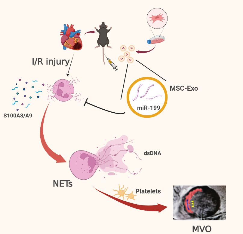
Introduction: Acute inflammatory storm is a major cause of myocardial ischemia/reperfusion (I/R) injury, with no effective treatment currently available. The excessive aggregation of neutrophils is correlated with an unfavorable prognosis in acute myocardial infarction (AMI) patients. Exosomes derived from mesenchymal stromal cells (MSC-Exo) have certain immunomodulatory potential and might be a therapeutic application. Therefore, we investigated the protective role of MSC-Exo in modulating neutrophil infiltration and formation of neutrophil extracellular traps (NETs) following myocardial I/R injury.
Methods: Exosomes were isolated from the supernatant of MSCs using a gradient centrifugation method. We used flow cytometry, histochemistry, and immunofluorescence to detect the changes of neutrophils post-intravenous MSC-Exo injection. Additionally, cardiac magnetic resonance (CMR) and thioflavin S experiments were applied to detect microvascular obstruction (MVO). The NLR family pyrin domain containing 3 (NLRP3) inflammasome was examined for mechanism exploration. Primary neutrophils were extracted for in vitro experiment. Antibody of Ly6G was given to depleting the neutrophils in mice for verification the effect of MSC-Exo. Finally, we analyzed the MiRNA sequence of MSC-Exo and verified it in vitro.
Results: MSC-Exo administration reduced neutrophil infiltration and NETs formation after myocardial I/R. MSC-Exo treatment also could attenuate the activation of NLRP3 inflammasome both in vivo and in vitro. At the same time, the infarction size and MVO following I/R injury were reduced by MSC-Exo. Moreover, systemic depletion of neutrophils partly negated the therapeutic effects of MSC-Exo. Up-regulation of miR-199 in neutrophils has been shown to decrease the expression of NETs formation after stimulation.
Discussion: Our results demonstrated that MSC-Exo mitigated myocardial I/R injury in mice by modulating neutrophil infiltration and NETs formation. This study provides novel insights into the potential therapeutic application of MSC-Exo for myocardial ischemia/reperfusion injury.

Keywords: myocardial ischemia/reperfusion injury, mesenchymal stromal cells, exosomes, neutrophil extracellular traps, mir-199

Graphical Abstract:

Introduction

Acute myocardial infarction (AMI) has been a major cause of death and heart failure (HF) worldwide in current. Percutaneous coronary intervention (PCI) to recanalize the artery in the early stage of AMI salvages the injured myocardium and improves survival. However, the rapid restoration of blood flow triggers ischemia/reperfusion (I/R) injury,1 which reduces the curative effect of PCI.

The inflammatory response is one of the important pathological mechanisms of myocardial I/R injury.2 Neutrophils play a pivotal role in mediating inflammation as the first immune cells recruited from the bloodstream into injured tissues.3 The number and proportion of neutrophils in the peripheral blood and myocardium tissue increase rapidly in the acute phase of I/R injury and the infiltration of neutrophils in the myocardium reaches the peak within 24 hours after reperfusion. Neutrophils remove necrotic debris at the injured site and recruit the mononuclear macrophage system for further repair.4 But the excessive aggregation and activation of neutrophils result in the release of a plethora of damage-associated molecular patterns (DAMPs) and inflammatory cytokines, which subsequently contribute to myocardial necrosis, aggravated ventricular remodeling, and reduced cardiac function.5

Activated neutrophils can release neutrophil extracellular traps (NETs), manifesting as weblike DNA structures decorated with histones and antimicrobial proteins.6 The released NETs bind to platelets and red blood cells and then finally form plugs. These plugs are dispersed by blood flow into microvessels, forming embolisms that impede tissue perfusion despite the restoration of large coronary arteries.7 This phenomenon, known as microvascular obstruction (MVO), is a major contributor to I/R injury and infarct size, and an increased risk of developing heart failure after AMI.8 Besides, NETs promote the formation of reactive oxygen species9 and the secretion of inflammatory factors and chemokines to aggravate the inflammatory response.10 Neutrophils have also been proven as the primary source of the proinflammatory alarmins, S100A8 and S100A9, after AMI.11 The release of S100A8 and S100A9 in the myocardium stimulates additional leukocyte recruitment and increases the secretion of pro-inflammatory cytokines.12 High plasma levels of S100A8 and S100A9 after PCI are associated with poor left ventricular ejection fraction (LVEF) and an increased incidence of HF13 in AMI patients. These pathological processes above finally result in a vicious circle of myocardium injury. Neutrophil influx and NETs are associated with poor outcomes after cardiac ischemia. However, there are currently not any clinically approved interventions able to modulate neutrophils after I/R injury.

Mesenchymal stromal cells (MSCs) have been demonstrated to have immunomodulatory properties14 and the therapeutic applications of MSCs are being explored in cardiovascular diseases.15 However, the risk of thrombosis formation and immunoreactivity have suggested that caution is still warranted in the clinical use of MSCs.16 The exosomes secreted by MSCs (MSC-Exo) have been known to mediate cell-to-cell communication, which has well-established anti-inflammatory effects.17 We have reported that MSC-Exo plays a protective effect on ischemic myocardium by polarizing inflammatory macrophage towards an anti-inflammatory macrophage population.18 In consideration that neutrophils infiltration into the myocardium peaks within 24 hours after reperfusion and they are prime drivers of the pro-inflammatory phase, it remains unknown whether MSC-Exo could inhibit neutrophils from mobilization, recruitment or MVO formation thus limiting inflammation.

In this study, we assessed whether MSC-Exo therapy in the early stage could suppress neutrophil infiltration and NETs formation to preserve cardiac function. Our research has shown the promising effect of MSC-Exo in myocardium I/R injury.

Friday, October 13, 2023

CD13 facilitates immune cell migration and aggravates acute injury but promotes chronic post-stroke recovery

 So the research was incomplete, since nothing tells us how this gets us recovered. Further research needed that your doctors and hospital should initiate. NO EXCUSES!

CD13 facilitates immune cell migration and aggravates acute injury but promotes chronic post-stroke recovery

Abstract

Introduction

Acute stroke leads to the activation of myeloid cells. These cells express adhesion molecules and transmigrate to the brain, thereby aggravating injury. Chronically after stroke, repair processes, including angiogenesis, are activated and enhance post-stroke recovery. Activated myeloid cells express CD13, which facilitates their migration into the site of injury. However, angiogenic blood vessels which play a role in recovery also express CD13. Overall, the specific contribution of CD13 to acute and chronic stroke outcomes is unknown.

Methods

CD13 expression was estimated in both mice and humans after the ischemic stroke. Young (8–12 weeks) male wild-type and global CD13 knockout (KO) mice were used for this study. Mice underwent 60 min of middle cerebral artery occlusion (MCAO) followed by reperfusion. For acute studies, the mice were euthanized at either 24- or 72 h post-stroke. For chronic studies, the Y-maze, Barnes maze, and the open field were performed on day 7 and day 28 post-stroke. Mice were euthanized at day 30 post-stroke and the brains were collected for assessment of inflammation, white matter injury, tissue loss, and angiogenesis. Flow cytometry was performed on days 3 and 7 post-stroke to quantify infiltrated monocytes and neutrophils and CXCL12/CXCR4 signaling.

Results

Brain CD13 expression and infiltrated CD13+ monocytes and neutrophils increased acutely after the stroke. The brain CD13+lectin+ blood vessels increased on day 15 after the stroke. Similarly, an increase in the percentage area CD13 was observed in human stroke patients at the subacute time after stroke. Deletion of CD13 resulted in reduced infarct volume and improved neurological recovery after acute stroke. However, CD13KO mice had significantly worse memory deficits, amplified gliosis, and white matter damage compared to wild-type animals at chronic time points. CD13-deficient mice had an increased percentage of CXCL12+cells but a reduced percentage of CXCR4+cells and decreased angiogenesis at day 30 post-stroke.

Conclusions

CD13 is involved in the trans-migration of monocytes and neutrophils after stroke, and acutely, led to decreased infarct size and improved behavioral outcomes. However, loss of CD13 led to reductions in post-stroke angiogenesis by reducing CXCL12/CXCR4 signaling.

Thursday, September 22, 2022

Association between neutrophil percentage-to-albumin ratio and 3-month functional outcome in acute ischemic stroke patients with reperfusion therapy

 So you described a problem; WHAT THE FUCK IS THE SOLUTION? No solution, useless research, I'd fire you all.

Association between neutrophil percentage-to-albumin ratio and 3-month functional outcome in acute ischemic stroke patients with reperfusion therapy

Ting Cui1, Changyi Wang2, Qiange Zhu3, Shucheng Li1, Yuan Yang1, Anmo Wang1, Xuening Zhang1, Wenzuo Shang1 and Bo Wu1*
  • 1Center of Cerebrovascular Diseases, Department of Neurology, West China Hospital, Sichuan University, Chengdu, China
  • 2Department of Rehabilitation Medicine Center, West China Hospital, Sichuan University, Chengdu, China
  • 3The Second Department of Neurology, Shanxi Provincial People's Hospital, Xi'an, China

Background: Neutrophils and albumin are associated with outcomes in patients with acute ischemic stroke (AIS). We aimed to explore the association between the neutrophil percentage-to-albumin ratio (NPAR), a novel marker of inflammation and oxidative stress, and the 3-month functional outcome in AIS patients with reperfusion therapy.

Methods: This single-center, retrospective cohort study consecutively enrolled AIS patients with reperfusion therapy. Neutrophils and albumin were collected on admission. The primary outcome was a poor functional outcome, which was defined as a modified Rankin scale score of 3–6 at 3 months.

Results: A total of 647 patients with AIS who received reperfusion therapy were analyzed. The mean age was 68.9 ± 13.9 years, and 358 (55.3%) of the patients were men. The median NPAR was 1.89 (interquartile range [IQR] 1.64–2.09). The percentage of patients with a 3-month poor functional outcome was 57.0% (369/647). NPAR was positively associated with a poor functional outcome (odds ratio [OR] 2.76, 95% CI: 1.52–5.03, p = 0.001). When patients were classified into tertiles, patients in the upper tertile (2.03–7.59) had a higher risk of poor outcome than patients in the lower tertile after adjusting for potential confounders (0.78–1.73) (OR 2.10, 95% CI: 1.28–3.42, p = 0.003). The risk of poor outcome increased with NPAR tertiles (p-trend = 0.003). The optimal cut-off value of the NPAR for predicting a poor outcome was 1.72, with a sensitivity of 0.75, and a specificity of 0.43.

Conclusion: Neutrophil percentage-to-albumin ratio was significantly associated with 3-month poor functional outcomes in patients with AIS who received reperfusion therapy.

Introduction

Reperfusion therapy has become the standard of care for patients with acute ischemic stroke (AIS) (1). However, approximately half of the patients with AIS suffer poor clinical outcomes after reperfusion therapy (2, 3). Inflammation and oxidative stress are two critical variables influencing the prognosis of patients with AIS after reperfusion therapy (4).

During the acute phase of AIS, neutrophils are the earliest inflammatory cells that are abundantly present in cerebral microvessels, and their subsequent release of reactive oxygen species (ROS) is thought to be the main cause of reperfusion injury after AIS (59). Some studies found that serum albumin played a key role in scavenging ROS (10, 11) and might exert an anti-inflammatory effect by inhibiting neutrophil spreading (12, 13). Neutrophils and albumin are associated with outcomes in patients with AIS (1418).

The neutrophil percentage-to-albumin ratio (NPAR) is an emerging marker of inflammation and oxidative stress. The NPAR has been reported to have prognostic significance in patients with cancer, spinal cord injury, acute kidney injury, acute myocardial infarction, and cardiogenic shock (1927). Recently, a retrospective study explored the association between NPAR and infection in patients with AIS (28). However, there is uncertainty regarding the association between NPAR and 3-month functional outcomes in AIS patients with reperfusion therapy. We hypothesized that NPAR may reflect the severity of inflammation and ROS damage in the acute phase of AIS. We sought to assess the association between NPAR and patient outcomes after reperfusion therapy.

More at link.

Friday, March 19, 2021

Tool predicts risk for severe illness, death from COVID-19

If the tool predicts severe illness or death your doctor has a lot of work to do to prevent that outcome. You can't let your doctor throw up their hands in defeat and say nothing can be done. Without seeing how this is built you can't tell what is the most important factor. If oxygen saturation maybe you want these:

Possible solutions: Obviously not vetted coming from me. Don't do them. 

Normobaric oxygen (10)

How to Improve Your Brain Function with An Oxygen Concentrator April 2018 

Or is it more important to increase the loading ability of red blood cells to carry more oxygen? 

Like this?

University of Glasgow Study Demonstrates the Ability of Oxycyte® to Supply Oxygen to Critical Penumbral Tissue in Acute Ischemic Stroke  August 2012

Or like this?

chronic cannabis users have higher cerebral blood flow and extract more oxygen from brain blood flow than nonusers. August 2017   

----------------------------------------------------------------------------------------

Or if neurophils maybe something in here: 

COVID-19 and Neutrophils: The Relationship between Hyperinflammation and Neutrophil Extracellular Traps

----------------------------------------------------------------------------------------------

The latest here:

Tool predicts risk for severe illness, death from COVID-19

The Severe COVID-19 Adaptive Risk Predictor, or SCARP, tool showed greater than 80% accuracy in predicting a patient’s progression from moderate to severe COVID-19 disease or death within 14 days of hospitalization, researchers wrote.

SCARP has an advantage over other clinical prediction tools for SARS-CoV-2, most of which rely solely on data that are collected at the time of hospital admission, according to Matthew Robinson, MD, assistant professor of medicine at Johns Hopkins University School of Medicine and a co-author of the study.

Patient variables that SCARP uses to predict COVID-19 outcomes include BMI, days since hospital admission, oxygen saturation rates, social history and absolute neutrophil counts
Reference: Wongvibulsin S, et al. Ann Intern Med. 2021;doi:10.7326/M20-6754.

“As patients improve or get worse, most existing tools do not have a way to update their predictions based on this new information,” he told Healio Primary Care. “Limitations of other tools in handling changing variables over time, arduous input requirements, performance caveats and uninterpretable logic inspired our efforts to create SCARP.”

Matthew Robinson

SCARP is an artificial intelligence tool that “dynamically updates” as physicians enter patient variables such as BMI level; C-reactive protein, supplemental oxygen delivery, oxygen saturation and respiratory rates; days since hospital admission; admission source; social history; oxygen level and amount of supplemental oxygen needed; and absolute neutrophil and lymphocyte counts, according to the researchers. They developed the tool using a machine-learning approach known as a random forest for survival, longitudinal and multivariate data analysis.

The researchers tested SCARP during a retrospective observational study of 3,163 patients (median age, 61 years) who tested positive for SARS-CoV-2 and were admitted to a five-hospital health system between March 5, 2020, and Dec. 4, 2020. Of that cohort, 7% became severely ill or died within 24 hours of admission and another 11% became severely ill or died during the next 7 days.

Robinson and colleagues reported that the area under the receiver-operating characteristic curve (AUC) for 1-day risk predictions for progression to severe COVID-19 disease or death was 0.89 (95% CI; 0.88-0.9) during the first week of hospitalization and 0.89 (95% CI; 0.87-0.91) during the second week. The AUC for 7-day risk predictions was 0.83 (95% CI; 0.83-0.84) during the first week and 0.87 (95% CI; 0.86-0.89) during the second week of hospitalization.

According to Robinson, unlike some other tools, SCARP provides useful information in “clinically meaningful timeframes.”

“For example, if a clinician is taking care of a patient with COVID‐19 in a hospital with limited remaining critical care capacity, and SCARP shows that the patient has a low risk of developing severe disease in the next 24 hours but a high risk in the next 7 days, then they may consider transferring the patient to another facility with more access to critical care safely before the patient becomes critically ill,” he said.

He added that his research team “included ‘adaptive’ in the name of the tool because the tool sequentially asks clinicians for the next most important variable the tool needs to improve the accuracy of the prediction, which is tailored to the information already inputted.”

References:

Johns Hopkins School of Medicine. Severe COVID-19 Adaptive Risk Predictor. https://rsconnect.biostat.jhsph.edu/covid_trajectory/. Accessed March 11, 2021.

Wongvibulsin S, et al. Ann Intern Med. 2021;doi:10.7326/M20-6754.


Sunday, January 19, 2020

Blue light reduces organ injury from ischemia and reperfusion

Would wearing blue light googles immediately upon entering the ambulance or hospital prevent some of the reperfusion injury? We'll never know because we have NO leadership and NO strategy in stroke.

Blue light reduces organ injury from ischemia and reperfusion

Significance

It is well established that light regulates mammalian biology. And yet, we have been unable to define and thus harness the underlying mechanisms so as to apply them to alter the course of human disease. In this study we determine that the spectrum of light is a critical determinant of its effect on critical illness. We show that an acute and short (24 h) exposure to high-illuminance (1,400 lx) blue spectrum (peak 442 nm) light prior to ischemia/reperfusion (I/R) significantly attenuates the degree of organ injury. Our characterization of the biological mechanisms through which blue light beneficially alters the cellular response to I/R provides an opportunity to develop novel therapeutics for the prevention and treatment of many diseases.
Keywords: blue light, ischemia, reperfusion, organ injury, circadian rhythms

Abstract

Evidence suggests that light and circadian rhythms profoundly influence the physiologic capacity with which an organism responds to stress. However, the ramifications of light spectrum on the course of critical illness remain to be determined. Here, we show that acute exposure to bright blue spectrum light reduces organ injury by comparison with bright red spectrum or ambient white fluorescent light in two murine models of sterile insult: warm liver ischemia/reperfusion (I/R) and unilateral renal I/R. Exposure to bright blue light before I/R reduced hepatocellular injury and necrosis and reduced acute kidney injury and necrosis. In both models, blue light reduced neutrophil influx, as evidenced by reduced myeloperoxidase (MPO) within each organ, and reduced the release of high-mobility group box 1 (HMGB1), a neutrophil chemotactant and key mediator in the pathogenesis of I/R injury. The protective mechanism appeared to involve an optic pathway and was mediated, in part, by a sympathetic (β3 adrenergic) pathway that functioned independent of significant alterations in melatonin or corticosterone concentrations to regulate neutrophil recruitment. These data suggest that modifying the spectrum of light may offer therapeutic utility in sterile forms of cellular injury.

Saturday, May 18, 2019

Harmful neutrophil subsets in patients with ischemic stroke Association with disease severity

I didn't understand what is going on here.  Are they trying to say neutrophils are bad? But they give no indication of how to reduce them, so this is useless.

Harmful neutrophil subsets in patients with ischemic stroke - Association with disease severity

David Weisenburger-Lile, Yuan Dong, Marion Yger, Gaëlle Weisenburger, Giulia Frasca Polara, Thomas Chaigneau, Riccardo Zapata Ochoa, Beatrice Marro, Bertrand Lapergue, Sonia Alamowitch, Carole Elbim

Abstract

Objective To better understand the functional state of circulating neutrophils in patients with ischemic stroke (IS) for planning future clinical trials.
Methods We analyzed by flow cytometry activation state of circulating neutrophils and the distribution of neutrophil peripheral subsets in 41 patients with acute IS less than 6 hours before admission and compared them with 22 age-matched healthy controls.
Results Our results demonstrated continuous basal hyperactivation of circulating neutrophils during acute IS, characterized by lower l-selectin expression and higher CD11b expression at the cell surface, increased ROS production by neutrophils, and greater circulating levels of neutrophil elastase. Neutrophil hyperactivation was associated with deregulation of the equilibrium between apoptotic and necrotic. Patients also had higher percentages than controls of the overactive senescent (CXCR4bright/CD62Ldim) neutrophil subset and increased percentage of neutrophils with a reverse transendothelial migration (CD54highCXCR1low) phenotype. Importantly, neutrophil alterations were associated with the clinical severity of the stroke, evaluated by its NIH Stroke Scale score.
Conclusion Altogether, our results indicate that during acute IS, the inflammatory properties of circulating neutrophils rise, associated with the expansion of harmful neutrophil subsets. These changes in neutrophil homeostasis, associated with disease severity, may play an instrumental role by contributing to systemic inflammation and to the blood-brain barrier breakdown. Our findings highlight new potential therapeutic approaches of stroke by rebalancing the ratio of senescent to immunosuppressive neutrophils or decreasing reverse neutrophil transmigration or both.

Glossary

AAD=
amino-actinomycin D;
ANOVA=
analysis of variance;
APC=
allophycocyanine;
BBB=
blood-brain barrier;
DAMP=
danger-associated molecular pattern;
fMLP=
N-formylmethionyl-leucyl-phenylalanine;
HC=
healthy control;
HE=
hydroethidine;
HMGB=
high-mobility group box;
IS=
ischemic stroke;
LPS=
lipopolysaccharide;
MMP=
matrix metalloproteinase;
MPO=
myeloperoxidase;
NET=
neutrophil extracellular trap;
NIHSS=
NIH Stroke Scale;
NLR=
Nod-like receptor;
PBS=
phosphate buffered saline;
PMN=
polymorphonuclear neutrophil;
ROS=
reactive oxygen species;
rTEM=
reverse transendothelial migration;
sJAM-C=
soluble JAM-C;
TNF=
tumor necrosis factor;
TLR=
Toll-like receptor

Footnotes

  • Go to Neurology.org/NN for full disclosures. Funding information are provided at the end of the article.
  • The Article Processing Charge was funded by the authors.
  • Editorial, page e570
  • Received December 19, 2018.
  • Accepted in final form March 12, 2019.
This is an open access article distributed under the terms of the Creative Commons Attribution-NonCommercial-NoDerivatives License 4.0 (CC BY-NC-ND), which permits downloading and sharing the work provided it is properly cited. The work cannot be changed in any way or used commercially without permission from the journal.
View Full Text