Have your competent? doctor summarize this INTO AN EXACT PROTOCOL! You don't want to do it wrong and your doctor, IF COMPETENT AT ALL, should easily be able to do that. But you don't have a functioning stroke doctor, do you? If your doctor doesn't know about the Merlin app for recognizing bird songs; that's even worse!
Birding Influences Brain Health and Mental Health
From the Summer 2026 issue of Living Bird magazine. Subscribe now.


I was 26, thriving in a corporate communications career, flying high. Then, like a bird’s nest in a windstorm, my life collapsed. Suddenly and without warning, I suffered a traumatic brain injury.
Not even a creative writer like me could have scripted a wild plot twist like this. And, little did I know, birds would become a big part of my comeback story.
I rolled my eyes when my neurosurgeon prescribed 90 minutes of walking daily. But walking around my suburban neighborhood in Phoenix, Arizona, became my new reality. So, I walked … and I walked … and I walked.
Behind the scenes, anxiety, stress, and sadness hovered like a cloud. It was hard to control my emotions.
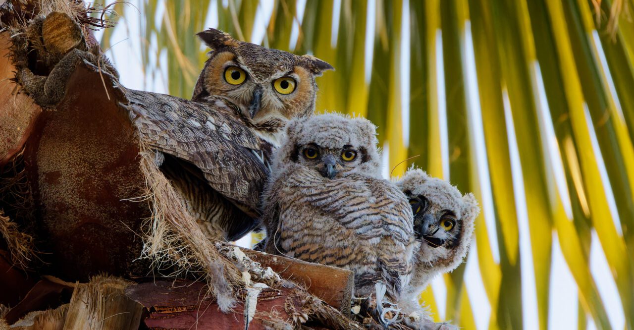
Then, one day on a walk, I spotted a pair of Great Horned Owls and three owlets in a palm tree. Seeing those downy, wide-eyed owlets nestled together stopped me in my tracks. And in that moment, I stopped worrying about my own life and started wondering about theirs.
Learning about the owl family added another purpose to my prescribed walking. I started visiting them every sunrise and sunset—curious, obsessed, and honestly, it just felt better when I was out there. It quickly became part of my daily routine.
Why do I feel so good? Why did I become so attached to birds? Could this be a form of therapy—owl therapy?
It turns out that birds and birding are emerging as a powerful, nature-based prescription for people’s mental well-being, capable of restoring a sense of purpose and inspiring a sense of connection. A new study suggests that building skills in birding may also yield structural benefits in a person’s brain. Research published in February 2026 in the Journal of Neuroscience showed that experienced birders had denser brain matter in the frontoparietal cortex—a region associated with cognition and working memory—than beginners. Birding, wrote the study authors in their research, “leaves a coherent imprint on the brain.”
“Birding strengthens cognitive function by transforming momentary personal experiences into lasting knowledge,” explains Dr. Erik Wing, lead author on the study and a neuroscientist at the Rotman Research Institute in Toronto, Canada.
His studies and work by other researchers in human psychology and social science are showing that birds do more than spark an interest in people’s lives. In fact, birds may help support a person’s cognitive function and mental health across their lifespan.
In his studies at the Rotman Research Institute, one of the world’s preeminent centers for understanding aging and human brain function, Wing explores how deep, accumulated knowledge—such as bird knowledge—changes the brain.
By accumulating sightings and experiences, birders develop complex mental networks as they connect species, behaviors, sounds, and habitats to identify birds. This form of knowledge compounds over time.
“You’re not only learning a new concept—you’re learning something that applies broadly to the entire network,” says Wing. “Everything you learn informs everything else.”
And the benefits to the brain multiply as birders level up their skills. For the study published in the Journal of Neuroscience, Wing and his coauthors conducted MRI brain scans on experienced and beginner birders. The experienced birders—defined primarily by their performance on bird ID tests rather than age alone—had a “tuned cortical network” in their brains, according to the study.
In other words, interconnected learning associated with birding may lead to structural and functional changes through neuroplasticity—the brain’s capacity to rewire and adapt with experience. Wing says that there are multiple routes through which birding can sharpen perception and calm the mind. Identifying subtle patterns—movement, shape, and sound—requires adaptive thinking and integrating our senses.
“Birding combines multiple factors we know support cognitive and mental health—like nature exposure, social interaction, and novelty—into one activity,” Wing says.
And though it demands high-level thinking, birding is also a form of mental rest.
“Birding helps interrupt cycles of stress and rumination by directing attention outward,” Wing says, referencing a 2022 study by German scientists published in the journal Scientific Reports that documented how listening to birdsong reduces anxiety in people. Birding, he says, offers “a moving, engaging focus in the natural world.”
In my case, daily walks for owl therapy were just what I needed to calm my anxious mind. Birding distracted me by giving me space to slow down, stay curious, and change my mental state. I began paying closer attention to what was happening outdoors in my neighborhood, with a growing awareness that deepened my connection to the owls and sparked curiosity about other birds nearby. What once felt like background noise now revealed a world I hadn’t noticed before.
According to Wing, this kind of rapid awakening is often most pronounced in the early stages of birding.
“You progress really fast because you know so little when you start,” he explains, noting that it’s not necessary to put in hundreds of hours up front to feel a sense of reward. Setting simple goals—like seeing 10 different birds in a week—can be both motivating and gratifying.
The benefits of birding experience may also endure across a person’s lifespan. The research published in the Journal of Neuroscience suggests that brain changes associated with birding persist in older experts, who continue to show differences in brain activity and organization compared to their age-matched counterparts. According to Wing, birding engages a variety of brain processes related to memory, attention, and flexible thinking—core components of cognition that are often associated with resilience in aging.
Scientists are also finding that, beyond possibly strengthening cognitive function, birding helps people experience joy, find meaning, and make connections.
Dr. Jenn Lodi-Smith, a professor of psychology at Canisius University in Buffalo, New York, is leading the Spark Bird Project, which collects people’s stories and survey data about how birding sparks transformative experiences that grow into lifelong passions. Since launching the project in 2022, Lodi-Smith has collected more than 500 stories from people about the moment they got hooked on birding. She says the Spark Bird stories are emotionally rich and overwhelmingly positive.
“People describe feelings of joy, curiosity, and pride,” she says. Yet some also reflect on grief or trauma, she says, suggesting that birding can help people process difficult emotions. Stories of recovery, she adds, may highlight the role of birding in emotional healing.
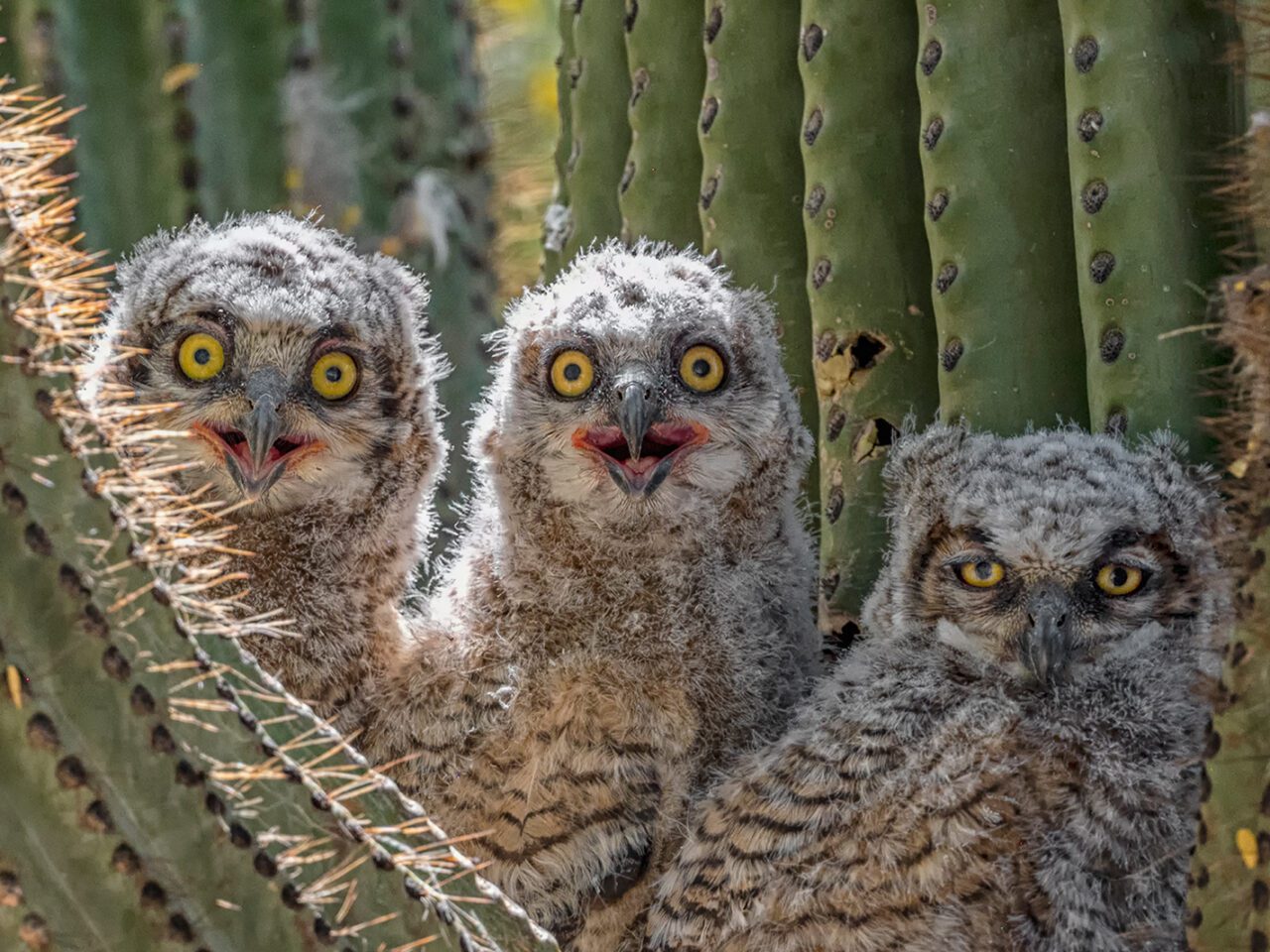
Some days I walked to the owl nest with sadness about my lost career and anxiety about my future. Witnessing the owlets’ wobbly dependence and shaky first flights taught me that recovery is slow and nonlinear—and that accepting support is okay. Their vulnerability and determination helped me let go of rigid expectations, slow down, and find purpose in recovering at my own pace. Gradually, I began to think more positively and see new ways I could be part of the world I shared with the birds—a world bigger than my own struggles.
In Lodi-Smith’s more than 20-year academic career studying identity development—including the psychological construct of Self-Concept Clarity, which describes how clearly defined, consistent, and stable a person’s self-beliefs are—she has found that human mental health isn’t just the absence of stress or illness. It’s the presence of meaning.
“Do people feel a sense of purpose? Do they have a sense of meaning in life?” she says. “One idea we study is generativity—the desire to give back and contribute to the broader world. Birds make us care about birds, but they also make us care about the world we share.”
Lodi-Smith says her research has also explored how the communities that people form can nurture a sense of belonging and identity.
“The birding community offers a space where people can express wonder, awe, and curiosity—emotions that aren’t always encouraged in everyday social roles,” she explains. “Whether a world-renowned ornithologist or a novice, birders share moments of excitement and connection that are deeply meaningful.”
And because birds can be found anywhere that people live, they are a great tool for boosting people’s mental health, says Dr. Katie Holland, a research scientist and social scientist at Virginia Tech University. Holland was a member of the science committee for the State of the Birds 2025 report, and the lead author on a section in that report about birds and mental health that cited studies on how birds ease symptoms of stress, anxiety, and depression in people. According to Holland, those mental health benefits are widely available across every possible demographic group of Americans.
Birding, she says, “can be adapted to diverse landscapes and participants—from those with minimal time or physical ability, to those seeking more elaborate experiences. This inclusivity means the mental health benefits of birding can reach many.”
Holland says the unique flexibility of birding makes it a potentially powerful wellness tool that can be scaled up across communities, at a time of rising need. According to data from the World Health Organization, more than 50% of the world’s population is expected to experience a mental health issue at some point in their lifetime.
“Colleagues of mine at Virginia Tech have also been exploring ways to make birding more accessible for diverse participants and those with disabilities,” she says. “This is important work, and can help make the well-being benefits of this activity available to many more people.”
I am living proof that birds and birding can be a multidimensional mental health tool—one that strengthens cognitive function, restores a sense of purpose, and inspires connection.
And I am continuing to recover. Six years after my traumatic brain injury, I feel more at ease, balanced, and emotionally stable as the clouds of anxiety, stress, and sadness begin to lift—clear signs of my improving mental health. With this progress, I am now working as a freelance writer, photographer, and speaker.
Hearing the owl parents hooting, watching the male deliver food, smiling at the owlets’ first awkward flights and early hunting attempts, and noticing the patterns on their wings as they preened were a few of my most treasured moments from owl therapy. It’s been a powerful force for good in my personal mental health journey—a force rooted in the simple acts of noticing, presence, reconnecting, rebuilding, recalibrating, and reclaiming power. And now when I go birding, I can feel it give me space when I feel stuck, confidence when I feel shattered, and curiosity when I feel uncertain.
It turns out the wildest plot twist in my story wasn’t the injury. It was the ease of recovery, sparked by owls.
I wonder what Great Horned Owls would charge per hour for human therapy? Whatever it is, I’d pay it.
About the Author
Emily Nichols is a bilingual English and Spanish freelance writer, as well as a photographer and speaker. She is a local captain in Phoenix, Arizona, for Birdability, a nonprofit organization that makes birding more accessible and inclusive for people with disabilities and other health concerns. Nichols holds a master’s degree in mass communications from Arizona State University’s Walter Cronkite School of Journalism.