Changing stroke rehab and research worldwide now.Time is Brain! trillions and trillions of neurons that DIE each day because there are NO effective hyperacute therapies besides tPA(only 12% effective). I have 523 posts on hyperacute therapy, enough for researchers to spend decades proving them out. These are my personal ideas and blog on stroke rehabilitation and stroke research. Do not attempt any of these without checking with your medical provider. Unless you join me in agitating, when you need these therapies they won't be there.

What this blog is for:

My blog is not to help survivors recover, it is to have the 10 million yearly stroke survivors light fires underneath their doctors, stroke hospitals and stroke researchers to get stroke solved. 100% recovery. The stroke medical world is completely failing at that goal, they don't even have it as a goal. Shortly after getting out of the hospital and getting NO information on the process or protocols of stroke rehabilitation and recovery I started searching on the internet and found that no other survivor received useful information. This is an attempt to cover all stroke rehabilitation information that should be readily available to survivors so they can talk with informed knowledge to their medical staff. It lays out what needs to be done to get stroke survivors closer to 100% recovery. It's quite disgusting that this information is not available from every stroke association and doctors group.

Showing posts with label optogenetics. Show all posts

Thursday, February 13, 2025

Korean researchers enhance stroke recovery by stimulating brain cells with light

 They never explain if this could possibly be expanded to humans.

  1. a technique in neuroscience in which genes for light-sensitive proteins are introduced into specific types of brain cells in order to monitor and control their activity precisely using light signals.
    "optogenetics allows researchers to control how nerve cells communicate"

With this definition, I wonder if human thick skulls would prevent the effects from occurring. Ask your competent? doctor.

Korean researchers enhance stroke recovery by stimulating brain cells with light

Korean study explores innovative ways to aid recovery from brain injuries using light technology
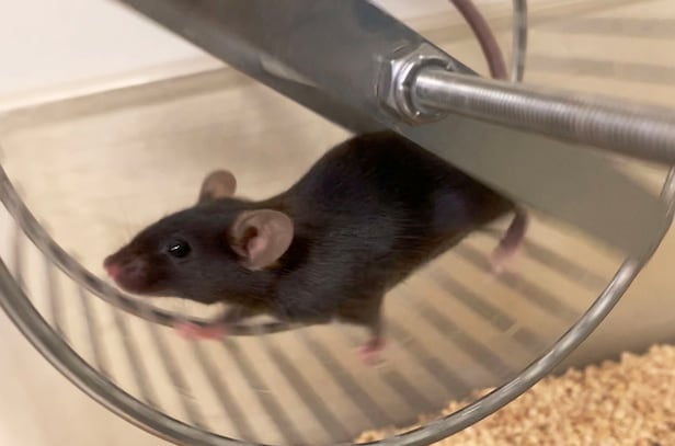
Domestic researchers successfully regulate the calcium signals of astrocytes in mice using optogenetic technology to promote the recovery of motor functions after chronic stroke./Courtesy of Univ. of Pennsylvania

Stroke is a disease that occurs when blood vessels in the brain become blocked or burst, causing damage to brain cells, and it can leave severe sequelae even if one survives. Prompt intervention, as well as treatment and rehabilitation, is important. Recently, domestic researchers have developed a treatment targeting astrocytes in the brain, successfully inducing the recovery of motor functions.

The research team led by Director General Lee Chang-jun from the Institute for Basic Science (IBS) collaborated with Professor Kim Hyung-il from the Gwangju Institute of Science and Technology (GIST) and Professor Heo Won-do from the Korea Advanced Institute of Science and Technology (KAIST) to demonstrate that they could enhance the recovery of motor functions after chronic stroke by regulating the calcium signals of astrocytes using optogenetic technology. The research results were published in the international journal Science Advances on the 31st of last month.

Stroke occurs due to various factors such as hypertension, diabetes, and hyperlipidemia, with ischemic stroke, which is caused by blockage or reduction of blood flow to the brain, accounting for the majority. In particular, subcortical strokes, which occur in deep structures below the cortex, represent about 30% of all ischemic strokes and have a poor prognosis.

Currently, neurorehabilitation therapy for stroke primarily uses methods that directly stimulate neurons. These include using strong magnetic fields or attaching electrodes to deliver electric currents. However, these methods have a non-selective effect on all cells in the stimulated area, and their mechanisms of action are unclear, making it difficult to predict treatment outcomes, with significant variability in treatment effects.

The research team attempted a new approach by regulating the calcium signals of astrocytes instead of directly stimulating neurons. Astrocytes are star-shaped non-neuronal cells that make up the majority of the cells in the brain. Researcher Lee Sang-kyu from IBS noted, "Astrocytes play a crucial role not only in supporting neurons but also in significantly affecting neuronal activation and synaptic plasticity," and he explained, "We aimed to reconstruct neural circuits and induce recovery of brain functions by regulating calcium signals in astrocytes."

When calcium signals in astrocytes increase, neurochemical regulators such as ATP and D-serine are secreted, which play important roles in regulating neuronal activation. ATP enhances neuronal excitability, while D-serine enhances synaptic plasticity. Synaptic plasticity is crucial for the recovery of damaged neural circuits after stroke, as it refers to the ability of neural connections to be strengthened and restructured. Additionally, astrocytes regulate glutamate, which is essential for maintaining the balance of neural circuits, preventing excessive neuronal excitation and promoting stable neural activity.

The research team used the optogenetic tool OptoSTIM1, jointly developed by IBS and KAIST in 2015, to regulate calcium signals in mouse brain astrocytes with light. As a result of activating the calcium signals in the astrocytes of the sensorimotor cortex region, which is closely related to motor function recovery, not only were fine motor skills using the forelimbs improved, but overall motor abilities were enhanced. Even low-intensity light stimulation for one hour a day over the course of two weeks led to recovery of motor abilities.

Director General Lee Chang-jun said, "We presented a precise and safe stroke treatment strategy targeting astrocytes," and noted, "This could lead to the development of drugs that regulate calcium signals in astrocytes, which may be applied not only to stroke but also to the treatment of various neurological diseases, including Alzheimer's disease."

References

Science Advances (2025), DOI: https://doi.org/10.1126/sciadv.adn7577

※ This article has been translated using the OpenAI translation tool.

Thursday, April 4, 2019

Genetic Variants May Influence Post-Stroke Recovery

So What? It is still the responsibility of our stroke doctors, therapists, hospitals and researchers to get all survivors 100% recovered. This will not be allowed as an excuse to not do that. Leaders solve difficult problems. ARE YOU LEADERS OR CHICKENSHITS?

Genetic Variants May Influence Post-Stroke Recovery

Genes may have a bearing not only on stroke risk, but also on how well patients recover after stroke, according to a study published in Neurology.
For the first time, researchers have identified common genetic variants that are associated with outcome after ischaemic stroke.
For the study, Martin Söderholm, MD, Lund University, Lund, Sweden, and colleagues conducted a meta-analysis of 12 international stroke studies that included 6,165 patients with ischaemic stroke.
The patients were divided into 2 groups depending on their outcome at 3 months after ischaemic stroke -- 1 group was composed of patients who died and those who were dependent on help from others to cope with activities of daily living. The other group consisted of patients who were able to cope unaided 3 months after stroke.
By comparing analyses of the patients’ genomes, the researchers were able to find several different genetic variants that appear to have played a part in the patients’ outcomes.
“One of the common genetic variants we found was significant -- that is, clearly associated with a worse outcome in the large volumes of data we were able to access,” said Annie Pedersen, University of Gothenburg, Gothenburg, Sweden.
That variation, identified as rs1842681 in the LOC105372028 gene, can be linked to another gene that is part of a major process involved in brain plasticity.
The study took into account several other factors -- age, sex, and the extent of each patient’s brain damage -- that can also affect outcome after stroke. After adjustment for these factors, the association remained between the genetic variant and elevated risk of being in the group of patients who still, 3 months after the stroke onset, were unable to manage without assistance.
“Even if 2 patients seem to have the same prospects of recovering, their outcomes can be different,” said Pederson.
“Our limited knowledge of why some patients recover well while others incur lasting functional impairments after the same type of stroke has made it difficult to develop new treatment methods,” added Christina Jern, MD, University of Gothenburg.
“In the long run, we hope the research may enable us to identify new targets for medication that might help to improve post-stroke outcome, but there is a lot of research to be done before we get there,” she concluded.
Reference: https://doi.org/10.1212/WNL.0000000000007138
SOURCE: University of Gothenburg

Tuesday, January 23, 2018

Methods from optogenetics, machine learning should help improve treatment options for stroke patients

Don't just lazily say should and then never followup with actual inteventional treatment options. I bet nothing will occur, NO leadership, NO strategy. 

Methods from optogenetics, machine learning should help improve treatment options for stroke patients


Heidelberg researchers develop computer vision technique to analyze rehabilitation process
Methods from optogenetics and machine learning should help improve treatment options for stroke patients. Researchers from Heidelberg University have developed a computer vision technique to analyze the changes in motor skills that result from targeted stimulation of healthy areas of the brain. Movements recorded with a video camera are automatically analyzed to monitor the rehabilitation process and evaluate and adjust the optogenetic stimulation. Researchers from the Interdisciplinary Center for Scientific Computing (IWR) in Heidelberg worked with neurobiologists from Switzerland to develop the method.
Along with speech and vision problems, motor paralyses are the most common symptoms post-stroke. According to lead author Dr Dr Anna-Sophia Wahl, a neuroscientist at the Swiss Federal Institute of Technology (ETH) in Zurich, neurorehabilitation is the only treatment option for the majority of stroke victims. "Many approaches in basic science and in the clinic aim to trigger regeneration processes post-stroke by stimulating healthy brain regions of indeterminate size. However, we use optogenetics to systematically stimulate certain unaffected areas of the brain so that they sprout connections into the damaged hemisphere in order to assume its functions." So-called corticospinal circuits from the cerebral cortex to the spinal cord are specifically activated.
In optogenetics, light is used to control genetically modified cells. The cooperation partners in Switzerland - researchers from the ETH and the University of Zurich - used optogenetic stimulation in combination with intensive rehabilitation training to restore the paralysed paw function in rats. "Using our automatic evaluation of the movement processes, we were able to demonstrate a full recovery," explains Prof. Dr Björn Ommer, IWR researcher and head of the Heidelberg team. The new computer vision technique is able to quantify even the slightest changes in motor functions. "By recording and analyzing the movements, we can objectively assess whether there was true restoration of the original function or merely compensation."

Friday, January 19, 2018

Researchers develop computer vision technique to analyse stroke rehabilitation process

Up to you to get your doctor and stroke hospital to do something with this.

Is it anything like this?

What Would Dean Do? - As head of a stroke hospital dept.

The latest here:

Researchers develop computer vision technique to analyse stroke rehabilitation process

January 17, 2018, Heidelberg University
Methods from optogenetics and machine learning should improve treatment options for stroke patients. Researchers from Heidelberg University have developed a computer vision technique to analyse the changes in motor skills that result from targeted stimulation of healthy areas of the brain. Movements recorded with a video camera are automatically analysed to monitor the rehabilitation process and evaluate and adjust the optogenetic stimulation. Researchers from the Interdisciplinary Center for Scientific Computing (IWR) in Heidelberg worked with neurobiologists from Switzerland to develop the method.
Along with speech and vision problems, motor paralyses are the most common symptoms post-stroke. According to lead author Dr. Dr. Anna-Sophia Wahl, a neuroscientist at the Swiss Federal Institute of Technology (ETH) in Zurich, neurorehabilitation is the only treatment option for the majority of stroke victims. "Many approaches in basic science and in the clinic aim to trigger regeneration processes post-stroke by stimulating healthy brain regions of indeterminate size. However, we use optogenetics to systematically stimulate certain unaffected areas of the brain so that they sprout connections into the damaged hemisphere in order to assume its functions." So-called corticospinal circuits from the cerebral cortex to the spinal cord are specifically activated.
In optogenetics, light is used to control . The cooperation partners in Switzerland – researchers from the ETH and the University of Zurich – used in combination with intensive rehabilitation training to restore the paralysed paw function in rats. "Using our automatic evaluation of the movement processes, we were able to demonstrate a full recovery," explains Prof. Dr. Björn Ommer, IWR researcher and head of the Heidelberg team. The new computer vision technique is able to quantify even the slightest changes in motor functions. "By recording and analysing the movements, we can objectively assess whether there was true restoration of the original function or merely compensation."
Prof. Ommer is a member of the Interdisciplinary Center for Scientific Computing of Heidelberg University. His Computer Vision research group is located at the Heidelberg Collaboratory for Image Processing. The latest results of the collaborative study with the researchers in Zurich were published in the journal Nature Communications.
More information: A. S. Wahl et al. Optogenetically stimulating intact rat corticospinal tract post-stroke restores motor control through regionalized functional circuit formation, Nature Communications (2017). DOI: 10.1038/s41467-017-01090-6

Saturday, August 26, 2017

CPSR researchers use light to restore function in brain circuits damaged by stroke

I would like to see a writeup of how this could possibly work in humans. Maybe shining light thru nanoneedles? Followup needed which will never occur.
http://www.canadianstroke.ca/en/news/cpsr-researchers-use-light-to-restore-function-in-brain-circuits-damaged-by-stroke/
A University of Victoria neuroscientist and his team has discovered that stimulating brain circuits with light can improve recovery from a stroke.
A major challenge in stroke research is to understand how stroke disrupts brain circuits that are crucial for sensation and movement. When these circuits are damaged by stroke, people experience profound difficulties in everyday life tasks such as lifting a fork, brushing their teeth, buttoning up a shirt or driving a car.
Dr. Brown’s neurobiology research lab is providing new clues in pre-clinical research as to what happens to these circuits after stroke and developed a treatment strategy for enhancing recovery.
“What we’ve found is that stroke makes certain circuits in a brain region called the thalamus, less active or excitable. These circuits are important for processing sensory information, for example allowing us to grasp an object in our hand. However when a stroke occurs, these circuits are disrupted and do not properly process sensory information in a normal way.”
In order to make these circuits work properly after a stroke, Dr. Brown’s team used an “optogenetic” strategy where brain cells that express an algae protein can be controlled with blue light. “When brain cells express this protein, we can flash light on them and make them excitable again”. As it turns out, stimulating these circuits with light for several weeks after stroke allowed experimental animals to regain better use of their paw.
“Although this is just a first step in developing a new approach for treating stroke, we are really excited about the possibility that one day it may be used in the clinic. The fact that clinical trials are in the works to use optogenetics to treat blindness and other neurological conditions, suggest it is possible.
The study’s results were published June 23, in Nature Communications.
For more information on Brown and his research is available here.
Pictured above: UVic Professor Dr. Craig Brown, right, and Dr. Kelly Tennant, left. Dr. Tennant is a former co-chair of the CPSR National Trainee Association. 
Link to publication: 
Nat Commun. 2017 Jun 23;8:15879. doi: 10.1038/ncomms15879.

Thursday, December 15, 2016

New approach uses silicon-based photonic probe to deliver light deep within brain tissues

So your doctor and researcher should now be able to stimulate deeper neurons using optogenetics.
http://www.news-medical.net/news/20161208/New-approach-uses-silicon-based-photonic-probe-to-deliver-light-deep-within-brain-tissues.aspx
The ability to stimulate neural circuits with very high precision light to control cells -- optogenetics -- is key to exciting advances in the study and mapping of the living brain. In the current state of the art, spatially patterned light projected via free-space optics stimulates small, transparent organisms and excites neurons within superficial layers of the cortex.
However, light scattering and absorption in neural tissue cause light penetration to be extremely short, making it impossible to employ free-space optical methods to probe brain regions deeper than about 2 mm.
In "Patterned photostimulation via visible-wavelength photonic probes for deep brain optogenetics," published today by SPIE, the international society for optics and photonics, in the journal Neurophotonics, principal author Eran Segev of professor Michael Roukes' group at Caltech, along with coauthors from Caltech, Baylor College of Medicine, and Stanford University, describe a solution. The article is available via open access.
Their approach combines nanophotonics and microelectromechanical systems (MEMS) in an implantable, ultra-narrow, silicon-based photonic probe to deliver light deep within brain tissues. This minimally invasive technique avoids major tissue displacement during implantation.
Using techniques of optogenetics, a protein in the brain serves as a sensory photoreceptor and can be controlled by specific wavelengths of light. These combined techniques provide a new approach to stimulation of brain circuits with remarkable resolution, enabling observation and control of individual neurons.
These breakthroughs present widespread and promising applications for the neuroscience and neuromedical research communities. From characterizing the role of specific neurons and identifying neural circuits responsible for behavior to enabling new methods of operant conditioning through reward-induced circuit activations, optogenetics has become a new path for neuroscientists seeking advances in research capabilities.
The article appears in a special section in Neurophotonics, Brain Mapping and Therapeutics, with Shouleh Nikzad, Jet Propulsion Laboratory, Caltech, serving as senior guest editor. The special section is part of an SPIE partnership with the Society for Brain Mapping and Therapeutics (SBMT), serving as a multidisciplinary approach for using advanced technology to solve neurological disorders and disease and to understand neuroscience. The effort was initiated during Nikzad's term as SBMT president in 2015.
David Boas of Massachusetts General Hospital, Harvard Medical School, is the editor-in-chief of Neurophotonics. Launched in 2014, Neurophotonics is published digitally in the SPIE Digital Library and in print. The journal covers advances in optical technology applicable to the study of the brain and their impact on basic and clinical neuroscience applications.
The SPIE Digital Library contains more than 458,000 articles from SPIE journals, proceedings, and books, with approximately 18,000 new research papers added each year. Abstracts are freely searchable, and a number of journal articles are published with open access.
Source:
SPIE--International Society for Optics and Photonics

Thursday, August 21, 2014

Scientists Discover Brain Area Responsible for Exercise Motivation

So your doctor will need to check this area for damage to see if you even have the motivation to exercise. And then have a stroke protocol to correct such damage and restore your motivation.
http://www.biosciencetechnology.com/news/2014/08/scientists-discover-brain-area-responsible-exercise-motivation?et_cid=4111840&
Scientists at Seattle Children’s Research Institute have discovered an area of the brain that could control a person’s motivation to exercise and participate in other rewarding activities – potentially leading to improved treatments for depression.
 
Dr. Eric Turner, a principal investigator in Seattle Children’s Research Institute’s Center for Integrative Brain Research, together with lead author Dr. Yun-Wei (Toni) Hsu, have discovered that a tiny region of the brain – the dorsal medial habenula– controls the desire to exercise in mice. The structure of the habenula is similar in humans and rodents and these basic functions in mood regulation and motivation are likely to be the same across species.  
 
Exercise is one of the most effective non-pharmacological therapies for depression. Determining that such a specific area of the brain may be responsible for motivation to exercise could help researchers develop more targeted, effective treatments for depression. 
 
“Changes in physical activity and the inability to enjoy rewarding or pleasurable experiences are two hallmarks of major depression,” Turner said. “But the brain pathways responsible for exercise motivation have not been well understood. Now, we can seek ways to manipulate activity within this specific area of the brain without impacting the rest of the brain’s activity.” 
 
Turner’s study, titled “Role of the Dorsal Medial Habenula in the Regulation of Voluntary Activity, Motor Function, Hedonic State, and Primary Reinforcement,” was published by the Journal of Neuroscience and funded by the National Institute of Mental Health and National Institute on Drug Abuse. The study used mouse models that were genetically engineered to block signals from the dorsal medial habenula. In the first part of the study, Turner’s team collaborated with Dr. Horacio de la Iglesia, a professor in University of Washington’s Department of Biology, to show that compared to typical mice, who love to run in their exercise wheels, the genetically engineered mice were lethargic and ran far less. Turner’s genetically engineered mice also lost their preference for sweetened drinking water. 
 
“Without a functioning dorsal medial habenula, the mice became couch potatoes,” Turner said. “They were physically capable of running but appeared unmotivated to do it.” 
 
In a second group of mice, Turner’s team activated the dorsal medial habenula using optogenetics– a precise laser technology developed in collaboration with the Allen Institute for Brain Science. The mice could “choose” to activate this area of the brain by turning one of two response wheels with their paws. The mice strongly preferred turning the wheel that stimulated the dorsal medial habenula, demonstrating that this area of the brain is tied to rewarding behavior.  
 
Past studies have attributed many different functions to the habenula, but technology was not advanced enough to determine roles of the various subsections of this area of the brain, including the dorsal medial habenula. 
 
“Traditional methods of stimulation could not isolate this part of the brain,” Turner said. “But cutting-edge technology at Seattle Children’s Research Institute makes discoveries like this possible.” 
 
As a professor in the University of Washington Department of Psychiatry and Behavioral Sciences, Turner treats depression and hopes this research will make a difference in the lives of future patients. 
 
“Working in mental health can be frustrating,” Turner said. “We have not made a lot of progress in developing new treatments. I hope the more we can learn about how the brain functions the more we can help people with all kinds of mental illness.”
 

Tuesday, August 19, 2014

Targeted stimulation of specific brain cells boosts stroke recovery in mice

So who is going to take charge of getting this into human clinical trials? We know it is not going to be the ASA, NSA or WSO because obviously their boards of directors have no intention of ever solving any stroke problem.
Only a GREAT STROKE ASSOCIATION will ever tackle all the hardest problems in stroke.
http://scopeblog.stanford.edu/2014/08/19/targeted-stimulation-of-specific-brain-cells-boosts-stroke-recovery-in-mice/ 

Monday, June 2, 2014

Self-Tuning Neurons Promote Resilience to Stress, Depression

What is your doctor going to do based on this to handle your stress/depression on not being told of your objective diagnosis or any way to get to 100% recovery?
http://www.nih.gov/researchmatters/may2014/05052014resilience.htm
Enhancing brain mechanisms triggered by stress raised the resilience of mice to stress and relieved depression-like behaviors. The surprising results suggest novel approaches to promoting mental health.
Some mice exposed to repeated encounters with a dominant animal develop depression-like behaviors, while others don’t. Sensitive mice avoid other animals and lose their preference for sugar. In past work, a group of researchers led by Dr. Ming-Hu Han of the Icahn School of Medicine at Mount Sinai found that neurons in the ventral tegmental area (VTA)—one of the “reward” areas deep in the brain—fire at higher rates in mice that are more susceptible to social stress. These neurons are known to secrete the chemical messenger dopamine.
The scientists later found that mouse susceptibility to social stress could be turned on and off by manipulating the firing rates of these neurons. To explore how this mechanism works at the cellular level, the researchers focused on electrical events within the neurons. The study, funded in part by NIH’s National Institute of Mental Health (NIMH), appeared on April 18, 2014, in Science.
The researchers found that while stress-resilient mice had VTA dopamine neurons with stable firing rates and normal dopamine activity, these neurons had higher levels of an excitatory electrical current than those of stressed mice. The higher activation currents were accompanied by higher inhibitory potassium channel currents. The researchers hypothesized that, in resilient animals, runaway excitatory currents trigger a boost in inhibitory currents, resulting in normal mood-related behaviors.
The team thus tested whether boosting excitatory currents could activate compensatory currents in susceptible mice. Over 5 days, the scientists repeatedly infused the VTA of susceptible mice with a drug called lamotrigine, which is known to increase excitatory currents. The treated mice socialized more and their characteristic rodent sweet tooth came back. At the cellular level, these mice showed a marked increase in both excitatory and inhibitory currents, resulting in normal neuron activity. This self-tuning balance of activity, common in other body systems, is called homeostasis.
The scientists achieved similar results using a technique called optogenetics to activate neuronal activity. Further experiments showed that the homeostatic mechanism worked specifically in the reward circuit running from the VTA to cells in a brain area called the nucleus accumbens.
“To our surprise, neurons in this circuit harbor their own self-tuning, homeostatic mechanism of natural resilience,” Han says. When an excitatory current develops in response to social stress—and is driven high enough for a sustained period—it triggers its own compensatory adaptation. Inhibitory currents correct out-of-balance electrical activity and thus produce resilience.
As counterintuitive as it seems, in this case, exaggerating an abnormality can be beneficial. Future strategies might harness this homeostatic mechanism to promote resilience to stress and combat depression.

Thursday, December 19, 2013

Shining a Light on Stroke

This should be continued as a test for the next week so the neuronal cascade of death could be observed and maybe figure out how long it lasts.
Shining a Light on Stroke

  1. Michael J. Minzenberg
  1. Department of Psychiatry, University of California School Of Medicine, Sacramento, CA 95817, USA. Email: michael.minzenberg{at}ucdmc.ucdavis.edu
Stroke remains a common and serious consequence of numerous underlying illnesses and risk factors, such as hypertension and diabetes. Understanding how the brain changes after stroke may help to advance treatments for this illness. To this end, Barth and Mody have developed an improved in vivo model of the anatomy and physiology of ischemic stroke that uses photothrombosis—occlusion of a blood vessel through injection of a dye followed by irradiation—combined with stereotaxic localization (the use of a three-dimensional coordinate system to place the optic fiber) to monitor neurological changes before and after ischemic stroke.
The researchers inserted the optic fiber into the hippocampal artery of mice to isolate blood flow to the hippocampus, a brain region that is important in learning and memory and is particularly vulnerable to ischemic stroke. Blood flow was then selectively blocked in the artery through light activation of a photosensitive dye, rose bengal, which induced blood clot formation. Measuring neuron population activity in the hippocampus before and after this procedure, the authors observed a massive, brief high-frequency epileptiform discharge (HFD) in affected neurons, followed by a negative shift in the baseline electrical potential, which is consistent with neuronal depolarization due to hypoxia (inadequate oxygen). This was followed by a long-lasting decrease in neuron oscillatory activity in the gamma range (30 to 119 Hz), which is generally important to complex cognitive processes such as memory. Interestingly, only the initial HFD was also observed in the contralateral hippocampus, which is often affected by the spread of seizure activity from the other side of the brain.
This work elegantly characterizes the neurophysiological changes that unfold in the wake of stroke, thus setting the stage for elaboration of the biochemical basis and time course of these changes. These findings also suggest that events, such as the HFD and subsequent negative electrical potential, might provide specific targets for therapeutic intervention aimed at attenuating epileptiform activity or other disturbances in electrical activity, ultimately to mitigate the deleterious effects of these processes on the brain. Such knowledge should aid in the development of new therapies designed to restore brain function in stroke survivors.

Sunday, November 10, 2013

The flexDrive: an ultra-light implant for optical control and highly parallel chronic recording of neuronal ensembles in freely moving mice

By following this to its logical conclusion we would be able to implant these into human subjects and listen in to the signals that neurons send to each other to enable neuroplasticity. With that knowledge we can make neuroplasticity repeatable and assured. And close to 100% recovery can be acheived. If you don't have big goals you will never get there. Or you can be like the pathetic stroke associations that just send out press releases and their board of directors that will not take a chance.  Bah humbug.
http://www.ncbi.nlm.nih.gov/pubmed/23717267

Source

Department of Brain and Cognitive Sciences, Massachusetts Institute of Technology Cambridge, MA, USA ; Department of Neuroscience, Brown University Providence, RI, USA.

Abstract

Electrophysiological recordings from ensembles of neurons in behaving mice are a central tool in the study of neural circuits. Despite the widespread use of chronic electrophysiology, the precise positioning of recording electrodes required for high-quality recordings remains a challenge, especially in behaving mice. The complexity of available drive mechanisms, combined with restrictions on implant weight tolerated by mice, limits current methods to recordings from no more than 4-8 electrodes in a single target area. We developed a highly miniaturized yet simple drive design that can be used to independently position 16 electrodes with up to 64 channels in a package that weighs ~2 g. This advance over current designs is achieved by a novel spring-based drive mechanism that reduces implant weight and complexity. The device is easy to build and accommodates arbitrary spatial arrangements of electrodes. Multiple optical fibers can be integrated into the recording array and independently manipulated in depth. Thus, our novel design enables precise optogenetic control and highly parallel chronic recordings of identified single neurons throughout neural circuits in mice.

Images at link.

Tuesday, September 3, 2013

'Brain window' implant devised

Maybe we could get laser treatments or see what optogenetics can do.
The BBC report here;

'Brain window' implant devised


the abstract and paper it is based upon here;
http://www.sciencedirect.com/science/article/pii/S1549963413003614

Or you could ask your doctor what use they will make of it to help your recovery.

Monday, May 13, 2013

A Bright Idea: Tiny Injectable LEDs Help Neuroscientists Study the Brain

Something our researchers should use to determine how a next door neuron goes to help its neighbor with a task. That understanding of neuroplasticity could make it repeatable within understood parameters.
http://livasperiklis.com/2013/05/12/httpwp-mep29tmj-3uu/
The researchers demonstrated the first application of their devices in optogenetics, a new area of neuroscience that uses light to stimulate targeted neural pathways in the brain. The procedure involves genetically programming specific neurons to respond to light. Optogenetics allows researchers to study precise brain functions in isolation in ways that are impossible with electrical stimulation, which affects neurons throughout a broad area, or with drugs, which saturate the whole brain.
Optogenetics experiments with mice illustrate the ability to train complex behaviors without physical reward, and to alleviate certain anxiety responses. Yet fundamental insights into the structure and function of the brain that emerge from such studies could have implications for treatment of Alzheimer’s, Parkinson’s, depression, anxiety and other neurological disorders.
While a number of important neural pathways now can be studied by optogenetics, researchers continue to struggle with the engineering challenge of delivering light to precise regions deep within the brain. The most widely used methods tether the animals to lasers with fiber-optic cables embedded in the skull and brain – an invasive procedure that also limits movements, affects natural behaviors and prevents study of social interactions.
The newly developed technologies bypass these limitations with specially designed powerful LEDs – among the world’s smallest, with sizes comparable to single cells – that are injected into the brain to provide direct illumination and precise control. The devices are printed onto the tip end of a thin, flexible plastic ribbon – thinner than a human hair and narrower than the eye of a needle – that can insert deep into the brain with very little stress to tissue.
More with pictures at the link.

Sunday, November 18, 2012

New way to stimulate transplanted human stem cell-derived neurons into a rodent hippocampus

Pieces of the stem cell puzzle are falling into place.
http://www.news-medical.net/news/20121117/New-way-to-stimulate-transplanted-human-stem-cell-derived-neurons-into-a-rodent-hippocampus.aspx
Researchers and patients look forward to the day when stem cells might be used to replace dying brain cells in Alzheimer's disease and other neurodegenerative conditions. Scientists are currently able to make neurons and other brain cells from stem cells, but getting these neurons to properly function when transplanted to the host has proven to be more difficult. Now, researchers at Sanford-Burnham Medical Research Institute (Sanford-Burnham) have found a way to stimulate stem cell-derived neurons to direct cognitive function after transplantation to an existing neural network. The study was published November 7 in the Journal of Neuroscience.
"We showed for the first time that embryonic stem cells that we've programmed to become neurons can integrate into existing brain circuits and fire patterns of electrical activity that are critical for consciousness and neural network activity," said Stuart A. Lipton, M.D., Ph.D., senior author of the study. Lipton is director of Sanford-Burnham's Del E. Webb Neuroscience, Aging, and Stem Cell Research Center and a clinical neurologist.
The trick turned out to be light. Lipton and his team-including Juan Pi-a-Crespo, Ph.D., Maria Talantova, Ph.D., and other colleagues at Sanford-Burnham and Stanford University-transplanted human stem cell-derived neurons into a rodent hippocampus, the brain's information-processing center. Then they specifically activated the transplanted neurons with optogenetic stimulation, a relatively new technique that combines light and genetics to precisely control cellular behavior in living tissues or animals.

Page 2 at the link.

Wednesday, May 9, 2012

Mapping the connectome: multi-level analysis of brain connectivity

I know connectome knowledge has little bearing on stroke rehab but if we don't know how the brain connects up neurons in 'good' brains, how will we ever figure out how to connect them up again in 'bad' brains?
So ask your doctor what their thoughts are on the using this knowledge to help your rehab. If it makes them uncomfortable all the better.
article here:
http://www.frontiersin.org/Neuroinformatics/10.3389/fninf.2012.00014/full
Blogger dissecting the article here:
http://brainslab.wordpress.com/2012/05/07/19832741923/

Saturday, April 28, 2012

Optogenetics: Stroke Rehabilitation

Interesting idea. I've always had at least 20 lbs. grip in my left hand., So I guess I wouldn't qualify. Would it work in reverse? Relaxing the spastic muscles?
http://rrg.utk.edu/resources/BME473/lectures/presentation_team_2.pdf
see page 6.

Tuesday, May 17, 2011

Optogenetics Control Desk for the Neural Switchboard

I could see using this to fire damaged neurons and speed up neuroplasticity.
http://www.nytimes.com/2011/05/17/science/17optics.html
Treating anxiety no longer requires years of pills or psychotherapy. At least, not for a certain set of bioengineered mice.
Science Times Podcast
Subscribe
This week: Manipulating the brain with light, and an organ transplant saga with a happy ending.


Multimedia
In a study recently published in the journal Nature, a team of neuroscientists turned these high-strung prey into bold explorers with the flip of a switch.
The group, led by Dr. Karl Deisseroth, a psychiatrist and researcher at Stanford, employed an emerging technology called optogenetics to control electrical activity in a few carefully selected neurons.
First they engineered these neurons to be sensitive to light. Then, using implanted optical fibers, they flashed blue light on a specific neural pathway in the amygdala, a brain region involved in processing emotions.
And the mice, which had been keeping to the sides of their enclosure, scampered freely across an open space.
While such tools are very far from being used or even tested in humans, scientists say optogenetics research is exciting because it gives them extraordinary control over specific brain circuits — and with it, new insights into an array of disorders, among them anxiety and Parkinson’s disease.
Mice are very different from humans, as Dr. Deisseroth (pronounced DICE-er-roth) acknowledged. But he added that because “the mammalian brain has striking commonalities across species,” the findings might lead to a better understanding of the neural mechanisms of human anxiety.
David Barlow, founder of the Center for Anxiety and Related Disorders at Boston University, cautions against pushing the analogy too far: “I am sure the investigators would agree that these complex syndromes can’t be reduced to the firing of a single small neural circuit without considering other important brain circuits, including those involved in thinking and appraisal.”
But a deeper insight is suggested by a follow-up experiment in which Dr. Deisseroth’s team directed their light beam just a little more broadly, activating more pathways in the amygdala. This erased the effect entirely, leaving the mouse as skittish as ever.
This implies that current drug treatments, which are far less specific and often cause side effects, could also in part be working against themselves.
David Anderson, a professor of biology at the California Institute of Technology who also does research using optogenetics, compares the drugs’ effects to a sloppy oil change. If you dump a gallon of oil over your car’s engine, some of it will dribble into the right place, but a lot of it will end up doing more harm than good.
“Psychiatric disorders are probably not due only to chemical imbalances in the brain,” Dr. Anderson said. “It’s more than just a giant bag of serotonin or dopamine whose concentrations sometimes are too low or too high. Rather, they likely involve disorders of specific circuits within specific brain regions.”
So optogenetics, which can focus on individual circuits with exceptional precision, may hold promise for psychiatric treatment. But Dr. Deisseroth and others caution that it will be years before these tools are used on humans, if ever.
For one, the procedure involves bioengineering that most people would think twice about. First, biologists identify an “opsin,” a protein found in photosensitive organisms like pond scum that allows them to detect light. Next, they fish out the opsin’s gene and insert it into a neuron within the brain, using viruses that have been engineered to be harmless —“disposable molecular syringes,” as Dr. Anderson calls them.
There, the opsin DNA becomes part of the cell’s genetic material, and the resulting opsin proteins conduct electric currents — the language of the brain — when they are exposed to light. (Some opsins, like channelrhodopsin, which responds to blue light, activate neurons; others, like halorhodopsin, activated by yellow light, silence them.)
Finally, researchers delicately thread thin optical fibers down through layers of nervous tissue and deliver light to just the right spot.
