Changing stroke rehab and research worldwide now.Time is Brain! trillions and trillions of neurons that DIE each day because there are NO effective hyperacute therapies besides tPA(only 12% effective). I have 523 posts on hyperacute therapy, enough for researchers to spend decades proving them out. These are my personal ideas and blog on stroke rehabilitation and stroke research. Do not attempt any of these without checking with your medical provider. Unless you join me in agitating, when you need these therapies they won't be there.

What this blog is for:

My blog is not to help survivors recover, it is to have the 10 million yearly stroke survivors light fires underneath their doctors, stroke hospitals and stroke researchers to get stroke solved. 100% recovery. The stroke medical world is completely failing at that goal, they don't even have it as a goal. Shortly after getting out of the hospital and getting NO information on the process or protocols of stroke rehabilitation and recovery I started searching on the internet and found that no other survivor received useful information. This is an attempt to cover all stroke rehabilitation information that should be readily available to survivors so they can talk with informed knowledge to their medical staff. It lays out what needs to be done to get stroke survivors closer to 100% recovery. It's quite disgusting that this information is not available from every stroke association and doctors group.

Showing posts with label microglia. Show all posts

Friday, May 22, 2026

Can microglial iron-driven ferroptosis be the key to post-hemorrhage neuroinflammation?

 Your competent doctor has been working on solving microbleeds  for over two decades, right? And ferroptosis for well over a decade? NO? So, INCOMPETENT THEN? 

Can microglial iron-driven ferroptosis be the key to post-hemorrhage neuroinflammation?

After a cerebral hemorrhage (CH), heme oxygenase-1 (HO-1) catalyzes the conversion of heme to release Fe2+. Microglia are the primary cells responsible for immune function in the brain. Upon the uptake of heme and iron ions, microglia are activated, leading to the subsequent release of inflammatory mediators and reactive oxygen species. Neuroinflammation and oxidative stress are common features of various brain diseases. Cerebral hemorrhage can trigger microglial iron accumulation and ferroptosis, which in turn leads to neuroinflammation and oxidative stress imbalance in the brain. Therefore, inhibiting microglial iron accumulation and ferroptosis alleviates cerebral hemorrhage-mediated neural damage and counteracts various brain diseases induced by it. We propose that the occurrence of many brain diseases is influenced by the location of cerebral microbleeds, suggesting that cerebral microbleeds may be a high-risk factor for inducing these diseases.

REFERENCES

  1. Neuroinflammatory diseases triggered by cerebral hemorrhage: microglial iron accumulation and ferroptosis.

    Gao X, Li H, Liang J, Liu S.

    Eur J Pharmacol. 2026 May 14; 1026 178972 [Epub ahead of print]

Thursday, June 20, 2024

Microglia protect against age-associated brain pathologies

 If you  have a competent? doctor they will get human testing going and create interventions that save these microglia! Do you have a competent doctor?

Microglia protect against age-associated brain pathologies

Open AccessPublished:June 18, 2024DOI:https://doi.org/10.1016/j.neuron.2024.05.01

Highlights

  • Lack of microglia leads to progressive brain calcifications and macroglial reactivity
  • scRNA-seq reveals brain cell profiles across the lifespan in the absence of microglia
  • The thalamus is particularly sensitive to the absence of microglia with aging
  • Transplanting microglia protects against pathology development

Summary

Microglia are brain-resident macrophages that contribute to central nervous system (CNS) development, maturation, and preservation. Here, we examine the consequences of permanent microglial deficiencies on brain aging using the Csf1rΔFIRE/ΔFIRE mouse model. In juvenile Csf1rΔFIRE/ΔFIRE mice, we show that microglia are dispensable for the transcriptomic maturation of other brain cell types. By contrast, with advancing age, pathologies accumulate in Csf1rΔFIRE/ΔFIRE brains, macroglia become increasingly dysregulated, and white matter integrity declines, mimicking many pathological features of human CSF1R-related leukoencephalopathy. The thalamus is particularly vulnerable to neuropathological changes in the absence of microglia, with atrophy, neuron loss, vascular alterations, macroglial dysregulation, and severe tissue calcification. We show that populating Csf1rΔFIRE/ΔFIRE brains with wild-type microglia protects against many of these pathological changes. Together with the accompanying study by Chadarevian and colleagues, our results indicate that the lifelong absence of microglia results in an age-related neurodegenerative condition that can be counteracted via transplantation of healthy microglia.

Graphical abstract


Friday, April 26, 2024

Study Suggests Treatments that Unleash Immune Cells in the Brain Could Help Combat Alzheimer’s

 Because of your risk of dementia post stroke; is your competent? doctor and hospital closely following this? NO? So you have INCOMPETENT MEDICAL 'PROFESSIONALS'?

Your risk of dementia, has your doctor told you of this?  Your doctor is responsible for preventing this!

1. A documented 33% dementia chance post-stroke from an Australian study?   May 2012.

2. Then this study came out and seems to have a range from 17-66%. December 2013.`    

3. A 20% chance in this research.   July 2013.

4. Dementia Risk Doubled in Patients Following Stroke September 2018 


The latest here:

Study Suggests Treatments that Unleash Immune Cells in the Brain Could Help Combat Alzheimer’s

Posted on by Dr. Monica M. Bertagnolli

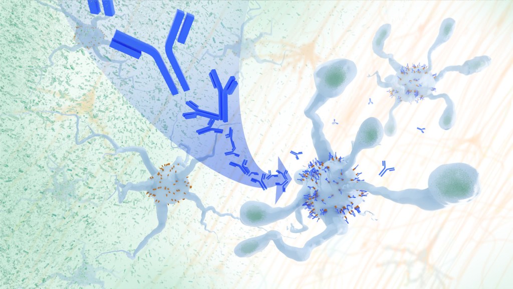
A cloud of particles is cleared as antibodies cover receptors on microglia.
In a study, an antibody treatment blocked interaction between APOE proteins and LILRB4 receptors in the brain, enabling microglia immune cells to clear amyloid plaques, a feature of Alzheimer’s. Credit: Donny Bliss/NIH

In Alzheimer’s disease, a buildup of sticky amyloid proteins in the brain clump together to form plaques, causing damage that gradually leads to worsening dementia symptoms. A promising way to change the course of this disease is with treatments that clear away damaging amyloid plaques or stop them from forming in the first place. In fact, the Food and Drug Administration recently approved the first drug for early Alzheimer’s that moderately slows cognitive decline by reducing amyloid plaques.1 Still, more progress is needed to combat this devastating disease that as many as 6.7 million Americans were living with in 2023.

Recent findings from a study in mice, supported in part by NIH and reported in Science Translational Medicine , offer another potential way to clear amyloid plaques in the brain. The key component of this strategy is using the brain’s built-in cleanup crew for amyloid plaques and other waste products: immune cells known as microglia that naturally help to limit the progression of Alzheimer’s. The findings suggest it may be possible to develop immunotherapies—treatments that use the body’s immune system to fight disease—to activate microglia in the brains of people with Alzheimer’s and clear amyloid plaques more effectively.2

In their report, the research team—including Marco Colonna , Washington University School of Medicine in St. Louis, and Jinchao Hou, now at Children’s Hospital of Zhejiang University School of Medicine in Zhejiang Province, China—wrote that microglia in the brain surround plaques to create a barrier that controls their spread. Microglia can also destroy amyloid plaques directly. But how microglia work in the brain depends on a fine-tuned balance of signals that activate or inhibit them. In people with Alzheimer’s, microglia don’t do their job well enough.  

The researchers suspected this might have something to do with a protein called apolipoprotein E (APOE). This protein normally helps carry cholesterol and other fats in the bloodstream. But the gene encoding the protein is known for its role in influencing a person’s risk for developing Alzheimer’s, and in the Alzheimer’s brain, the protein is a key component of amyloid plaques. The protein can also inactivate microglia by binding to a receptor called LILRB4 found on the immune cells’ surfaces.

Earlier studies in mouse models of Alzheimer’s showed that the LILRB4 receptor is expressed at high levels in microglia when amyloid plaques build up. This suggested that treatments targeting this receptor on microglia might hold promise for treating Alzheimer’s. In the new study, the research team looked for evidence that an increase in LILRB4 receptors on microglia plays an important role in the brains of people with Alzheimer’s.

To do this, the researchers first studied brain tissue samples from people who died with this disease and discovered unusually high amounts of the LILRB4 receptor on the surfaces of microglia, similar to what had been seen in the mouse models. This could help explain why microglia struggle to control amyloid plaques in the Alzheimer’s brain.

Next, the researchers conducted studies of mouse brains with accumulating amyloid plaques that express the LILRB4 receptor to see if an antibody targeting the receptor could lower amyloid levels by boosting activity of immune microglia. Their findings suggest that the antibody treatment blocked the interaction between APOE proteins and LILRB4 receptors and enabled microglia to clear amyloid plaques. Intriguingly, the team’s additional studies found that this clearing process also changed the animals’ behavior, making them less likely to take risks. That’s important because people with Alzheimer’s may engage in risky behaviors as they lack memories of earlier experiences that they could use to make decisions.

There’s plenty more to learn. For instance, the researchers don’t know yet whether this approach will affect the tau protein, which forms damaging tangles inside neurons in the Alzheimer’s brain. They also want to investigate whether this strategy of clearing amyloid plaques might come with other health risks.

But overall, these findings add to evidence that immunotherapies of this kind could be a promising way to treat Alzheimer’s. This strategy may also have implications for treating other neurodegenerative conditions characterized by toxic debris in the brain, such as Parkinson’s disease, amyotrophic lateral sclerosis (ALS), and Huntington’s disease. The hope is that this kind of research will ultimately lead to more effective treatments for Alzheimer’s and other conditions affecting the brain.

References:

[1] FDA Converts Novel Alzheimer’s Disease Treatment to Traditional Approval. U.S. Food and Drug Administration (2023).

[2] Hou J, et al. Antibody-mediated targeting of human microglial leukocyte Ig-like receptor B4 attenuates amyloid pathology in a mouse model . Science Translational Medicine. DOI: 10.1126/scitranslmed.adj9052 (2024).

NIH Support: National Institute of General Medical Sciences, National Institute on Aging

Wednesday, March 13, 2024

Omega-3 polyunsaturated fatty acids ameliorate neuroinflammation and mitigate ischemic stroke damage through interactions with astrocytes and microglia


Did your doctor instruct the dietician to get these into your hospital meals? NO? Then you don't have a functioning stroke doctor!

What foods provide omega-3s?

  • Fish and other seafood (especially cold-water fatty fish, such as salmon, mackerel, tuna, herring, and sardines)

  • Nuts and seeds (such as flaxseed, chia seeds, and walnuts)

  • Plant oils (such as flaxseed oil, soybean oil, and canola oil)

Omega-3 polyunsaturated fatty acids ameliorate neuroinflammation and mitigate ischemic stroke damage through interactions with astrocytes and microglia

, , , , , ,
https://doi.org/10.1016/j.jneuroim.2014.11.007Get rights and content

Highlights

  • PUFA n3 reduce stroke damage.

  • PUFA n3 attenuate hypoxia-induced inflammation.

  • PUFA n3 interact with microglia and astroglia.

Abstract

Omega-3 polyunsaturated fatty acids (PUFA n3) provide neuroprotection due to their anti-inflammatory and anti-apoptotic properties as well as their regulatory function on growth factors and neuronal plasticity. These qualities enable PUFA n3 to ameliorate stroke outcome and limit neuronal damage. Young adult male rats received transient middle cerebral artery occlusion (tMCAO). PUFA n3 were intravenously administered into the jugular vein immediately after stroke and 12 h later. We analyzed stroke volume and behavioral performance as well as the regulation of functionally-relevant genes in the penumbra. The extent of ischemic damage was reduced and behavioral performance improved subject to applied PUFA n3. Expression of Tau and growth-associated protein-43 genes were likewise restored. Ischemia-induced increase of cytokine mRNA levels was abated by PUFA n3. Using an in vitro approach, we demonstrate that cultured astroglial and microglia directly respond to PUFA n3 administration by preventing ischemia-induced increase of cyclooxygenase 2, hypoxia-inducible factor 1alpha, inducible nitric oxide synthase, and interleukin 1beta. Cultured cortical neurons also appeared as direct targets, since PUFA n3 shifted the Bcl-2-like protein 4 (Bax)/B-cell lymphoma 2 (Bcl 2) ratio towards an anti-apoptotic constellation. Thus, PUFA n3 reveal a high neuroprotective and anti-inflammatory potential in an acute ischemic stroke model by targeting astroglial and microglial function as well as improving neuronal survival strategies. Our findings signify the potential clinical feasibility of PUFA n3 therapeutic treatment in stroke and other acute neurological diseases.

Introduction

Stroke is the result of a permanent or transient focal occlusion of major brain arteries or their branches and represents a main cause of death and disability in the industrialized civilization. Brain damage and neuronal cell death following acute ischemia result from a series of complex pathophysiological processes that evolves in time and space beginning a few minutes after stroke onset and lasting for hours and days including secondary damage due to edema spreading even if reperfusion has already been revived. Cell dysfunction and tissue destruction are accompanied by local blood–brain barrier (BBB) breakdown followed by the invasion of peripheral immune cells, i.e. T-lymphocytes, macrophages and polymorph nuclear granulocytes. Beforehand, a massive early disturbance of ion homeostasis, calcium dysregulation, excitotoxicity, mitochondrial impairment together with reactive oxygen species (ROS) formation can be observed (Iadecola and Anrather, 2011, Dirnagl, 2012). The described pathomechanisms coincide with the activation, attraction and proliferation of astroglial and microglial cells. Astrogliosis and microgliosis are prevailing incidents in the penumbra during the initial stage of ischemia. Both glial cell types control and tune early and late neuroinflammatory responses resulting from oxygen and nutrient deprivation soon after the beginning of the ischemic phase (Dang et al., 2011). Although microglia is believed to play the most prominent role in the shaping of inflammatory responses after stroke, latterly astrocytes in the center of ischemic tissue disintegration are considered to actively sense hypoxia and trigger a battery of anti-inflammatory reactions (Ronaldson and Davis, 2012, Habib and Beyer, 2014, Habib et al., 2014). Importantly, both types of glial cells, the adjacent extracellular matrix, the endothelium and neurons form a “neurovascular unit” that represents a dynamic entity which shapes neuroinflammation in the setting of stroke (Dirnagl, 2012).

Recent studies have shown that omega-3 essential polyunsaturated fatty acids (PUFA n3) and in particular docosahexaenoic acid (DHA, 22:6, n-3) and to a lesser extent eicosapentanaenoic acid (EPA, 20:5, n-3) exert profound anti-inflammatory effects on the brain and protect brain tissue in experimental models of acute stroke in neonatal and adult animals and neuroinflammatory challenges besides being beneficial for brain development and cognitive function (Bazan, 2007, Belayev et al., 2009, Hoffman et al., 2009, Cole et al., 2010, Orr et al., 2013). Following short-term transient middle cerebral artery occlusion (tMCAO), rodents with DHA substitution and higher brain DHA levels revealed reduced infarct areas and cellular inflammatory responses as well as attenuated leukocyte infiltration and concomitantly fewer microglial cells (Belayev et al., 2009, Lalancette-Hebert et al., 2011, Orr et al., 2013). Several hours after stroke, the resident microglial cells become activated, accumulate in the vicinity of the lesion site and in the penumbra region and start proliferating (Kriz and Lalancette-Hebert, 2009, Dang et al., 2011, Lalancette-Hebert et al., 2011). This defines the post-ischemic treatment window with DHA as 3–5 h. There is also good evidence that consumption of fish and fish products (fish oil contains large amounts of DHA) is positively associated with a reduced risk of ischemic events in the CNS and cardiovascular disease (Pascoe et al., 2014). There are several proposed cellular mechanisms which could explain the safeguarding role of PUFA n3 under neuropathological conditions in the brain (Orr et al., 2013). DHA affects growth factor regulation which may be responsible for increased neurite growth and synapse formation (Kim et al., 2011). Coevally, DHA is anti-inflammatory in non-neuronal and neural tissues targeting for instance cyclooxygenases (COX) and cytosolic phospholipase A2 (cPLA2) as well as leukocyte infiltration and nuclear factor kappa-light-chain-enhancer of activated B cells (NF-κB) activation (Marcheselli et al., 2003, Orr et al., 2013). Such effects occur brain-intrinsically but it has also been shown that DHA dampens systemic inflammatory responses (Sijben and Calder, 2007).

In the present study, we aimed at demonstrating the neuroprotective potency of PUFA n3 in an experimental stroke rat model (transient middle cerebral artery occlusion, tMCAO), its efficacy in restoring motoric and sensory behavioral defects as well as morphological injury, and analyzing its influence on the expression of stroke-associated inflammatory gene markers. By adopting an in vitro hypoxia approach, we intended to curtail cell type-specific effects which might explain neuroprotective mechanisms at the subcellular level.

More at link.

Saturday, May 13, 2023

Human Brain Organoids Reveal How Microglia Develop and Function

And since microglia are incredibly important to our recovery it's good thing brain organoids are not considered human. We need this research.  

Tiny Human Brain Tissue Organoids Can’t Be Legally Considered “a Person”

The latest here: 

Human Brain Organoids Reveal How Microglia Develop and Function

Summary: Researchers have developed human brain organoids that contain microglia, the immune cells of the brain. These organoids allow researchers to study how microglia develop and function in a more realistic setting than previous models.

The researchers found that microglia are influenced by the environment in which they develop and that they play a role in both development and disease. Their findings could lead to new treatments for neurological disorders.

Key Facts:

  • Microglia are responsible for clearing away cellular debris and pathogens, and they also play a role in neuroprotection.
  • The researchers found that microglia from individuals with autism spectrum disorder were more reactive to damage or intruders.
  • The researchers hope that their findings will lead to new treatments for neurological disorders such as autism spectrum disorder and Alzheimer’s disease.

Source: Salk Institute

Situated at the intersection of the human immune system and the brain are microglia, specialized brain immune cells that play a crucial role in development and disease. Although the importance of microglia is undisputed, modeling and studying them has remained a difficult task.

Unlike some human cells that can be studied outside of the body or in nonhuman models, human microglia are difficult to study when removed from the human-brain-like environment.

To overcome this barrier, Salk scientists developed an organoid model—a three-dimensional collection of cells that mimics features of human tissues. This model allows researchers to study human microglial development and function for the first time in living human-derived tissue.

Further, the scientists examined patient-derived microglia from children with macrocephalic autism spectrum disorder (a condition where infant head circumference is greater than 97 percent of other infants’) to determine whether brain environment influences the development of more reactive microglia.

The findings, published in Cell on May 11, 2023, highlight the importance of immune cell and brain interaction, and improve the understanding of neurodegenerative and developmental diseases, such as autism spectrum disorder and Alzheimer’s disease.

“Outside of the brain environment, microglia lose almost all function and meaning,” says Professor Rusty Gage, senior author and holder of the Vi and John Alder Chair for Research on Age-Related Neurodegenerative Disease.

“We knew that if we found a way to replicate the human brain environment in an organoid in order to study human microglia, then we would finally have a tool for examining how the heathy and diseased brain influence microglia and, reciprocally, how healthy and diseased microglia influence the brain.”

Emerging roughly 10 years ago, organoids have become a prevalent tool to bridge the gap between cell and human studies. Organoids can mimic human development and organ generation better than other laboratory systems, allowing researchers to study how drugs or diseases affect human cells in a more realistic setting.

Brain organoids are typically grown in culture dishes, but the organoids are structurally and functionally limited by the lack of blood vessels, short survival time, and inability to sustain diverse cell types (like microglia).

“To create a brain organoid model that contains mature microglia and enables us to research them, we used a novel transplantation technique to create a human-brain-like environment” says co-first author Abed Mansour, a former postdoctoral researcher in Gage’s lab and now an assistant professor at the Hebrew University of Jerusalem.

“So we could finally make a human brain organoid that had all the features necessary to orchestrate human microglia growth, behavior, and function.”

Unlike previous models, the researchers created a human brain organoid that had microglia and a human-brain-like environment, which finally allowed them to look at environmental influences on microglia throughout brain development.

They found that a characteristic protein called SALL1 appeared as early as eleven weeks into development and served to confirm microglial identity and promote mature function. Additionally, they found that brain environment-specific factors, like the proteins TMEM119 and P2RY12, were necessary for microglia to function.

“Creating a human brain model that can effectively replicate the human brain environment is very exciting,” says Associate Professor Axel Nimmerjahn, another author of the study.

“With this model, we can finally investigate how human microglia function within the human brain environment.”

As the team learned more about microglia, the importance of the relationship between brain environment and microglia became clear—especially in disease scenarios.

The lab previously examined neurons derived from people with autism spectrum disorder and found their neurons grew faster and had more complex branches than neurotypical counterparts.

With the new organoid model, the team could ask whether those neuronal differences altered the brain environment and influenced microglia development.

To do so, they compared microglia derived from skin samples from three individuals with macrocephalic autism spectrum disorder versus three neurotypical individuals with macrocephaly.

The researchers found that individuals with autism spectrum disorder exhibited the neuronal differences the team had previously noted, and that the microglia were influenced by those differences in their growth environment.

Because of this neuron-dependent environmental change, the microglia became more reactive to damage or intruders—a finding that may explain the brain inflammation observed in some individuals with autism spectrum disorder.

Since this was a preliminary study with a small sample size, the team plans to examine more microglia from additional people in the future to verify their findings. They also aim to expand their research to study other developmental and neurodegenerative diseases to see how microglia are contributing to disease onset.

“Rather than deconstruct the brain, we decided to construct it ourselves,” says co-first author Simon Schafer, a former postdoctoral researcher in Gage’s lab and now an assistant professor at Technical University of Munich.

“By building our own brain model we can work from the bottom up and see solutions that may be impossible to see from the top down. We are eager to continue improving on our model and unravelling the relationship between the brain and immune system.”

Other authors include Monique Pena, Saeed Ghassemzadeh, Lisa Mitchell, Amanda Mar, Daphne Quang, Sarah Stumpf, and Clara Baek of the Salk Institute; Johannes C. M. Schlachetzki, Addison J. Lana, and Christopher K. Glass of UC San Diego; Irene Santisteban of the Technical University of Munich; and Raghad Zaghal of the Hebrew University of Jerusalem.

Funding: The work was supported by the National Institutes of Health (R01 AG056306, R01 AG057706, R01 AG056511, R01 AG061060, R01 NS108034, U19 NS123719, NCI CCSG: P30 014195, NCI CCSG: P30 014195), the American Heart Association and Paul G. Allen Frontiers Group (grant 19PABHI34610000), the Brain and Behavior Research Foundation (27685 and 30421), the German Research Foundation (500300695), the Milky Way Research Foundation, Annette C. Merle-Smith and the Robert and Mary Jane Engman Foundation, the European Molecular Biology Organization (ALTF 1214-2014), the Human Frontier Science Program (LT001074/2015), the European Research Council, the Chapman Foundation, the JBP Foundation and the Helmsley Charitable Trust.

About this neuroscience research news

Author: Salk Communications
Source: Salk Institute
Contact: Salk Communications – Salk Institute
Image: The image is credited to Neuroscience News

Original Research: Open access.
An in vivo neuroimmune organoid model to study human microglia phenotypes” by Rusty Gage et al. Cell

Monday, March 27, 2023

The Implications of Microglial Regulation in Neuroplasticity-Dependent Stroke Recovery

FYI.

The Implications of Microglial Regulation in Neuroplasticity-Dependent Stroke Recovery 

Department of Rehabilitation, Beijing Rehabilitation Hospital, Capital Medical University, Beijing 100144, China
*
Authors to whom correspondence should be addressed.
Biomolecules 2023, 13(3), 571; https://doi.org/10.3390/biom13030571
Received: 17 January 2023 / Revised: 23 February 2023 / Accepted: 14 March 2023 / Published: 21 March 2023

Abstract

Stroke causes varying degrees of neurological deficits, leading to corresponding dysfunctions. There are different therapeutic principles for each stage of pathological development. Neuroprotection is the main treatment in the acute phase,(Except that it doesn't exist and should be called the neuronal cascade of death signifying extreme urgency while neuroprotection means nothing to survivors and doctor use that to bamboozle patients; 'We didn't get neuroprotection to work'.  As compared to the statement; 'We failed at stopping the neuronal cascade of death  thus allowing millions to billions of your neurons to die'  

WHICH STATEMENT WILL GET YOUR DOCTORS TO SOLVE STROKE? )

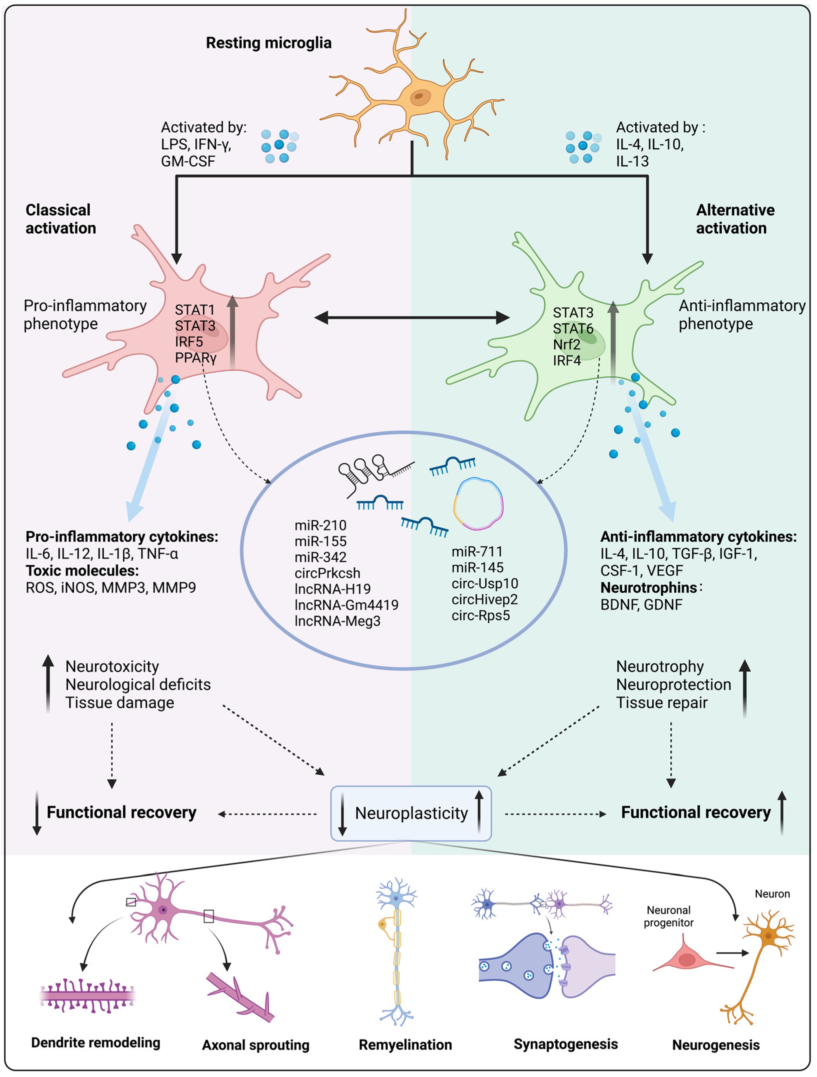
and functional recovery becomes primary in the subacute and chronic phases. Neuroplasticity is considered the basis of functional restoration(But your doctor knows nothing on how to make it repeatable on demand.) and neurological rehabilitation after stroke, including the remodeling of dendrites and dendritic spines, axonal sprouting, myelin regeneration, synapse shaping, and neurogenesis. Spatiotemporal development affects the spontaneous rewiring of neural circuits and brain networks. Microglia are resident immune cells in the brain that contribute to homeostasis under physiological conditions. Microglia are activated immediately after stroke, and phenotypic polarization changes and phagocytic function are crucial for regulating focal and global brain inflammation and neurological recovery. We have previously shown that the development of neuroplasticity is spatiotemporally consistent with microglial activation, suggesting that microglia may have a profound impact on neuroplasticity after stroke and may be a key therapeutic target for post-stroke rehabilitation. In this review, we explore the impact of neuroplasticity on post-stroke restoration as well as the functions and mechanisms of microglial activation, polarization, and phagocytosis. This is followed by a summary of microglia-targeted rehabilitative interventions that influence neuroplasticity and promote stroke recovery.

1. Introduction

Stroke is a major cause of death and long-term disability, worldwide. Despite constant incidence and declining mortality rates over the past 20 years, the number of stroke survivors continues to decrease [1,2,3]. They are unable to live independently and are more likely to experience subsequent neurological sequelae [4,5]. Stroke can cause focal and global neurological deficits. Different therapeutic principles are adopted in different periods. In the acute stage of stroke, neuroprotection is the main treatment [6]; reducing cerebral ischemia-reperfusion injury (IRI) is also crucial. In the subacute and chronic stages, functional recovery becomes the primary objective. Neuroplasticity is recognized as the basis of functional restoration and neurological rehabilitation after stroke, including remodeling of dendrites and dendritic spines, axonal sprouting, synapse shaping, and neurogenesis. Spontaneous neuroplasticity begins immediately after stroke, reaches a plateau in three to four weeks, and can be sustained in the chronic phase [7]. Spatiotemporal development profoundly affects the reconstruction of neural circuits and brain networks.
Microglia, the resident immune cells of the central nervous system (CNS), play a key role in brain development, homeostasis maintenance, and the disease response of the CNS through phenotypic polarization, morphological changes, and functional transformation. They participate in a variety of pathophysiological processes in the brain, including the promotion of neuronal survival, induction of programmed cell death, immune monitoring and antigen presentation, inflammation regulation, modulation of synaptic activity, synaptic pruning, remodeling, etc. [8,9,10,11,12].
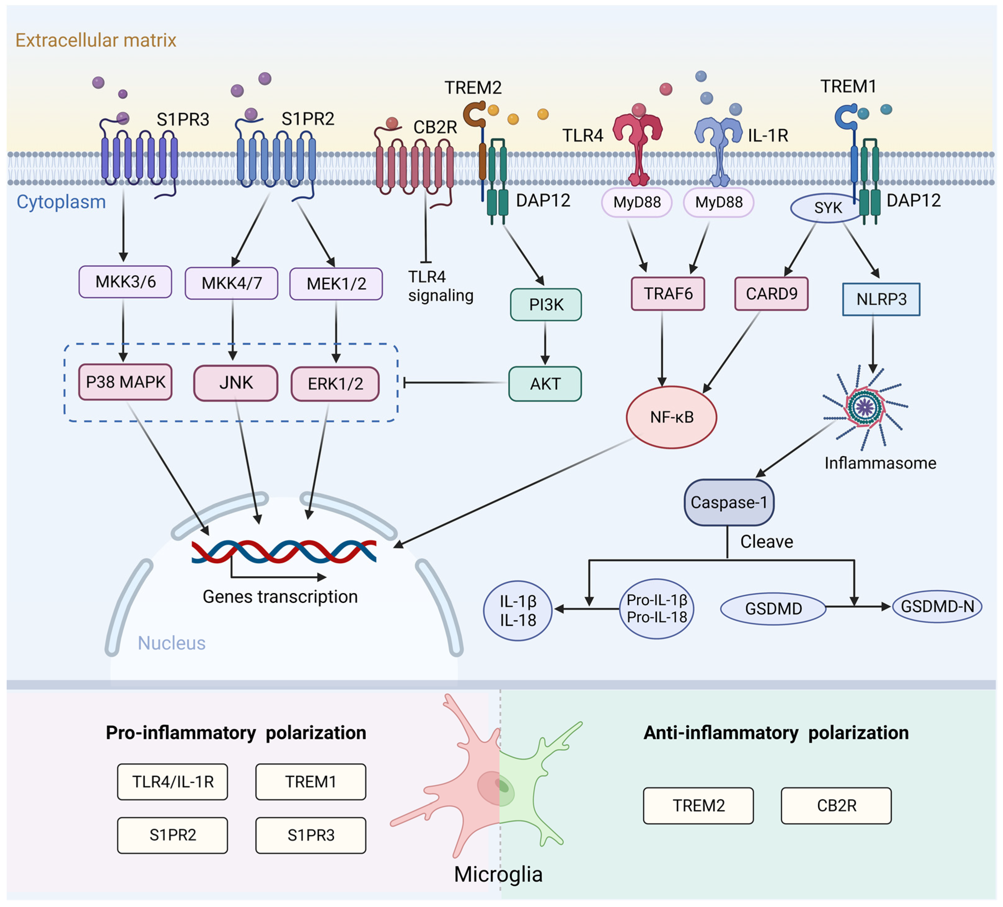
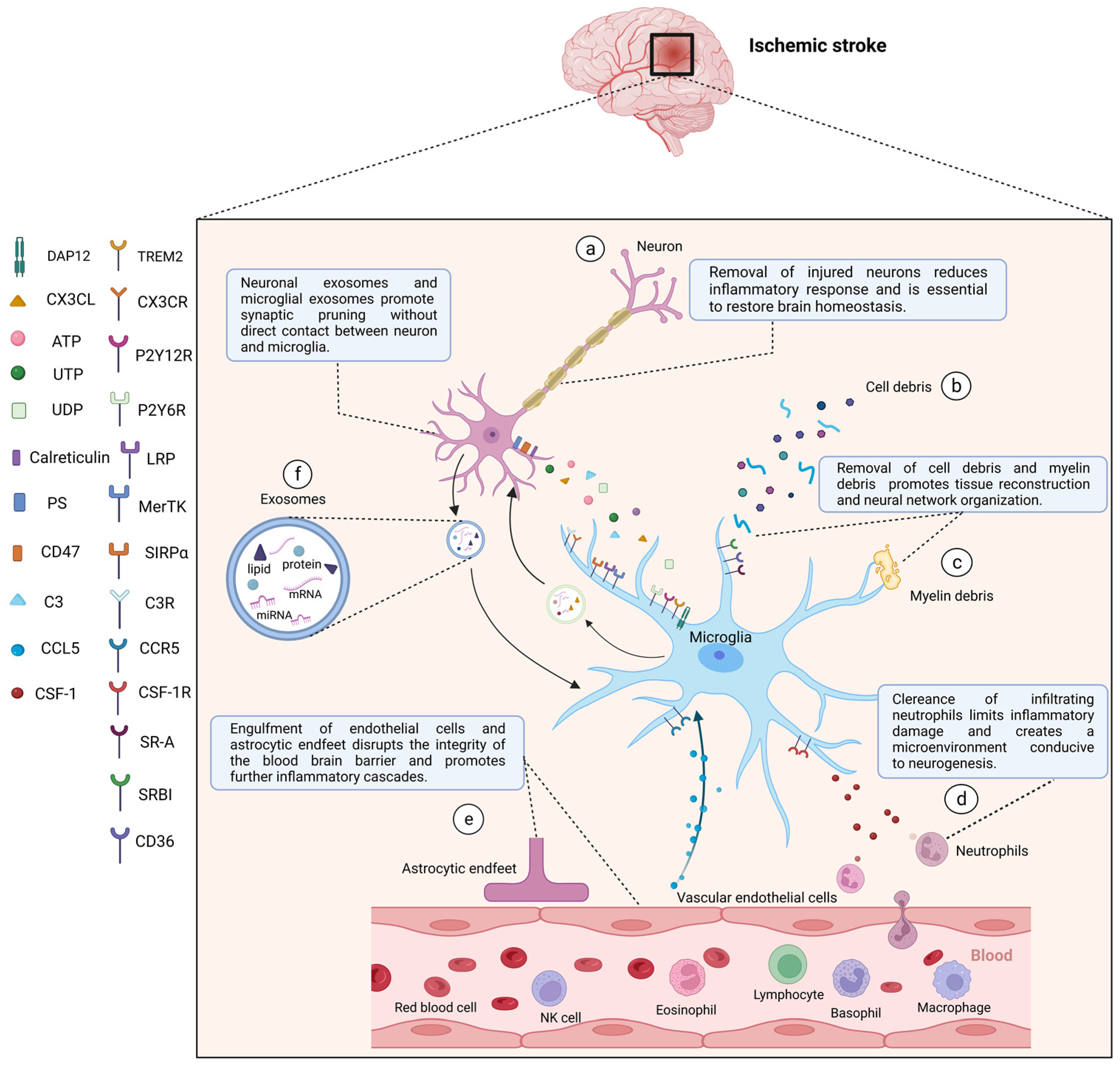
After stroke, the activation, polarization, and phagocytosis of microglia are crucial for regulating the neuroinflammatory microenvironment and enhancing neuroplasticity. Our previous study presented that the development of neuroplasticity overlaps both temporally and spatially with microglial activation [7], suggesting that microglia may have a profound impact on neuroplasticity following stroke and that they may be key therapeutic targets for stroke rehabilitation. In this review, we explore therapeutic targeting at different stages after stroke and the impact of neuroplasticity during this process. We then discuss the functions and mechanisms of microglial activation, polarization, and phagocytosis under physiological and pathological conditions. Finally, we provide a summary of microglia-targeted therapeutic interventions for promoting stroke recovery.

2. Pathophysiology and Therapeutic Target of Stroke Recovery

2.1. Pathophysiology of Stroke in Different Phases

Stroke commonly comprises two pathological subtypes. Hemorrhagic stroke accounts for approximately 10–15% of stroke cases. During this process, stress in the brain and internal injury cause the rupture of blood vessel [13]. Hematomas compressing brain tissue form for blood leakage into the brain parenchyma. The mass effect of the hematoma combined with neurotoxic effects further causes increased intracranial pressure, cerebral herniation, or death [14,15].
Ischemic stroke is caused by abrupt occlusion of the cerebral artery. The consequent interruption of blood flow and obstruction of the supply of oxygen lead to glutamate excitotoxicity, calcium overload, oxidative and nitrosative stress, and the release of inflammatory mediators, thereby activating a series of detrimental signaling cascades that induce neuronal injury or death [1,2,16,17]. Reversible neuronal impairment occurs after an ischemic attack, leading not only to relevant symptoms but also functional deficits corresponding to the location of the ischemia [18]. The progression of brain damage involves irreversibly injured necrotic tissue in the ischemic core, followed by injury development in the penumbral area, and then expanding to the entire ischemic territory [1,19]. Due to focal and global brain neurological damage following stroke, patients have different degrees of neurological deficits after stroke, such as dyskinesia, sensory dysfunction, swallowing dysfunction, dysarthria, aphasia, cognitive impairment, impaired cardiopulmonary function, mental disorders, and many complications, which further leads to a decline in quality of life and social participation [3,20].
Aside from revascularization therapy(thrombolysis and thrombectomy) and neuroprotective therapies (non-pharmaceutical and pharmaceutical therapies) for managing stroke in different phases [21], rehabilitative therapy helps to alleviate disability by promoting the recovery of impairment, activity, or participation after stroke [22] and is formally associated with a “time frame”, which coincides with the development of stroke and the period of maximal spontaneous recovery [23]. Thus, although rehabilitation plays a key role after stroke, not all stages are suitable for rehabilitative interventions [24]. According to both animal models and human trials, intensive rehabilitation within 24 h is potentially harmful [23]. In a clinical trial, a four-week intervention of physical fitness training did not result in an improvement in activities during the subacute period (days 5–45 after stroke) [25].
The therapeutic targets of stroke recovery vary according to the developmental pathophysiological process (Table 1). In the acute phase (minutes to days), a series of detrimental events occur after acute ischemic injury, including infiltration of peripheral immune cells, activation of resident glial cells, disturbance of ionic homeostasis, oxidative stress, mitochondrial dysfunction, and DNA damage. These processes involve cell necrosis within the lesion core and peri-infarct area. Therapeutic strategies have focused on neuroprotection to prevent neuronal injury and death, reduce infarct volume, and limit the decrease in neuronal density in the penumbra [16,26,27,28,29,30,31]. In addition, reducing IRI is critical. During the restoration of blood perfusion, IRI can lead to cerebral edema and even hemorrhage, thereby exacerbating the detrimental biological cascade response and causing irreversible tissue damage [21,32]. Therefore, besides neuroprotection, effective reduction of IRI is also a key target in the treatment of the acute phase of ischemic stroke [33].
Table 1. Pathophysiology and therapeutic targets of ischemic stroke in the acute, subacute, and chronic phases. BBB, blood-brain barrier; ROS, reactive oxygen species.
In the subacute phase (days to weeks), the mechanisms are more complicated than in the acute phase and include amplification of local and systemic immune responses, increased cytokine and reactive oxygen species (ROS) production, cell edema, and ion imbalances [28,34]. The activation of several protective mechanisms triggers beneficial repair processes, including neurogenesis and angiogenesis [27]. In addition, many endogenous processes are active, including axonal sprouting, dendrite remodeling, increased levels of growth factors, and altered synaptic and cortical excitability. Some of these processes have been demonstrated to mediate behavioral changes [35].
In the chronic phase (weeks to months), the end of spontaneous structural recovery is marked by stabilization of the post-stroke neurological deficits [35]. The therapeutic priorities should shift from neuroprotection to functional rehabilitation. Post-ischemic inflammatory responses appear to exacerbate tissue damage at an early stage, whereas they are assumed to promote tissue repair and functional restoration during the chronic phase [36]. During this stage, excitotoxicity decreases and the brain milieu becomes primarily inhibitory, and neural repair and excitability enhancement come to the forefront of post-stroke intervention [22,35].
 
More at link.

Saturday, October 29, 2022

Integrin β1 and the Repair after Nervous System Injury

You tell us about the central role of integrin β1 but nothing on how it can be used to recover. Useless.

Integrin β1 and the Repair after Nervous System Injury

Abstract

Background: 

Integrin β1, as a member of the adhesion molecule family, is widely distributed in many kinds of cells and participates in multiple biological functions of the nervous system, including cytoskeleton reorganization, axon growth, and inflammatory injury.  

Summary: 

After nervous system injury, integrin β1 expressed by microglia is mainly involved in promoting inflammatory damage; integrin β1 expressed by astrocytes plays an important role in axon regeneration; integrin β1 expressed by endothelial cells mainly participates in vascular remodeling. We concluded that the function of integrin β1 depends on the location of the receptor cells. The mechanism of integrin β1, which is involved in the inflammatory response of immune regulatory cells and affects the axonal regeneration of neuronal cells, is the key to explore the repair after nervous system injury. The development of drugs targeting integrin β1 is expected to bring a breakthrough in the treatment of nervous system injury.  

Key Messages: 

This paper expounds the important role of microglia in neurons of the nervous system and emphasizes the central role of integrin β1 in regulating non-neuronal cells after nervous system damage.

© 2022 The Author(s). Published by S. Karger AG, Basel

Yan L. · Cui Z.

Author affiliations

Corresponding Author


Tuesday, June 21, 2022

Particle Hydrogels Decrease Cerebral Atrophy and Attenuate Astrocyte and Microglia/macrophage Reactivity After Stroke

 WHOM will be doing the followup human testing? This sounds great but since we have fucking failures of stroke associations  and NO stroke leadership, nothing will occur.  You better plan on hiring your own researchers to solve this for your children and grandchildren.

Particle Hydrogels Decrease Cerebral Atrophy and Attenuate Astrocyte and Microglia/macrophage Reactivity After Stroke

First published: 17 June 2022

This article has been accepted for publication and undergone full peer review but has not been through the copyediting, typesetting, pagination and proofreading process, which may lead to differences between this version and the Version of Record. Please cite this article as https://doi.org/10.1002/adtp.202200048

Abstract

Increasing numbers of individuals live with stroke related disabilities. Following stroke, highly reactive astrocytes and pro-inflammatory microglia can release cytokines and lead to a cytotoxic environment that causes further brain damage and prevents endogenous repair. Paradoxically, these same cells also activate pro-repair mechanisms that contribute to endogenous repair and brain plasticity. Here, we show that the direct injection of a hyaluronic acid based microporous annealed particle (MAP) hydrogel into the stroke core in mice reduces the percent of highly reactive astrocytes, increases the percent of alternatively activated microglia, decreases cerebral atrophy and preserves NF200 axonal bundles. Further, we show that MAP hydrogel promotes reparative astrocyte infiltration into the lesion, which directly coincides with axonal penetration into the lesion. This work shows that the injection of a porous scaffold into the stroke core can lead to clinically relevant decrease in cerebral atrophy and modulates astrocytes and microglia towards a pro-repair phenotype.

This article is protected by copyright. All rights reserved


Monday, July 12, 2021

Glial Cells as Therapeutic Approaches in Brain Ischemia-Reperfusion Injury

 What is your stroke hospital doing with this?

Glial Cells as Therapeutic Approaches in Brain Ischemia-Reperfusion Injury

Ivó H. Hernández 1,2,3 , 
Mario Villa-González 2,4 , 
Gerardo Martín 4, 
Manuel Soto 2,5
and María José Pérez-Álvarez 2,4,*


Citation: Hernández, I.H.;
Villa-González, M.; Martín, G.; Soto,
M.; Pérez-Álvarez, M.J. Glial Cells as
Therapeutic Approaches in Brain
Ischemia-Reperfusion Injury. Cells
2021, 10, 1639. https://doi.org/
10.3390/cells10071639
Academic Editors:
Antonio Rodríguez-Sinovas,
Marisol Ruiz-Meana and
Javier Inserte
Received: 29 May 2021
Accepted: 26 June 2021
Published: 30 June 2021
Publisher’s Note: MDPI stays neutral
with regard to jurisdictional claims in
published maps and institutional affiliations.
Copyright: © 2021 by the authors.
Licensee MDPI, Basel, Switzerland.
This article is an open access article
distributed under the terms and
conditions of the Creative Commons
Attribution (CC BY) license (https://
creativecommons.org/licenses/by/
4.0/).
1 Genomic Instability Group, Spanish National Cancer Research Centre (CNIO), 28029 Madrid, Spain;
ihernandezh@cnio.es
2 Center for Molecular Biology “Severo Ochoa” (CBMSO) UAM/CSIC, 28049 Madrid, Spain;
mario.villa@uam.es (M.V.-G.); msoto@cbm.csic.es (M.S.)
3 Networking Research Center on Neurodegenerative Diseases (CIBERNED), Instituto de Salud Carlos III,
28031 Madrid, Spain
4 Departamento de Biología (Fisiología Animal), Facultad de Ciencias, Universidad Autónoma de Madrid,
28049 Madrid, Spain; gerardo.martin@estudiante.uam.es
5 Departamento de Biología Molecular, Facultad de Ciencias, Universidad Autónoma de Madrid,
28049 Madrid, Spain
* Correspondence: mj.perez@uam.es; Tel.: +34-91-497-2819

Abstract: 

Ischemic stroke is the second cause of mortality and the first cause of long-term disability constituting a serious socioeconomic burden worldwide. Approved treatments include thrombectomy and rtPA intravenous administration, which, despite their efficacy in some cases, are not suitable for a great proportion of patients. Glial cell-related therapies are progressively overcoming inefficient neuron-centered approaches in the preclinical phase. Exploiting the ability of microglia to naturally switch between detrimental and protective phenotypes represents a promising therapeutic treatment, in a similar way to what happens with astrocytes. However, the duality present in many of the roles of these cells upon ischemia poses a notorious difficulty in disentangling the precise pathways to target. Still, promoting M2/A2 microglia/astrocyte protective phenotypes and inhibiting M1/A1 neurotoxic profiles is globally rendering promising results in different in vivo models of stroke. On the other hand, described oligodendrogenesis after brain ischemia seems to be strictly beneficial, although these cells are the less studied players in the stroke paradigm and negative effects could be described for oligodendrocytes in the next years. Here, we review recent advances in understanding the precise role of mentioned glial cell types in the main pathological events of ischemic stroke, including inflammation, blood brain barrier integrity, excitotoxicity, reactive oxygen species management, metabolic support, and neurogenesis, among others, with a special attention to
tested therapeutic approaches.


Monday, January 18, 2021

Unique Subtype of Microglia in Degenerative Thalamus After Cortical Stroke

 Useless, you described Neurodegeneration from PD7 to PD28 but provided NOTHING that will prevent those problems.

Unique Subtype of Microglia in Degenerative Thalamus After Cortical Stroke

Originally publishedhttps://doi.org/10.1161/STROKEAHA.120.032402Stroke. ;0

Background and Purpose:

Stroke disrupts neuronal functions in both local and remotely connected regions, leading to network-wide deficits that can hinder recovery. The thalamus is particularly affected, with progressive development of neurodegeneration accompanied by inflammatory responses. However, the complexity of the involved inflammatory responses is poorly understood. Herein we investigated the spatiotemporal changes in the secondary degenerative thalamus after cortical stroke, using targeted transcriptome approach in conjunction with histology and flow cytometry.

Methods:

Cortical ischemic stroke was generated by permanent occlusion of the left middle cerebral artery in male C57BL6J mice. Neurodegeneration, neuroinflammatory responses, and microglial activation were examined in naive and stroke mice at from poststroke days (PD) 1 to 84, in both ipsilesional somatosensory cortex and ipsilesional thalamus. NanoString neuropathology panel (780 genes) was used to examine transcriptome changes at PD7 and PD28. Fluorescence activated cell sorting was used to collect CD11c+ microglia from ipsilesional thalamus, and gene expressions were validated by quantitative real-time polymerase chain reaction.

Results:

Neurodegeneration in the thalamus was detected at PD7 and progressively worsened by PD28. This was accompanied by rapid microglial activation detected as early as PD1, which preceded the neurodegenerative changes. Transcriptome analysis showed higher number of differentially expressed genes in ipsilesional thalamus at PD28. Notably, neuroinflammation was the top activated pathway, and microglia was the most enriched cell type. Itgax (CD11c) was the most significantly increased gene, and its expression was highly detected in microglia. Flow-sorted CD11c+ microglia from degenerative thalamus indicated molecular signatures similar to neurodegenerative disease–associated microglia; these included downregulated Tmem119 and CX3CR1 and upregulated ApoE, Axl, LpL, CSF1, and Cst7.

Conclusions:

Our findings demonstrate the dynamic changes of microglia after stroke and highlight the importance of investigating stroke network-wide deficits. Importantly, we report the existence of a unique subtype of microglia (CD11c+) with neurodegenerative disease–associated microglia features in the degenerative thalamus after stroke.