Showing posts with label don't care. Show all posts
Showing posts with label don't care. Show all posts
Friday, August 7, 2026


Thursday, June 18, 2026
Scientists find way to continue natural method to repair brains
How long will it take for your incompetent? doctor to drive the research that solves this for humans and creates protocols that deliver this solution? Just why can't your doctor accomplish that?
Laziness? Incompetence? Or just don't care? NO leadership? NO strategy? Not my job? Not my Problem!
Scientists find way to continue natural method to repair brains
A team of Japanese and German scientists said its recent discoveries could lead to medicine that promotes and prolongs reparations to brain functions among victims of strokes and other diseases.
The researchers uncovered a mechanism that prompts recovery of brain functions in the early stages after the onset of the disease. They also identified a protein that causes the reparability to wane and eventually halt.
And they developed a drug that suppresses the workings of that protein in mice.
The group, including researchers from the Institute of Science Tokyo and the Tokyo Metropolitan Institute of Medical Science, plans to apply the findings to humans to ease the aftereffects of brain diseases.
Their research results were published online May 13 in Nature, a British science journal.
Infarction and other diseases of the brain cause neurological symptoms, such as loss of limb mobility and speech impairment.
Dead brain cells cannot be restored.
However, part of the lost brain functions can be recovered through rehabilitation, although the reparability is lost after a while. The science world does not understand exactly why this occurs.
The researchers on the team set their sights on microglia, a class of immune cells that work in the brain.
In mouse experiments, they found a mechanism whereby a brain injury prompts the microglia to begin generating nourishing substances that encourage brain tissue repair.
The microglia continued making the nourishing substances for about a month in mice that suffered a brain stroke. But production of the substances eventually stopped, and the reparative functions were lost in two months.
The researchers found that a protein called ZFP384 works to prevent the microglia from generating those nourishing substances.
In mice disabled from making ZFP384, the nourishing substances continued to be produced one month following the stroke, and the neurological symptoms improved, according to the team.
The researchers developed a drug that inhibits the workings of ZFP384 and administered it to mice one week and one month following the onset of a stroke.
They found the neurological symptoms improved and the brain functions continued to recover.
The scientists said the microglia-created nourishing substances encouraged the recovery of synaptic connections and other features of the nervous system.
SAME MECHANISM IN HUMANS?
Among humans with brain infarction, the scholars found abundant microglia of the type that generates the nourishing substances one week following the disease.
Similar microglia, however, decreased with time in terms of abundance, leading to a loss of reparative functions. The researchers also found that more ZFP384 was being generated.
The scientists said this likely represents the same sort of change that took place in mice, and they are hoping to develop therapeutic drugs that work in humans as well.
“We have presented this novel therapeutic concept of relying on the natural reparability that is inherent in the brain and making it last longer,” said Jun Tsuyama, a Science Tokyo junior associate professor of neuroscience, one of the leading members of the research team.
“We hope to help realize medical treatment where you don’t have to give up hopes for post-stroke functional recovery with no lingering aftereffects,” he said.
The research article can be viewed at (https://www.nature.com/articles/s41586-026-10480-0).
Monday, June 1, 2026
Scientists found a protein that drives brain aging — and how to stop it
How long will it take for your incompetent? doctor to drive the research that solves this for humans and creates protocols that deliver this solution? Just why can't your doctor accomplish that?
Laziness? Incompetence? Or just don't care? NO leadership? NO strategy? Not my job? Not my Problem!
Your doctor has known of this for almost a year and should have preparations ready for this research NOW!
- FTL1 protein(2 posts to August 2025)
Scientists found a protein that drives brain aging — and how to stop it
FULL STORY
A protein called FTL1 was found to drive brain aging — and
lowering it restored memory and neural connections in mice. Credit:
Shutterstock
Aging takes a serious toll on the hippocampus, the part of the brain that plays a central role in learning and memory.
Scientists at UC San Francisco have now pinpointed a protein that appears to drive much of this decline.
FTL1 Emerges as a Key Driver of Brain Aging
To understand what changes with age, the researchers tracked shifts in genes and proteins in the hippocampus of mice over time. Among everything they examined, only one stood out as consistently different between young and old animals. That protein is called FTL1.

Older mice showed higher levels of FTL1. At the same time, they had fewer connections between neurons in the hippocampus and performed worse on cognitive tests.
How FTL1 Alters Brain Function
When the team boosted FTL1 levels in young mice, the effects were striking. Their brains began to look and function more like those of older mice, and their behavior reflected this shift.
Lab experiments revealed more detail. Nerve cells engineered to produce high amounts of FTL1 developed simplified structures, forming short, single extensions instead of the complex, branching networks seen in healthy cells.
Reversing Memory Decline by Lowering FTL1
The most surprising result came when researchers reduced FTL1 in older mice. The animals showed clear signs of recovery. Connections between brain cells increased, and their performance on memory tests improved.
"It is truly a reversal of impairments," said Saul Villeda, PhD, associate director of the UCSF Bakar Aging Research Institute and senior author of the paper, which was published in Nature Aging. "It's much more than merely delaying or preventing symptoms."
Metabolism Link Points to New Treatments
Further experiments showed that FTL1 also affects how brain cells use energy. In older mice, higher levels of the protein slowed cellular metabolism in the hippocampus. However, when researchers treated these cells with a compound that boosts metabolism, the negative effects were prevented.
Hope for Future Brain Aging Therapies
Villeda believes these findings could pave the way for treatments that target FTL1 and counter its effects in the brain.
"We're seeing more opportunities to alleviate the worst consequences of old age," he said. "It's a hopeful time to be working on the biology of aging."
Authors and Funding
Other UCSF authors are Laura Remesal, PhD, Juliana Sucharov-Costa, Karishma J.B. Pratt, PhD, Gregor Bieri, PhD, Amber Philp, PhD, Mason Phan, Turan Aghayev, MD, PhD, Charles W. White III, PhD, Elizabeth G. Wheatley, PhD, Brandon R. Desousa, Isha H. Jian, Jason C. Maynard, PhD, and Alma L. Burlingame, PhD. For all authors see the paper.
Association between uric acid to lymphocyte ratio and poor functional outcomes in acute ischemic stroke patients
Why are your predicting failure to recover RATHER THAN DELIVERING RECOVERY?
Laziness? Incompetence? Or just don't care? NO leadership? NO strategy? Not my job? Not my Problem!
You're all fired! Biomarkers do nothing for recovery unless you are mapping EXACT RECOVERY PROTOCOLS to them!
Association between uric acid to lymphocyte ratio and poor functional outcomes in acute ischemic stroke patients


Abstract
Background:
Inflammation has an important impact on the pathological progression associated with ischemic stroke. Serum uric acid (UA) to lymphocyte ratio (ULR) is a biomarker that responds to the level of inflammation but is not definitively associated with the clinical outcomes in patients with acute ischemic stroke (AIS).
Methods:
The data were obtained from the Third China National Stroke Registry (CNSR-III). Enrolled AIS patients were grouped by ULR quartiles at admission. The outcomes were poor functional outcomes (modified Rankin Scale [mRS] score of 3–6 or 2–6) and all-cause mortality at 3 months and 1 year. The associations of ULR with the risk of poor functional outcome and all-cause mortality were analyzed by multivariable logistic regression and Cox proportional hazards regression.
Results:
A total of 8,241 patients were included from the CNSR-III study. After adjusting for confounders, it was found that patients in the highest ULR quartile had higher mRS scores of 2–6 (odds ratio [OR], 1.33; 95% confidence interval [CI], 1.15–1.53) and 3–6 (OR, 1.35; 95% CI, 1.16–1.57) at the 3-month follow-up. Additionally, the highest ULR quartile was associated with an increased risk of all-cause mortality at the 3-month follow-up (hazard ratio [HR], 1.97; 95%CI, 1.22–3.18). Similar results were observed at the 1-year follow-up.
Conclusion:
Elevated ULR increased the risks of poorer functional outcomes and all-cause mortality in the AIS patients. However, this observational study was limited by potential unmeasured confounders, selection bias, residual confounding, and restricted generalizability to other populations.
Sunday, May 17, 2026
Comparison of PNI, HALP, and modified HALP scores in predicting 90-day mortality in elderly patients with acute ischemic stroke
Why are your predicting failure to recover RATHER THAN DELIVERING RECOVERY?
Laziness? Incompetence? Or just don't care? NO leadership? NO strategy? Not my job? Not my Problem!
Comparison of PNI, HALP, and modified HALP scores in predicting 90-day mortality in elderly patients with acute ischemic stroke

Abstract
Introduction:
Acute ischemic stroke (AIS) remains a major cause of mortality and disability, with older adults disproportionately affected. We aimed to evaluate and compare the prognostic utility of the Prognostic Nutritional Index (PNI), the Hemoglobin–Albumin–Lymphocyte–Platelet (HALP) score, and the modified HALP (mHALP) index for predicting 90-day mortality after AIS in a cohort predominantly composed of elderly patients.
Methods:
We conducted a single-center retrospective cohort study including 151 adult patients with radiologically confirmed AIS admitted to the emergency department between January 2021 and December 2024. Demographics, comorbidities, and laboratory parameters obtained within 24 hours of admission were recorded, and baseline stroke severity was assessed using the National Institutes of Health Stroke Scale (NIHSS). PNI, HALP, and mHALP were calculated from routine blood tests. The primary outcome was all-cause 90-day mortality. Associations with mortality were examined using univariate and pre-specified multivariable logistic regression models adjusted for age and NIHSS. Discriminatory performance was assessed using receiver operating characteristic (ROC) curve analysis.
Results:
Overall, 26 patients (17.2%) died within 90 days. Non-survivors were older and had significantly lower albumin, hemoglobin, lymphocyte counts, PNI, and HALP values than survivors, and higher NIHSS at presentation. In univariate analyses, PNI, HALP, and mHALP were significantly associated with 90-day mortality. In multivariable models adjusted for age and NIHSS, both PNI and HALP remained independently associated with 90-day mortality, whereas the association for mHALP was attenuated and did not reach conventional statistical significance. ROC analyses indicated fair discrimination for PNI and HALP, modest-to-fair performance for mHALP, and NIHSS performance comparable to a clinical reference.
Discussion:
In this AIS cohort, simple immunonutritional indices—particularly PNI and HALP—were independently associated with 90-day mortality and demonstrated fair discriminative ability, supporting their potential role as adjunctive tools for early risk stratification. These findings warrant validation in larger prospective multicenter cohorts and further clarification of the prognostic contribution of mHALP in AIS.
Comparative effectiveness of neuromuscular electrical stimulation-based combination interventions for post-stroke lower limb rehabilitation: a systematic review and network meta-analysis
Why didn't you write protocols for the effective ones?
Laziness? Incompetence? Or just don't care? NO leadership? NO strategy? Not my job? Not my Problem!
Comparative effectiveness of neuromuscular electrical stimulation-based combination interventions for post-stroke lower limb rehabilitation: a systematic review and network meta-analysis
Abstract
Objective
This study aimed to comparatively evaluate the efficacy of different neuromuscular electrical stimulation (NMES)-based combination protocols for improving lower limb dysfunction in patients with stroke, in order to provide evidence-based support for clinical rehabilitation practice.
Methods
Randomized controlled trials (RCTs) investigating NMES-based combination interventions for lower limb dysfunction in patients with stroke published up to September 2, 2025 were systematically identified through searches of PubMed, Embase, the Cochrane Library, and Web of Science. The risk of bias of eligible studies was evaluated using the Cochrane Risk of Bias 2.0 (ROB 2.0) tool. Network meta-analysis (NMA) and subgroup analyses were conducted using R software (version 4.5.1) and Stata (version 15.0).
Results
In total, 37 RCTs were incorporated into the present NMA, involving 1,764 patients with stroke and encompassing nine distinct NMES-based combination intervention protocols. Regarding balance ability, core stability training (CST) combined with NMES (SMD = 0.99, 95% CrI: 0.33–1.67), gait training (GT) combined with NMES (SMD = 0.83, 95% CrI: 0.35–1.27), and NMES alone (SMD = 0.71, 95% CrI: 0.21–1.22) demonstrated significant therapeutic effects. CST+NMES (SUCRA = 76.02%) ranked highest among the NMES-based combined interventions for balance ability in patients with stroke. With respect to walking ability, no statistically significant differences were observed among the NMES-based intervention comparisons. Nevertheless, cycling exercise (CE) combined with NMES (SUCRA = 77.46%) received the highest ranking probability for walking ability, although this exploratory finding should be interpreted with caution given the absence of statistically significant differences among interventions and the lack of closed loops in the network. In terms of lower limb functional mobility, augmented reality technology (ART) combined with GT and NMES (SMD = 1.38, 95% CrI: 0.25–2.52), GT+NMES (SMD = 0.85, 95% CrI: 0.39–1.36), and NMES alone (SMD = 0.55, 95% CrI: 0.06–1.05) showed significant improvements. ART+GT+NMES (SUCRA = 84.23%) ranked highest for lower limb functional mobility. The results of the two-dimensional cluster ranking indicated that GT+NMES (SUCRA = 65.22%/49.29%/62.22%) demonstrated relatively stable ranking performance across multiple domains of lower limb dysfunction in patients with stroke. Subgroup analyses further indicated that CST+NMES appeared to be more effective for patients in the acute phase, GT+NMES for those in the subacute phase, and ART+GT+NMES for patients in the chronic phase. These subgroup-level rankings were based on limited evidence and should be considered exploratory.
Conclusions
NMES-based combination strategies have demonstrated beneficial effects on the recovery of lower limb function among individuals after stroke. Based on the results of this NMA, GT+NMES demonstrated relatively stable ranking performance across multiple outcomes, including balance, walking ability, and lower limb functional mobility, suggesting that it warrants further investigation(Why? You incompetently didn't do your job correctly?) as a combined intervention for clinical application. In addition, patients at different stages of stroke showed variable responses to NMES-based combined interventions, highlighting the importance of stage-specific and individualized rehabilitation strategies.
Trial registration: PROSPERO CRD420251229835.
Friday, May 15, 2026
Single dose of magic mushroom psychedelic can cause anatomical brain changes, study finds
Have your competent? doctor and hospital get research going on possible benefits to stroke recovery! Of course, they being up-to-date on all things stroke already have stuff in the works for over a decade, right?
Oh NO, NOTHING WAS DONE! WHY?
Laziness? Incompetence? Or just don't care? NO leadership? NO strategy? Not my job? Not my Problem!
magic mushrooms (40 posts to October 2014)
psilocybin (54 posts to May 2014)
Single dose of magic mushroom psychedelic can cause anatomical brain changes, study finds
Tuesday, May 12, 2026
Scientists Identify Diet That May Trigger Fat Burning Without Exercise
Will your competent? doctor and hospital GUARANATEE that human testing occurs? Why not?
Laziness? Incompetence? Or just don't care? NO leadership? NO strategy? Not my job? Not my Problem!
Scientists Identify Diet That May Trigger Fat Burning Without Exercise
Researchers at the University of Southern Denmark have discovered that reducing two specific amino acids in the diet may trigger the body to burn
More at link.
Friday, March 20, 2026
Gut microbes may drive memory decline during aging by disrupting vagal brain signaling
Will your competent? doctor and hospital GUARANATEE that human testing occurs AND AN EXACT PREVENTIVE PROTOCOL IS CREATED? Why not?
Laziness? Incompetence? Or just don't care? NO leadership? NO strategy? Not my job? Not my Problem!
Gut microbes may drive memory decline during aging by disrupting vagal brain signaling
A new study reveals how age-related microbiome shifts can disrupt gut–brain signaling, uncovering a microbial pathway that may accelerate memory decline during aging.
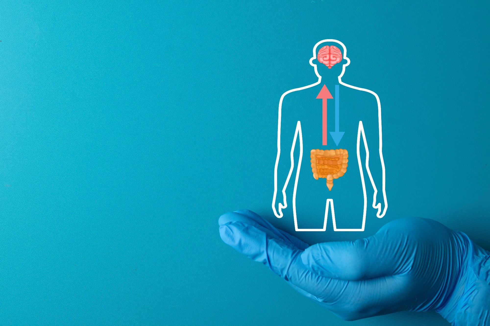
Study: Intestinal interoceptive dysfunction drives age-associated cognitive decline. Image Credit: Toey Andante / Shutterstock
In a recent study published in the journal Nature, researchers identified in mice a microbiome–gut–brain pathway in which impaired intestinal interoceptive signaling contributes to age-associated memory decline and may influence the rate of cognitive decline during aging.
Age-related memory decline impacts the quality of life of older adults. Neuronal engrams in the hippocampus are involved in memory formation, storage, and recall. The gut microbiome has been implicated as a modifiable factor contributing to age-related memory loss. Nonetheless, the circuits that transmit microbial signals to the brain for modulating memory remain largely unknown.
Scientists uncover a gut–brain signaling pathway linked to age-related memory decline
In the present study, researchers mapped the microbiome–gut–brain pathway influencing cognitive decline in mice. First, they investigated the consequences of age-related changes in the microbiome on cognitive aging by uncoupling microbiome age from host age.
Accelerated microbiome aging was achieved in young mice by co-housing them with 18-month-old mice. This resulted in microbiome equilibration between young and old mice resembling an old-like state.
One month of co-housing impaired short-term memory in young mice during the novel object recognition (NOR) task. Next, co-housing young mice with different young mice did not impact their cognitive behavior. Colonizing young germ-free (GF) mice with fecal microbiome from aged or young donors reproduced donor microbiomes in recipients, establishing age-related microbiome changes without co-housing.
Further, gut microbiota was ablated using antibiotics before, during, and after co-housing. Co-housing young mice with old mice lacking microbiota did not impair performance on the NOR task. Antibiotic treatment concurrent with co-housing also protected young mice from the detrimental effects of co-housing. Treating young mice with antibiotics after they developed co-housing-related cognitive deficits restored memory performance.
A specific gut bacterium emerges as a driver of age-associated cognitive decline
Next, the team explored the microbial drivers of cognitive decline. Microbiome changes were characterized over the lifespan in mice. The animals showed signs of frailty and mortality at two years of age.
Parabacteroides goldsteinii was identified as the top candidate, transmissible to young mice by transplantation and co-housing, and whose abundance increased with age. Colonizing antibiotic-treated or GF young mice with P. goldsteinii resulted in cognitive impairment.

The researchers then examined whether P. goldsteinii outgrowth and microbiome aging affected hippocampal function. They found reduced hippocampal neurogenesis and enhanced inflammation in old mice but not in co-housed young mice.
Young mice showed robust activation of immediate-early genes, reflecting neuronal activation, in response to exposure to a novel object. This response was blunted in old mice and co-housed young mice.
FOS staining showed that hippocampal regions CA1, CA3, and the dentate gyrus had impaired activation to novelty exposure in co-housed young mice. GF mice receiving microbiota from young mice showed elevated dentate gyrus activity to novelty exposure, while responses were blunted in those receiving microbiota from old mice. Colonization of young mice with P. goldsteinii also inhibited hippocampal responses to novelty exposure.
Impaired gut sensory signaling appears to disrupt memory-related brain circuits
Several other brain areas, including the entorhinal and somatosensory cortices and the nucleus tractus solitarii, exhibited reduced neuronal activation in old mice and co-housed young mice. Given their role in sensory processing, the researchers speculated that aging may reduce the transmission of interoceptive information through vagal sensory neurons innervating the gut rather than through spinal sensory afferents.
The researchers assessed cognitive performance in mice lacking neurons expressing the vanilloid receptor (TRPV1). Young Trpv1DTA mice phenocopied old mice in the NOR task and showed decreased hippocampal activation. The same effect was reproduced through chemogenetic silencing of TRPV1-positive neurons.
In contrast, chemogenetic activation of TRPV1-positive neurons in co-housed young mice restored their cognitive performance and hippocampal FOS activity. Capsaicin, a TRPV1 agonist, produced similar effects by restoring memory function and hippocampal responses in both old mice and co-housed young mice. The detrimental effects of P. goldsteinii were also negated by capsaicin treatment.
Microbial metabolites trigger inflammatory signaling that disrupts gut–brain communication
Additional experiments suggested that impaired vagal afferent function may mediate intestinal regulation of age-related cognitive decline. Oral administration of size-filtered fractions of P. goldsteinii culture supernatants resulted in cognitive decline. Untargeted metabolomics revealed that 3-hydroxyoctanoic acid (3-HOA), a medium-chain fatty acid (MCFA), was the most strongly enriched metabolite.
Oral administration of 3-HOA impaired object recognition and reduced hippocampal responses. Other MCFAs, such as dodecanoic and decanoic acids, produced similar effects. These metabolites increased in abundance in the intestinal lumen with age but not in GF or antibiotic-treated mice.
Treatment with a bacteriophage targeting Parabacteroides distasonis altered microbial transcriptional programs and reduced intestinal MCFA levels in old mice.
The researchers investigated whether MCFA effects on memory were mediated by G protein–coupled receptor 84 (GPR84) signaling. GPR84-deficient mice were protected from reduced hippocampal responses and memory dysfunction. A GPR84 agonist reproduced the effects of MCFAs, whereas an inhibitor counteracted the effects of 3-HOA.
Because GPR84 expression is largely restricted to myeloid cells, the team explored the role of the immune system. Their findings suggest that aging alters the gut environment, including P. goldsteinii outgrowth and MCFA accumulation.
These metabolites trigger pro-inflammatory myeloid responses through GPR84 signaling, including cytokine pathways involving TNF and IL-1β acting on PHOX2B-expressing sensory neurons. This process impairs vagal activity, hippocampal responses, and memory.
Findings highlight interoceptive gut–brain pathways as potential targets for cognitive aging
The results suggest that interoceptive communication pathways between the gut and brain may deteriorate over the lifespan, with reduced sensory input contributing to brain aging.
Interoceptive dysfunction may therefore contribute to age-associated cognitive decline in mice and influence the rate of memory deterioration. Pharmacological activation of interoceptive pathways may represent a potential therapeutic strategy, although whether similar mechanisms operate in human cognitive aging remains uncertain.
Journal reference:
- Cox TO, Devason AS, de Araujo A, et al. (2026). Intestinal interoceptive dysfunction drives age-associated cognitive decline. Nature. DOI: 10.1038/s41586-026-10191-6, https://www.nature.com/articles/s41586-026-10191-6
Wednesday, March 11, 2026
Scientists Identify Diet That May Trigger Fat Burning Without Exercise
Will your competent? doctor and hospital GUARANATEE that human testing occurs? Why not?
Laziness? Incompetence? Or just don't care? NO leadership? NO strategy? Not my job? Not my Problem!
Scientists Identify Diet That May Trigger Fat Burning Without Exercise
Friday, March 6, 2026
Single-Injection Immunotherapy That Halts Alzheimer’s
Will your competent? doctor and hospital GUARANATEE that human testing occurs? Why not?
Laziness? Incompetence? Or just don't care? NO leadership? NO strategy? Not my job? Not my Problem!
Your risk of dementia, has your doctor
told you of this? Your doctor is responsible for preventing this! Is
s/he willing to try this on you?
1. A documented 33% dementia chance post-stroke from an Australian study? May 2012.
2. Then this study came out and seems to have a range from 17-66%. December 2013.`
3. A 20% chance in this research. July 2013.
4. Dementia Risk Doubled in Patients Following Stroke September 2018
The latest here:
Single-Injection Immunotherapy That Halts Alzheimer’s
Summary: Current Alzheimer’s treatments typically require frequent, high-dose infusions of monoclonal antibodies to slow cognitive decline. But researchers have engineered a potentially game-changing alternative: CAR-astrocytes. Taking inspiration from CAR-T cell therapy used in cancer, scientists used a harmless virus to “reprogram” astrocytes—the most abundant cells in the brain—with a specialized “homing device.”
These modified cells, dubbed “super cleaners,” are designed to specifically target and engulf amyloid-beta plaques. In a study published in Science, a single injection of these CAR-astrocytes completely prevented plaque development in young mice and cut existing plaque levels in half in older mice.
Key Facts
- The “Homing Device”: Researchers equipped astrocytes with a Chimeric Antigen Receptor (CAR) that allows them to identify and “grab” amyloid-beta proteins for destruction.
- Single-Shot Potential: Unlike monthly antibody infusions, this cellular immunotherapy showed massive effectiveness with just a single injection in mouse models.
- Prevention & Treatment: When given before plaques formed, the therapy kept the brain plaque-free. In brains already saturated with amyloid, it reduced the plaque load by 50% within three months.
- Reducing the Burden: Traditionally, microglia (the brain’s immune cells) handle “trash removal,” but they become overwhelmed in Alzheimer’s. CAR-astrocytes provide much-needed reinforcement.
- Beyond Alzheimer’s: The team believes this technology could be adapted to target brain tumors by switching the CAR device to recognize cancer markers instead of amyloid.
Source: WUSTL
The new generation of Alzheimer’s disease drugs — the first proven to change the course of the disease — typically extend independent living for patients by 10 months. Called monoclonal antibodies, they reduce the accumulations of a harmful protein, amyloid, in the brain and require high-dose, once- or twice-monthly infusions of the medication.
Now, to reduce the frequency of treatment and potentially improve the efficacy of an anti-amyloid therapy, researchers at Washington University School of Medicine in St. Louis have engineered a new cellular immunotherapy that requires just a single injection to prevent amyloid plaques from developing when given before plaques start to form in mice. Furthermore, a single treatment in mice that had already developed plaques cut the amount of amyloid plaques in half.

The study was published March 5 in Science.
Like CAR-T cell therapies used for cancer treatment, in which T cells of the immune system are genetically modified to attack cancer cells, this new approach equips cells — in this case, brain cells called astrocytes — with a CAR homing device to grab onto a target for destruction. These new CAR-astrocyte cells have features that transform them into super cleaners that remove damaging proteins from the brain that play a role in cognitive decline.
“This study marks the first successful attempt at engineering astrocytes to specifically target and remove amyloid beta plaques in the brains of mice with Alzheimer’s disease,” said the study’s senior author, Marco Colonna, MD, the Robert Rock Belliveau, MD, Professor of Pathology at WashU Medicine.
“Although more work needs to be done to optimize the approach and address potential side effects, these results open up an exciting new opportunity to develop CAR-astrocytes into an immunotherapy for neurodegenerative diseases and even brain tumors.”
Removing brain waste
Alzheimer’s disease starts with a sticky protein called amyloid beta that builds up into plaques in the brain, setting off a chain of events that results in brain atrophy and cognitive decline.
Microglia, immune cells that reside in the brain, are responsible for removing brain waste but can become dysfunctional when overwhelmed in the context of neurodegenerative disease.
To reduce the cleaning burden on microglia, first author Yun Chen, PhD, then a graduate student in the labs of Colonna and David M. Holtzman, MD, the Barbara Burton and Reuben M. Morriss III Distinguished Professor of Neurology at WashU Medicine, transformed astrocytes, the most abundant cell type in the brain, into amyloid-cleaning machines.
He custom-designed and delivered a gene to astrocytes that codes for the chimeric antigen receptor (CAR) via a harmless virus injected into mice. The CAR, now present on the surface of astrocytes, enabled the cells to capture and engulf amyloid beta proteins.
With their newly acquired ability, the astrocytes — generally responsible for keeping the brain tidy — concentrated their efforts on only cleaning amyloid beta plaques in mice prone to its buildup.
Mice carrying genetic mutations that increase people’s risk of developing Alzheimer’s disease develop amyloid beta plaques that saturate the brain by six months of age. Chen, now a postdoctoral researcher in the Holtzman lab, injected two groups of mice with the virus carrying the CAR-expressing gene: young mice before they developed plaques and older mice with brains saturated with plaques. Then he waited three months.
As the younger mice aged, the CAR-astrocytes prevented amyloid beta plaque development. At nearly six months of age, when untreated mice normally have brains saturated with harmful plaques, brains of treated mice were plaque-free. Meanwhile, older mice with plaque-saturated brains at the time of treatment saw a 50% reduction in the amount of amyloid beta plaques compared to mice receiving an injection of a virus lacking the CAR gene.
The researchers have filed a patent, with help from the Office of Technology Management at WashU, related to the approach used to engineer CAR-astrocytes.
“Consistent with the antibody drug treatments, this new CAR-astrocyte immunotherapy is more effective when given in the earlier stages of the disease,” said Holtzman, who is a co-author on the paper.
“But where it differs, and where it could make a difference in clinical care, is in the single injection that successfully reduced the amount of harmful brain proteins in mice.”
In future studies, the authors aim to continue improving their CAR-astrocyte immunotherapy by fine-tuning its design to better target harmful proteins, while ensuring no harmful effects on normal brain cell functions.
Additionally, by adjusting the CAR homing device to recognize specific markers on brain tumors, they could potentially switch astrocytes’ function from cleaning up debris to directly killing tumor cells. Such an approach could offer a promising new way to treat brain tumors and other central nervous system diseases.
Key Questions Answered:
Q: Is this like the “cancer-killing” T-cell therapy but for the brain?
A: Exactly! It uses the same core technology (CAR). Instead of teaching immune cells to find and eat cancer, scientists are teaching brain cells (astrocytes) to find and eat the “sticky” plaques that cause Alzheimer’s.
Q: Why use astrocytes instead of just giving more antibodies?
A: Antibodies have to be pumped into the body constantly and don’t always cross into the brain efficiently. By “upgrading” the cells already inside your brain, you create a permanent cleaning crew. This study showed that one single “upgrade” was more effective and lasted longer than current methods in mice.
Q: When will this be available for humans?
A: We are still in the early stages. While the results in mice are “plaque-free” spectacular, the researchers need to ensure these super-cleaners don’t accidentally eat healthy brain parts or cause inflammation. Human trials are the next major hurdle.
Editorial Notes:
- This article was edited by a Neuroscience News editor.
- Journal paper reviewed in full.
- Additional context added by our staff.
About this neurology and Alzheimer’s disease research news
Author: Jessica Church
Source: Washington University
Contact: Jessica Church – Washington University
Image: The image is credited to Neuroscience New
Subscribe to:
Posts (Atom)