Changing stroke rehab and research worldwide now.Time is Brain! trillions and trillions of neurons that DIE each day because there are NO effective hyperacute therapies besides tPA(only 12% effective). I have 523 posts on hyperacute therapy, enough for researchers to spend decades proving them out. These are my personal ideas and blog on stroke rehabilitation and stroke research. Do not attempt any of these without checking with your medical provider. Unless you join me in agitating, when you need these therapies they won't be there.

What this blog is for:

My blog is not to help survivors recover, it is to have the 10 million yearly stroke survivors light fires underneath their doctors, stroke hospitals and stroke researchers to get stroke solved. 100% recovery. The stroke medical world is completely failing at that goal, they don't even have it as a goal. Shortly after getting out of the hospital and getting NO information on the process or protocols of stroke rehabilitation and recovery I started searching on the internet and found that no other survivor received useful information. This is an attempt to cover all stroke rehabilitation information that should be readily available to survivors so they can talk with informed knowledge to their medical staff. It lays out what needs to be done to get stroke survivors closer to 100% recovery. It's quite disgusting that this information is not available from every stroke association and doctors group.

Showing posts with label in vivo. Show all posts

Friday, March 15, 2024

Oriented Graphene Oxide Scaffold Promotes Nerve Regeneration in vitro and in vivo

Yeah, this is for peripheral nerves, but doesn't your competent? doctor have enough functioning brain cells to test this out for stroke recovery?

Oriented Graphene Oxide Scaffold Promotes Nerve Regeneration in vitro and in vivo

Authors Zhou X , Tang A, Xiong C , Zhang G , Huang L, Xu F

Received 24 October 2023

Accepted for publication 14 February 2024

Published 13 March 2024 Volume 2024:19 Pages 2573—2589

DOI https://doi.org/10.2147/IJN.S439656

Checked for plagiarism Yes

Review by Single anonymous peer review

Peer reviewer comments 3

Editor who approved publication: Professor Lijie Grace Zhang


Xu Zhou,1,2,* Aolin Tang,2,3,* Chengjie Xiong,2,* Guoquan Zhang,2 Liangliang Huang,1,2 Feng Xu1,2

1The First School of Clinical Medicine, Southern Medical University, Guangzhou, 510515, People’s Republic of China; 2Department of Orthopaedics, General Hospital of Central Theater Command, Wuhan, 430070, People’s Republic of China; 3Department of Orthopaedics, Minda Hospital of Hubei Minzu University, Enshi, 445000, People’s Republic of China

*These authors contributed equally to this work

Correspondence: Liangliang Huang; Feng Xu, Department of Orthopaedics, General Hospital of Central Theater Command, Wuhan, 430070, People’s Republic of China, Email hll666789@163.com; fengxu1969@163.com

Background: Treating peripheral nerve injuries (PNI) with defects remains challenging in clinical practice. The commercial conduits have shown suboptimal nerve regeneration and functional recovery due to their basic tubular design without electroactive and oriented topographical cues.
Purpose: To develop a new scaffold with oriented microstructure and electroactive Graphene oxide (GO) and investigate its’ therapeutic effect on nerve regeneration in vitro and in vivo.
Methods: This study employed a straightforward approach to co-spin PCL and GO, yielding an oriented hybrid nanofibrous scaffold known as the O-GO/PCL scaffold. The physical and chemical properties of nanofibrous scaffold were tested by scanning electron microscopy (SEM), transmission electron microscope (TEM), tensile test and so on. Primary Schwann cells (SCs) and dorsal root ganglia (DRG) were used to investigate the impact of the newly developed scaffolds on the biological behavior of neural cells in vitro. Transcriptome sequencing (mRNA-seq) was employed to probe the underlying mechanisms of the synergistic effect of electroactive GO and longitudinal topographic guidance on nerve regeneration. Furthermore, the developed O-GO/PCL scaffold was utilized to bridge a 10-mm sciatic nerve defect in rat, aiming to investigate its therapeutic potential for peripheral nerve regeneration in vivo.
Results and discussion: The SEM and TEM revealed that the newly developed O-GO/PCL scaffold showed longitudinally oriented microstructure and GO particles were homogenously and uniformly distributed inside the nanofibers. Primary SCs were utilized to assess the biocompatibility of the GO-based scaffold, revealing that negligible cytotoxicity when GO concentration does not exceed 0.5%. In vitro analysis of nerve regeneration demonstrated that axons in the O-GO/PCL group exhibited an average length of 1054.88 ± 161.32 μm, significant longer than those in the other groups (P < 0.05). Moreover, mRNA sequencing results suggested that the O-GO/PCL scaffold could enhance nerve regeneration by upregulating genes associated with neural regeneration, encompassing ion transport, axon guidance and cell–cell interactions. Most importantly, we employed the O-GO/PCL scaffold to repair a 10-mm sciatic nerve defect in rat, resulting in augmented nerve regeneration, myelination, and functional recovery.
Conclusion: The O-GO/PCL scaffold with oriented microstructure and electroactive GO represents a promising heral nerve reconstruction.

Keywords: nanofibers, topographical guidance, electroactive, graphene oxide, nerve regeneration

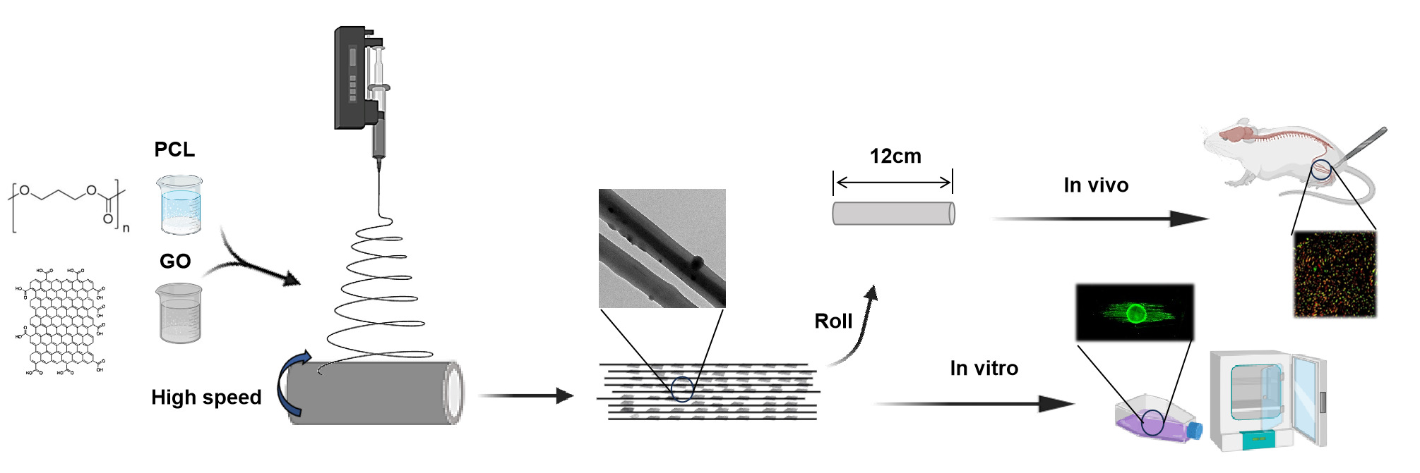
Graphical Abstract:
Introduction

Peripheral nerve injury represents a prevalent global health issue, impacting over 5 million individuals worldwide.1,2 Despite significant advancements in microsurgical techniques in recent years, effectively addressing nerve defects remains a formidable challenge. While autografts have stood as the gold standard for nerve defect repair, they are plagued by issues like donor nerve scarcity, secondary trauma, and neuroma formation.3 In recent years, artificial nerve conduits, including NeuroTube, NeuraGen, NeuroMatrix, and others, have been developed as an alternative approach.4 However, these commercial conduits have shown suboptimal nerve regeneration and functional recovery due to their basic tubular design that offers only general guidance.5,6 Consequently, there is a pressing need for innovative solutions with enhanced effectiveness for nerve repair in clinical scenarios.

It is now understood that following peripheral nervous system (PNS) injuries, the distal nerve stump undergoes Wallerian degeneration and forms aligned Büngner bands, facilitating and promoting axon growth. Inspired by the organized structures of the PNS, longitudinal microstructures such as aligned fibers, microgrooves, and microchannels have been introduced into nerve guidance scaffolds (NGSs) using various methods like electrospinning, phase separation, microstereolithography, template thermo-crosslinking, and mold freeze-drying.7–11 Electrospinning offers numerous advantages, including ease of use, customizable scaffold structure, adjustable fiber diameter, and suitability for various materials. State-of-the-art techniques have enabled the production of highly aligned fibers that mimic tissue structures, thereby improving regeneration.12,13 For instance, Wang et al demonstrated that highly aligned electrospun poly-L-lactic acid (PLLA) fibers with appropriate diameters encouraged neurite extension and Schwann cell (SC) migration.14 Besides, Zhang et al reported that aligned nanofibers offered optimal topographical cues for guiding cell and neurite growth in specific orientations.15

Beyond topographical cues defined by the scaffold’s microstructure, the significance of electroactive functional materials has gained prominence.16–18 Among these conductive materials, graphene oxide (GO) stands out due to its exceptional electrical conductivity, biocompatibility, and favorable interactions with cell interfaces. The negative carboxylate groups on GO enhance colloidal stability and hydrophilicity, making it suitable for surface attachment, proliferation, and differentiation of nerve cells.19 Li et al demonstrated that GO significantly stimulates neural cell proliferation and differentiation.20 Importantly, SCs exhibited improved adhesion, proliferation, and neurotrophic factor secretion on GO-based substrates. GO has also been shown to enhance neuro-specific gene expression, neurite outgrowth, and sprouting in neuronal-like cells.21

Recently, a significant amount of effort has been taken in developing nerve scaffolds with oriented microstructures and electroactive GO to promote nerve regeneration. Most of these strategies have successfully coated GO onto 2D surfaces. However, the coated GO is easy to detach from the 2D surfaces. Thus, it is challenging to translate them into 3D implantable scaffolds for clinical use.15,22 Furthermore, most of these studies have just investigated the effect of oriented microstructures and electroactive GO on nerve regeneration using cell lines (such as PC12 and RSC96) in vitro. Notably, the cell lines are tumor-like cells, which show different biological characteristics compared with primary cells and in vivo situations. Moreover, the mechanisms underlying the synergistic impact of electroactive GO and oriented topographic guidance on nerve regeneration remains largely unexplored. Thus, for potential clinical applications, it is imperative to adopt a more straightforward approach to fabricate nerve scaffolds with electroactive GO and oriented microstructures topography, assess the therapeutic effect of the scaffolds using primary cell cultures and in vivo animal models, and comprehend the underlying mechanisms.

In the present study, we employed a straightforward technique to co-spin polycaprolactone (PCL) and GO, resulting in an oriented GO/PCL hybrid nanofibrous scaffold termed the O-GO/PCL scaffold. The physical and chemical properties of nanofibrous scaffold were tested by scanning electron microscopy (SEM), transmission electron microscope (TEM), tensile properties, 4-point probe method, Fourier transform infrared (FTIR), and contact angle analysis. The impact of these developed scaffolds on the biological properties and behavior of neural cells was examined through co-culturing with primary SCs and dorsal root ganglia (DRG) in vitro. Transcriptome sequencing (mRNA-seq) was employed to probe the underlying mechanisms of the synergistic effect of electroactive GO and longitudinal topographic guidance on nerve regeneration. Furthermore, the developed O-GO/PCL NGS was utilized to bridge a 10 mm sciatic nerve defect in rats, aiming to investigate its therapeutic potential for peripheral nerve regeneration in vivo.

Thursday, June 16, 2022

Click chemistry extracellular vesicle/peptide/chemokine nanomissiles for treating central nervous systems injuries

 Now if we only had ANY stroke leadership at all we could contact them to get this tested in humans. But nothing will occur, nobody has any intention of actually solving stroke.

Click chemistry extracellular vesicle/peptide/chemokine nanomissiles for treating central nervous systems injuries

YaohuiTangdWenguoCuiab
https://doi.org/10.1016/j.apsb.2022.06.007Get rights and content
Under a Creative Commons license
Open access

Abstract

Central nervous system (CNS) injuries, including stroke, traumatic brain injury, and spinal cord injury, are essential causes of death and long-term disability and difficult to cure, mainly due to the limited neuron regeneration and the formation of the glial scar. Herein, we apply extracellular vesicles (EVs) secreted by M2 microglia to improve the differentiation of neural stem cells (NSCs) at the injured site, and simultaneously modify them with the injured vascular targeting peptide (DA7R) and the stem cell recruiting factor (SDF-1) on their surface via copper-free click chemistry to recruit NSCs, inducing their neuronal differentiation, and serving as the nanomissiles at the injured site (Dual-EV). Results prove that the Dual-EV holds the ability to target human umbilical vascular endothelial cells (HUVECs), recruit NSCs, and promote the neuronal differentiation of NSCs in vitro. 10 miRNAs were found to be upregulated in Dual-M2-EVs compared to Dual-M0-EVs via bioinformatic analysis, and further NSC differentiation experiment by flow cytometry revealed that among these miRNAs, miR30b-3p, miR-222-3p, miR-129-5p, and miR-155-5p may exert effect of inducing NSC to differentiate into neurons. In vivo experiments show that Dual-EV nanomissiles achieve improved accumulation in the ischemic area of stroke model mice, potentiate NSCs recruitment, and increase neurogenesis. This work provides new insights for the treatment of neuronal regeneration after CNS injuries as well as endogenous stem cells, and the click chemistry EV/peptide/chemokine and related nanomissiles for improving human health.

Graphical abstract

Click chemistry extracellular vesicle/peptide/chemokine nanomissiles repair central nervous systems (CNS) injuries by targeting blood vessels, recruiting neural stem cells (NSCs) and inducing their differentiation into neurons.

Image 1


Saturday, September 11, 2021

Overexpression of EphB4 promotes neurogenesis, but inhibits neuroinflammation in mice with acute ischemic stroke

I'm sure there is something in here but not written in any way that survivors can use. 

Overexpression of EphB4 promotes neurogenesis, but inhibits neuroinflammation in mice with acute ischemic stroke

JIN WANG1
, ZUN ZHANG2
, SHAOJING FU1
, XIAOJIE LI1
, XINHUI LI3
, SHAOBIN WANG1
 and LIHE YUAN1
Departments of 1
Neurology and 2
Orthopedics, Inner Mongolia Baogang Hospital, Baotou, Inner Mongolia 014010;
3
Department of Neurology, First Affiliated Hospital of Baotou Medical College, Baotou, Inner Mongolia 014016, P.R. China
Received January 22, 2021; Accepted June 10, 2021
DOI: 10.3892/mmr.2021.12396

Abstract. 

Ischemic stroke is one of the most common diseases that has a high rate of mortality, and has become a burden to the healthcare system. Previous research has shown that EPH receptor B4 (EphB4) promotes neural stem cell proliferation and differentiation in vitro. However, little is known regarding
its role in the neurogenesis of ischemic stroke in vivo. Thus, the present study aimed to verify whether EphB4 was a key regulator of neurogenesis in ischemic stroke in vivo. Cerebral ischemia was induced in C57BL/6J mice via middle cerebral artery occlusion (MCAO), followed by reperfusion.
Immunofluorescence staining was performed to evaluate the effect of EphB4 on the neurogenesis in cerebral cortex. The levels of inflammatory cytokines were determined using an ELISA kit. The expression levels of ABL proto‑oncogene 1, non‑receptor tyrosine kinase (ABL1)/Cyclin D1 signaling
pathway‑related proteins were detected via western blotting. The current findings indicated that EphB4 expression was significantly increased in the cerebral cortex of MCAO model mice in comparison with sham‑operated mice. Moreover, EphB4 appeared to be expressed in neural stem cells (Nestin+),
and persisted as these cells became neuronal progenitors (Sox2+), neuroblasts [doublecortin (DCX)+], and eventually mature neurons [neuronal nuclei (NeuN)+]. Overexpression of EphB4 elevated the number of proliferating (bromodeoxyuridine+, Ki67+) and differentiated cells (Nestin+, Sox2+,
DCX+  and NeuN+), indicating the promoting effect of EphB4 on the neurogenesis of ischemic stroke. Furthermore, EphB4 overexpression alleviated the inflammation injury in MCAO model mice. The expression levels of proteins‑related to the ABL1/Cyclin D1 signaling pathway were significantly
increased by the overexpression of EphB4, which suggested that restoration of EphB4 promoted the activation of the ABL1/Cyclin D1 signaling pathway. In conclusion, this study contributes to the current understanding of the mechanisms of EphB4 in exerting neurorestorative effects and may recommend a potential new strategy for ischemic stroke treatment.

Introduction

Stroke remains the second major cause of mortality worldwide and is the leading cause of death in China (1). According to a Global Burden of Disease study, there were 5.5 million deaths
and 116.4 million disability adjusted life years due to stroke in 2017 (2). Although rapid progress has been achieved in the understanding of the pathogenesis of ischemic stroke, there is currently no effective therapeutic approach for nerve repair (3). Accumulating evidence has shown that cerebral ischemia can stimulate the activation of quiescent neural stem cells (NSCs) in the subventricular zone (SVZ) into transient amplifying progenitors, which become neuroblasts after several divisions (4,5). The neuroblasts migrate into the damaged cortex and differentiate into mature neurons to promote the recovery of the neurological function (6). The growth peak of NSCs in the adult brain after ischemia is 7‑10 days (7). However, this internal response cannot functionally compensate for the ischemic damage, and the migrating cells do not differentiate into mature neurons in the cortex (8,9). Thus, it is critical to search for therapeutic approaches and specific targets to augment the differentiation of endogenous NSCs into neurons. Neuroinflammation participates in the pathophysiological progress of secondary brain injury after ischemic stroke by increasing pro‑inflammatory cytokines and neuronal apoptosis (10). A previous study has shown that the levels of inflammatory cytokines gradually increase within 0‑24 h of cerebral ischemia reperfusion in rats, and reach a peak at 24 h (11). The interaction between neuroinflammation and subsequent neurogenesis remains unknown, and thus, has gained significant interest in recent years. Moreover, further understanding of neuroinflammation and its association with neurogenesis could provide a novel approach for brain repair. Previous studies have reported that ephrin (Eph) receptors can regulate the proliferation of stem cells and progenitor cells in the central nervous system (CNS) (12). The Eph receptor family is the largest known receptor tyrosine kinase family. The members of this family bind their ligand ephrins to initiate bidirectional signaling and regulate different physiological activities, which have become popular research subjects internationally (13). Previous studies have shown that ephrinB/EphB signaling served an early role in the regulation of stem cell behavior. For instance, ephrinB3/EPH receptor B3 (EphB3) signaling exhibited an inhibitory effect on NSC proliferation in the developing SVZ (14). In the adult CNS, overexpression of ephrinB2 or EphB2 promotes NSC proliferation and represses neuroblast migration (15). However, research regarding EphB4 focuses on tumor cells. Restoration of EphB4 promotes tumor cell proliferation, migration and angiogenesis (16,17). Moreover, a previous study revealed that EphB4 regulated the self‑renewal, proliferation and neuronal differentiation of human embryonic NSCs in vitro (18).The present study aimed to investigate the role of EphB4 in the neurogenesis and neuroinflammation of ischemic stroke in vivo.

Wednesday, July 7, 2021

Psilocybin induces rapid and persistent growth of dendritic spines in frontal cortex in vivo

 But this has been known for years, isn't your doctor already prescribing psilocybin for you?

Psilocybin: Magic mushrooms have been found to boost neurogenesis. August 2013

Psilocybin induces time-dependent changes in global functional connectivity: Psi-induced changes in brain connectivity February 2020 

The latest here:

Psilocybin induces rapid and persistent growth of dendritic spines in frontal cortex in vivo

Highlights

  • Psilocybin ameliorates stress-related behavioral deficit in mice
  • Psilocybin increases spine density and spine size in frontal cortical pyramidal cells
  • Psilocybin-evoked structural remodeling is persistent for at least 1 month
  • The dendritic rewiring is accompanied by elevated excitatory neurotransmission

Summary

Psilocybin is a serotonergic psychedelic with untapped therapeutic potential. There are hints that the use of psychedelics can produce neural adaptations, although the extent and timescale of the impact in a mammalian brain are unknown. In this study, we used chronic two-photon microscopy to image longitudinally the apical dendritic spines of layer 5 pyramidal neurons in the mouse medial frontal cortex. We found that a single dose of psilocybin led to ∼10% increases in spine size and density, driven by an elevated spine formation rate. The structural remodeling occurred quickly within 24 h and was persistent 1 month later. Psilocybin also ameliorated stress-related behavioral deficit and elevated excitatory neurotransmission. Overall, the results demonstrate that psilocybin-evoked synaptic rewiring in the cortex is fast and enduring, potentially providing a structural trace for long-term integration of experiences and lasting beneficial actions.

Graphical abstract

Keywords

To read this article in full you will need to make a payment
 

Monday, May 24, 2021

Antioxidant and Anti-Inflammatory Profiles of Spent Coffee Ground Extracts for the Treatment of Neurodegeneration

 Not sure how this can translate from in vitro(Outside a living organism) to in vivo(Inside a living organism).

Antioxidant and Anti-Inflammatory Profiles of Spent Coffee Ground Extracts for the Treatment of Neurodegeneration

Academic Editor: Alessandro Attanzio
Received23 Dec 2020
Revised06 Apr 2021
Accepted29 Apr 2021
Published20 May 2021

Abstract

Spent coffee grounds (SCGs), waste products of coffee beverage production, are rich in organic compounds such as phenols. Different studies have demonstrated phenol beneficial effects in counteracting neurodegenerative diseases. These diseases are associated with oxidative stress and neuroinflammation, which initiates the degeneration of neurons by overactivating microglia. Unfortunately, to date, there are no pharmacological therapies to treat these pathologies. The aim of this study was to evaluate the phenolic content of 4 different SCG extracts and their ability to counteract oxidative stress and neuroinflammation. Caffeine and 5-O-caffeoylquinic acid were the most abundant compounds in all extracts, followed by 3-O-caffeoylquinic acid and 3,5-O-dicaffeoylquinic acid. The four extracts demonstrated a different ability to counteract oxidative stress and neuroinflammation in vitro. In particular, the methanol extract was the most effective in protecting neuron-like SH-SY5Y cells against H2O2-induced oxidative stress by upregulating endogenous antioxidant enzymes such as thioredoxin reductase, heme oxygenase 1, NADPH quinone oxidoreductase, and glutathione reductase. The water extract was the most effective in counteracting lipopolysaccharide-induced neuroinflammation in microglial BV-2 cells by strongly reducing the expression of proinflammatory mediators through the modulation of the TLR4/NF-κB pathway. On these bases, SCG extracts could represent valuable nutraceutical sources for the treatment of neurodegeneration.

1. Introduction

The food industry generates considerable amounts of waste products that require to be appropriately managed to reduce their negative sustainability impacts. An appropriate waste management helps to reduce not only the negative effects on the environment but also has got an important economic impact, since there is less production of nonrenewable resources and less energy is used in the production of new goods. Among food industry wastes, coffee by-products have been extensively taken into consideration for recycle [15]. Coffee is made by roasting and grinding coffee beans to produce a powder that is extracted with hot water or brewed. During the preparation of coffee beverages, a solid residue known as spent coffee grounds (SCG) is produced and this is the most abundant coffee waste (55−67%) [6].

About 650 kg of SCG are produced from 1000 kg of green coffee beans, and nearly 2 kg of wet SCG are obtained by the preparation of 1 kg of soluble coffee [7]. SCG is a nonedible resource, which is not entering into the food chain, and its disposal in the environment is dangerous since SCG contains caffeine, tannins, and polyphenols that make it a toxic residue [6, 7]. On these bases, numerous authors have suggested different ways to recycle SCG, to manage and reduce its disposal [810]. SCG can be used as a source of oil for biodiesel production [1113] or as a source of recoverable sugars which can be employed as food addictive or for bioethanol production [1316]. Moreover, different papers focused on SCG constituents and their application in the food and nutraceutical industry [1, 1719]. The main constituents of SCG are polysaccharides, proteins, and lipids, as well as minerals, caffeine, melanoidins, and phenols [20]. Phenols of SCG are mainly represented by different highly bioavailable and bioactive phenolic acids such as chlorogenic, caffeic, ellagic, trans-ferulic, gallic, p-hydroxybenzoic, p-coumaric, protocatechuic and tannic acids, and flavonoids such as catechin, epicatechin, rutin, and quercetin [1, 21, 22]. Phenolic compounds are well known for their beneficial effects on human health, e.g., in the prevention of different chronic degenerative diseases such as cancer, cardiovascular, and neurodegenerative diseases [2325]. Neurodegenerative diseases, mainly including Parkinson’s and Alzheimer’s diseases, are a health problem primarily affecting the elderly. These disorders share common cellular and molecular events such as oxidative stress, abnormal protein deposition, damaged mitochondrial function, induction of apoptosis, impairment of proteostasis, and neuroinflammation [26]. Neuron cells are particularly vulnerable to oxidative damage due to their high polyunsaturated fatty acid content in membranes, high oxygen consumption, and weak antioxidant defenses [27]. Oxidative damage results in an increase in reactive oxygen species (ROS), which leads to further oxidative damage and feeds this self-propagating cycle. ROS may also trigger protein misfolding, potentially leading to protein aggregation, which is a classical hallmark of neurodegenerative diseases such as Alzheimer’s and Parkinson’s diseases [28].

In addition to oxidative damage, in recent years, the immune system is emerging as a key determinant in the onset and progression of neurodegeneration [29, 30] as it triggers modification of cytokine signaling, immune cell proliferation and migration, impaired phagocytosis, and reactive gliosis [31]. Neuroinflammation, caused by the activation into proinflammatory states of the brain immune cells, namely, microglia and astrocytes, represents a fundamental defense system that protects neurons from toxic substance and microorganisms. In normal physiological conditions, this is commonly a positive mechanism aimed at preserving the brain integrity by removing threats and reestablishing homeostasis [32]. However, chronic neuroinflammation can stimulate a series of events that induce progressive neuronal damage that characterizes many neurodegenerative disorders [33]. Unfortunately, currently, no drugs capable of slowing down or blocking the progression of these debilitating pathologies have been identified. This is why the research is turning its attention to the identification of natural compounds with a preventive/protective activity against neurodegenerative disorders. As we previously demonstrated that extracts obtained by coffee silverskin, another coffee by-product, are rich in bioactive compounds with antioxidant and antibacterial activities, we assumed that also SCG could be rich in bioactive phytochemicals with potential neuroprotective activity [5, 34].

The present study was undertaken to evaluate the phenolic content of 4 different SCG extracts and their ability to counteract oxidative stress and neuroinflammation in neuron-like SH-SY5Y and microglial BV-2 cells.

Monday, July 18, 2016

PDK1 Determines Collagen-Dependent Platelet Ca2+ Signaling and Is Critical to Development of Ischemic Stroke In Vivo

Ask your doctor what the hell this means for your recovery.
http://atvb.ahajournals.org/content/early/2016/06/23/ATVBAHA.115.307105.abstract
  1. Oliver Borst
+ Author Affiliations
  1. From the Department of Cardiology and Cardiovascular Medicine (P.M., B.W.-A., S.G., D.R., D.E., T.G., M.G., O.B.), Department of Physiology (B.W.-A., F.L.), Department of Evolutionary Biology of Invertebrates, Institute for Evolution and Ecology (M.M.), Department of Dermatology (M.S.), and Department of Clinical Pharmacology (M.S.), University of Tübingen, Tübingen, Germany; Department of Neurology, University of Würzburg, Würzburg, Germany (E.G., C.K.); Institute for Immunology and Transfusion Medicine, University of Greifswald, Greifswald, Germany (G.H., A.G.); and Dr Margarete Fischer-Bosch Institute of Clinical Pharmacology, Stuttgart and University of Tübingen, Tübingen, Germany (S.W., E.S., M.S.).
  1. Correspondence to Oliver Borst, MD, Department of Cardiology and Cardiovascular Medicine, University of Tuebingen, Otfried Mueller-Str.10, 72076 Tuebingen, Germany. E-mail oliver.borst@med.uni-tuebingen.de; or Meinrad Gawaz, MD, Department of Cardiology and Cardiovascular Medicine, University of Tuebingen, Otfried Mueller-Str.10, 72076 Tuebingen, Germany. E-mail meinrad.gawaz@med.uni-tuebingen.de

Abstract

Objective—Activation of platelets by subendothelial collagen results in an increase of cytosolic Ca2+ concentration ([Ca2+]i) and is followed by platelet activation and thrombus formation that may lead to vascular occlusion. The present study determined the role of phosphoinositide-dependent protein kinase 1 (PDK1) in collagen-dependent platelet Ca2+ signaling and ischemic stroke in vivo.
Approach and Results—Platelet activation with collagen receptor glycoprotein VI agonists collagen-related peptide or convulxin resulted in a significant increase in PDK1 activity independent of second-wave signaling. PDK1 deficiency was associated with reduced platelet phospholipase Cγ2–dependent inositol-1,4,5-trisphosphate production and intracellular [Ca2+]i in response to stimulation with collagen-related peptide or convulxin. The defective increase of [Ca2+]i resulted in a substantial defect in activation-dependent platelet secretion and aggregation on collagen-related peptide stimulation. Furthermore, Rac1 activation and spreading, adhesion to collagen, and thrombus formation under high arterial shear rates were significantly diminished in PDK1-deficient platelets. Mice with PDK1-deficient platelets were protected against arterial thrombotic occlusion after FeCl3-induced mesenteric arterioles injury and ischemic stroke in vivo. These mice had significantly reduced brain infarct volumes, with a significantly increased survival of 7 days after transient middle cerebral artery occlusion without increase of intracerebral hemorrhage. Tail bleeding time was prolonged in pdk1−/− mice, reflecting an important role of PDK1 in primary hemostasis.
Conclusions—PDK1 is required for Ca2+-dependent platelet activation on stimulation of collagen receptor glycoprotein VI, arterial thrombotic occlusion, and ischemic stroke in vivo.

Tuesday, May 31, 2016

Recent progress in tracking the viability of transplanted stem cells in vivo

If the stem cell company or researchers you are following don't mention tracking of stem cells then they have NO fucking clue if they survive or not. You can attribute that to laziness or fraud, but regardless not doing so is incompetent.
http://phys.org/news/2016-05-tracking-viability-transplanted-stem-cells.html
Noninvasive cell-tracking methods are indispensable for assessing the safety and efficacy of stem-cell based therapy. Thus, the research of noninvasive cell-tracking methods for determining in vivo the translocation and long-term viability of the transplanted stem cells have received considerable attention. A recent review article summarized the recent progress in tracking the viability of the transplanted stem cells in vivo.

In the article coauthored with S. Lin, G. Chen, D. Huang, C. Meng, and Q. Wang, scholars at Suzhou Institute of Nano-Tech and Nano-Bionics, Chinese Academy of Sciences, and College of Biological Science and Technology, Fuzhou University summarized the current methods for tracking the viability of the transplanted stem cells in vivo, including reporter-gene based methods, exogenous contrast label-based methods and multimodel imaging methods.

In recent decades, stem cell-based regenerative medicine has attracted intense attention and extraordinary expectation due to its potentials in the treatment of numerous major diseases, such as hepatic, cardiac, pulmonary, renal and neurological diseases.

Knowing the viability, distribution and differentiation of the transplanted stem cells in vivo is a prerequisite for better understanding the role of stem cells playing in the therapeutic process, in which the survival report of the transplanted stem cells in vivo is particularly crucial in determining the success of stem cell-based regenerative medicine. Therefore, the development of non-invasive imaging methods that can monitor the viability of the transplanted stem cells in situ is urgently needed.

In this article, the authors summarized the development history of stem cell-tracking imaging techniques, explained the imaging principles, pros and cons underlying these techniques, and provided an overview of the applications of these techniques in animal models or humans. Furthermore, this review provided a guideline for researchers to select the right tracking method for the right study. Finally, this review discussed the current challenges in tracking the viability of transplanted stem cells, and emphasized the promise of the combined NIR-II fluorescence imaging/BLI method and MRI/PET method for further applications in high-throughput cell therapy screening in animal models and safe imaging in clinical trials, respectively.

Explore further: Researchers track neural stem cells by coloring chicken eggs from the inside

More information: SuYing LIN et al. Progress of tracking the viability of transplanted stem cells, Chinese Science Bulletin (Chinese Version) (2016). DOI: 10.1360/N972015-01404


Image at link.


Noninvasive cell-tracking methods are indispensable for assessing the safety and efficacy of stem-cell based therapy. Thus, the research of noninvasive cell-tracking methods for determining in vivo the translocation and long-term viability of the transplanted stem cells have received considerable attention. A recent review article summarized the recent progress in tracking the viability of the transplanted stem cells in vivo.
In the article coauthored with S. Lin, G. Chen, D. Huang, C. Meng, and Q. Wang, scholars at Suzhou Institute of Nano-Tech and Nano-Bionics, Chinese Academy of Sciences, and College of Biological Science and Technology, Fuzhou University summarized the current methods for tracking the of the in vivo, including reporter-gene based methods, exogenous contrast label-based methods and multimodel imaging methods.
In recent decades, stem cell-based regenerative medicine has attracted intense attention and extraordinary expectation due to its potentials in the treatment of numerous major diseases, such as hepatic, cardiac, pulmonary, renal and neurological diseases.
Knowing the viability, distribution and differentiation of the transplanted stem cells in vivo is a prerequisite for better understanding the role of stem cells playing in the therapeutic process, in which the survival report of the transplanted stem cells in vivo is particularly crucial in determining the success of stem cell-based regenerative medicine. Therefore, the development of non-invasive imaging methods that can monitor the viability of the transplanted stem cells in situ is urgently needed.
In this article, the authors summarized the development history of stem cell-tracking imaging techniques, explained the imaging principles, pros and cons underlying these techniques, and provided an overview of the applications of these techniques in animal models or humans. Furthermore, this review provided a guideline for researchers to select the right tracking method for the right study. Finally, this review discussed the current challenges in tracking the viability of transplanted , and emphasized the promise of the combined NIR-II fluorescence imaging/BLI method and MRI/PET method for further applications in high-throughput cell therapy screening in animal models and safe imaging in clinical trials, respectively.
More information: SuYing LIN et al. Progress of tracking the viability of transplanted stem cells, Chinese Science Bulletin (Chinese Version) (2016). DOI: 10.1360/N972015-01404


Read more at: http://phys.org/news/2016-05-tracking-viability-transplanted-stem-cells.html#jCp

Friday, March 13, 2015

Aromatic-turmerone induces neural stem cell proliferation in vitro and in vivo

How many decades before your doctor puts something like this into a stroke protocol? Do not self medicate. 

Aromatic-turmerone induces neural stem cell proliferation in vitro and in vivo


Joerg Hucklenbroich1,2, Rebecca Klein2,3, Bernd Neumaier3, Rudolf Graf3, Gereon Rudolf Fink1,2,
Michael Schroeter1,2,3 and Maria Adele Rueger1,2,3*
Abstract
Introduction: Aromatic (ar-) turmerone is a major bioactive compound of the herb Curcuma longa. It has been suggested that ar-turmerone inhibits microglia activation, a property that may be useful in treating neurodegenerative disease. Furthermore, the effects of ar-turmerone on neural stem cells (NSCs) remain to be investigated.
Methods: We exposed primary fetal rat NSCs to various concentrations of ar-turmerone. Thereafter, cell proliferation and differentiation potential were assessed. In vivo, naïve rats were treated with a single intracerebroventricular (i.c.v.) injection of ar-turmerone. Proliferative activity of endogenous NSCs was assessed in vivo, by using noninvasive positron emission tomography (PET) imaging and the tracer [18F]-fluoro-L-thymidine ([18F]FLT), as well as ex vivo.
Results: In vitro, ar-turmerone increased dose-dependently the number of cultured NSCs, because of an increase in NSC proliferation (P < 0.01). Proliferation data were supported by qPCR-data for Ki-67 mRNA. In vitro as well as in vivo, ar-turmerone promoted neuronal differentiation of NSCs. In vivo, after i.c.v. injection of ar-turmerone, proliferating NSCs were mobilized from the subventricular zone (SVZ) and the hippocampus of adult rats, as demonstrated by both [18F]
FLT-PET and histology (P < 0.05).
Conclusions: Both in vitro and in vivo data suggest that ar-turmerone induces NSC proliferation. Ar-turmerone thus constitutes a promising candidate to support regeneration in neurologic disease.

Tuesday, September 9, 2014

Xenon Improves Neurologic Outcome and Reduces Secondary Injury Following Trauma in an In Vivo Model of Traumatic Brain Injury.

Of course this is in mice so your doctor will never take a chance on giving it to you post-stroke.
This from 2011 tested xenon gas for heart patients.

xenon gas and stroke rehab

Whom is going to run a clinical test in humans?  Possible use for concussions?

The mouse test here:

 Xenon Improves Neurologic Outcome and Reduces Secondary Injury Following Trauma in an In Vivo Model of Traumatic Brain Injury.

Campos-Pires, Rita MD; Armstrong, Scott P. PhD; Sebastiani, Anne MD; Luh, Clara PhD; Gruss, Marco MD; Radyushkin, Konstantin MD; Hirnet, Tobias; Werner, Christian MD, PhD; Engelhard, Kristin MD, PhD; Franks, Nicholas P. PhD; Thal, Serge C. MD; Dickinson, Robert PhD

Published Ahead-of-Print
Collapse Box

Abstract

Objectives: To determine the neuroprotective efficacy of the inert gas xenon following traumatic brain injury and to determine whether application of xenon has a clinically relevant therapeutic time window.
Design: Controlled animal study.
Setting: University research laboratory.
Subjects: Male C57BL/6N mice (n = 196).
Interventions: Seventy-five percent xenon, 50% xenon, or 30% xenon, with 25% oxygen (balance nitrogen) treatment following mechanical brain lesion by controlled cortical impact.
Measurements and Main Results: Outcome following trauma was measured using 1) functional neurologic outcome score, 2) histological measurement of contusion volume, and 3) analysis of locomotor function and gait. Our study shows that xenon treatment improves outcome following traumatic brain injury. Neurologic outcome scores were significantly (p < 0.05) better in xenon-treated groups in the early phase (24 hr) and up to 4 days after injury. Contusion volume was significantly (p < 0.05) reduced in the xenon-treated groups. Xenon treatment significantly (p < 0.05) reduced contusion volume when xenon was given 15 minutes after injury or when treatment was delayed 1 or 3 hours after injury. Neurologic outcome was significantly (p < 0.05) improved when xenon treatment was given 15 minutes or 1 hour after injury. Improvements in locomotor function (p < 0.05) were observed in the xenon-treated group, 1 month after trauma.
Conclusions: These results show for the first time that xenon improves neurologic outcome and reduces contusion volume following traumatic brain injury in mice. In this model, xenon application has a therapeutic time window of up to at least 3 hours. These findings support the idea that xenon may be of benefit as a neuroprotective treatment in patients with brain trauma.

Thursday, December 19, 2013

Shining a Light on Stroke

This should be continued as a test for the next week so the neuronal cascade of death could be observed and maybe figure out how long it lasts.
Shining a Light on Stroke

  1. Michael J. Minzenberg
  1. Department of Psychiatry, University of California School Of Medicine, Sacramento, CA 95817, USA. Email: michael.minzenberg{at}ucdmc.ucdavis.edu
Stroke remains a common and serious consequence of numerous underlying illnesses and risk factors, such as hypertension and diabetes. Understanding how the brain changes after stroke may help to advance treatments for this illness. To this end, Barth and Mody have developed an improved in vivo model of the anatomy and physiology of ischemic stroke that uses photothrombosis—occlusion of a blood vessel through injection of a dye followed by irradiation—combined with stereotaxic localization (the use of a three-dimensional coordinate system to place the optic fiber) to monitor neurological changes before and after ischemic stroke.
The researchers inserted the optic fiber into the hippocampal artery of mice to isolate blood flow to the hippocampus, a brain region that is important in learning and memory and is particularly vulnerable to ischemic stroke. Blood flow was then selectively blocked in the artery through light activation of a photosensitive dye, rose bengal, which induced blood clot formation. Measuring neuron population activity in the hippocampus before and after this procedure, the authors observed a massive, brief high-frequency epileptiform discharge (HFD) in affected neurons, followed by a negative shift in the baseline electrical potential, which is consistent with neuronal depolarization due to hypoxia (inadequate oxygen). This was followed by a long-lasting decrease in neuron oscillatory activity in the gamma range (30 to 119 Hz), which is generally important to complex cognitive processes such as memory. Interestingly, only the initial HFD was also observed in the contralateral hippocampus, which is often affected by the spread of seizure activity from the other side of the brain.
This work elegantly characterizes the neurophysiological changes that unfold in the wake of stroke, thus setting the stage for elaboration of the biochemical basis and time course of these changes. These findings also suggest that events, such as the HFD and subsequent negative electrical potential, might provide specific targets for therapeutic intervention aimed at attenuating epileptiform activity or other disturbances in electrical activity, ultimately to mitigate the deleterious effects of these processes on the brain. Such knowledge should aid in the development of new therapies designed to restore brain function in stroke survivors.

Monday, November 25, 2013

Brain Targetting through Intranasal Route

A 10 page paper on what I've been suggesting the last couple of years. 

Brain Targeting through Intranasal Route


Abstract:

The blood brain barrier (BBB) represents one of the strictest barriers of in vivo therapeutic drug delivery. The barrier is an restricted exchange of hydrophilic compounds, small proteins and charged molecules
between the plasma and central nervous system (CNS). For decades, the B
BB has prevented the use of many therapeutic agents for treating Alzheimer’s disease, stroke, Brain tumour, head injury, depression, anxiety and other CNS disorders. Various techniques and Attempts were made to deliver the drug across the BBB such as modification of therapeutic agents, Altering the barrier integrity, carrier mediated transport, invasive techniques,etc. However, opening the barrier by such means allows entry of toxins and undesirable molecules to the CNS, resulting in potentially significant damage. Many advanced and effective approaches to brain delivery of drugs have emerged in recent years. Intranasal drug delivery is one of the focused delivery options for brain targeting, as the brain and nose compartments are connected to each other via the olfactory route and via peripheral circulation. In this review we discuss the effects of microspheres and other Bioadhesive drug delivery systems on nasal drug absorption. Drug delivery systems, such as microspheres, liposomes, Microemulsion, Nano emulsion and gels have been demonstrated to have good Bioadhesives characteristics and that swell easily when in contact with the nasal mucosa.

Introduction:
Nasal drug delivery is used for various kinds of diseases. It is not only used recently but it recognised form oftreatment in the Ayurvedic system of Indian medicine called “nasal karma”.
1. in recent year growing interest has focused on the use of nasal route for systemic delivery & Brain targeting. Drug which undergoes first pass
metabolism to avoid this and increases there bioavailabili
ty of drug nasal route is preferred
2.  It is useful for the drug which are active at low doses & show very less oral bioavailability such as Protein and peptide
3. central nervous system diseases such as Epilepsies, meningitis, migraine, Parkinson diseases, Alzheimer diseases has difficulty In targeting because of the transport through Blood Brain Barrier
4.  From literature it shows that such diseases can be treated by transporting exogenous material to brain by nose or it’s an effective route bypassing BBB
5. The result of concentration time Profile of intranasal administration drug is similar to the Intravenous route
6.  The pathway employed for the delivery of particular drug from the nose to brain is highly dependent on various factors, such as existence of specific receptor on the olfactory neurons, the lipophilicity and molecular weight of the drug
7.  Intra nasal delivery is non invasive& painless delivery and it does not required sterile preparation & it is easy method of drug administration for patient or physician. The nasal route offers improve delivery for “non
-
Lipinski” drug
8.  Lipophilic drug can easy cross BBB by traveling throw Transcellular
pathway. Hydrophilic drug transport throw paracellular pathway so they have very less chance to pass BBB.
Polar molecule have very less chance to pass from respiratory region to blood stream so they have some chances to reach brain by passing or travelling throw olfactory mucosa in nose
9.  Although many novel nasal product for systemic delivery on various diseases are launched in market but still no drug exploiting the nasal route to treat CNS diseases. Development of drug delivery throw nose to enable rapid & effective concentration in Brain is challenges for Researchers
7. Advantages of nasal drug delivery:
10,11,12
1)  Drug degradation that is observed in the gastrointestinal track is absent.
2) Hepatic first pass metabolism is avoided.
3) The nasal bioavailability for smaller drug molecule is good.
4) Studies so far indicates that the nasal route is an alternative to parenteral route, especially for protein’sand peptide drug.
5) Convenient for the patient especially for those on long term therapy, when compared with parenteral medication.
6) Polar compound exhibiting poor oral absorption may be particularly studies for this route of delivery.
7) Large nasal mucosa surface area for dose absorption.
8) Ease of administration, non-invasive.
9) Lower dose reduced side effects.
10) Self-administration