Changing stroke rehab and research worldwide now.Time is Brain! trillions and trillions of neurons that DIE each day because there are NO effective hyperacute therapies besides tPA(only 12% effective). I have 523 posts on hyperacute therapy, enough for researchers to spend decades proving them out. These are my personal ideas and blog on stroke rehabilitation and stroke research. Do not attempt any of these without checking with your medical provider. Unless you join me in agitating, when you need these therapies they won't be there.

What this blog is for:

My blog is not to help survivors recover, it is to have the 10 million yearly stroke survivors light fires underneath their doctors, stroke hospitals and stroke researchers to get stroke solved. 100% recovery. The stroke medical world is completely failing at that goal, they don't even have it as a goal. Shortly after getting out of the hospital and getting NO information on the process or protocols of stroke rehabilitation and recovery I started searching on the internet and found that no other survivor received useful information. This is an attempt to cover all stroke rehabilitation information that should be readily available to survivors so they can talk with informed knowledge to their medical staff. It lays out what needs to be done to get stroke survivors closer to 100% recovery. It's quite disgusting that this information is not available from every stroke association and doctors group.

Showing posts with label CSF. Show all posts

Thursday, October 30, 2025

Impaired movement of cerebrospinal fluid predicts dementia risk later in life

 With your risk of dementia post stroke is your competent? doctor testing for this so EXACT PREVENTION PROTOCOLS can be implemented? 

Your risk of dementia, has your doctor told you of this?  Your doctor is responsible for preventing this!

1. A documented 33% dementia chance post-stroke from an Australian study?   May 2012.

2. Then this study came out and seems to have a range from 17-66%. December 2013.`    

3. A 20% chance in this research.   July 2013.

4. Dementia Risk Doubled in Patients Following Stroke September 2018 

Impaired movement of cerebrospinal fluid predicts dementia risk later in life

Problems with the brain's waste clearance system could underlie many cases of dementia and help explain why poor sleep patterns and cardiovascular risk factors such as high blood pressure increase the risk of dementia.

A study led by researchers at the University of Cambridge found that impaired movement of cerebrospinal fluid (CSF) – the clear liquid that cushions and cleans the brain – predicted risk of dementia later in life among 40,000 adults recruited to the UK Biobank. Their findings are published today in Alzheimer's & Dementia: The Journal of the Alzheimer's Association.

In the healthy brain, the so-called glymphatic system serves to clear out toxins and waste materials, keeping the brain healthy. Only discovered as recently as 2012, this system functions by flushing CSF through the brain along tiny channels around blood vessels known as perivascular spaces. It collects waste then drains out of the brain, helping keep it clean and healthy.

The glymphatic system is thought to be important in protecting against many of the common forms of dementia, which are often characterised by the build-up of toxic substances in the brain – for example, Alzheimer's disease sees amyloid 'plaques' and tau 'tangles accumulate in brain tissue.

One of the most common forms of dementia is vascular dementia, caused by reduced blood flow to the brain. The most common cause of this type of dementia is cerebral small vessel disease, which affects the small blood vessels in the brain. But the impact of cerebral small vessel disease is even greater because it also interacts with other dementias making them worse; for example, a study of nuns in the US found that among those nuns whose brains showed signs of Alzheimer's disease post mortem, only around a half exhibited symptoms of dementia – but this increased to around nine in 10 if they also had cerebral small vessel disease.

Professor Hugh Markus and colleagues at the University of Cambridge wanted to see whether cerebral small vessel disease and other cardiovascular risk factors damage the glymphatic system – and whether this in turn increases the risk of dementia.

Until recently, it has only been possible to study glymphatic function in mice, but recent advances in MRI scanning have made it possible to study it indirectly in humans. Even so, it was only possible to do this practically in relatively small numbers, but Yutong Chen, while a medical student at the University of Cambridge, developed machine learning algorithms capable of assessing glymphatic functions from MRI scans at scale.

The team applied the algorithm to MRI scans taken from around 40,000 adults in UK Biobank. They found three biomarkers – biological signatures – associated with impaired glymphatic function assessed at baseline, predicted the risk of dementia occurring over the subsequent decade. One of these was DTI-ALPS, a measure of the diffusion of water molecules along the perivascular spaces. Another was the size of the choroid plexus, where the CSF is produced. The third measure reflected the flow velocity of CSF into the brain.

Although we have to be cautious about indirect markers, our work provides good evidence in a very large cohort that disruption of the glymphatic system plays a role in dementia. This is exciting because it allows to ask: how can we improve this?"

Yutong Chen, Department of Clinical Neurosciences at Cambridge

Further analysis showed that several cardiovascular risk factors impaired glymphatic function – and hence increased dementia risk, and that this was partly via causing cerebral small vessel disease, which is visible in the MRI scans.

First author Hui Hong, now a radiologist at the Second Affiliated Hospital of Zhejiang University, Hangzhou, China, said: "We already have evidence that small vessel disease in the brain accelerates diseases like Alzheimer's, and now we have a likely explanation why. Disruption to the glymphatic system is likely to impair our ability to clear the brain of the amyloid and tau that causes Alzheimer's disease."

The research suggests possible approaches for reducing dementia risk. One is to look at strategies for improving glymphatic function. Sleep plays an important role in glymphatic function, and so disrupted sleep patterns are likely to impair its ability to clear toxins. Alternatively, there may be existing medicines that could be repurposed, or new ones that could be developed, to improve glymphatic function.

Another possible approach is to treat vascular risk factors such as high blood pressure. This is supported by recent studies: the SPRINT MIND trial, for example, showed that intensive blood pressure control (maintaining a systolic blood pressure of less than 120 mm Hg) led to a 20% reduction in cognitive decline or dementia compared to participants in the standard treatment group.

Professor Markus, who leads the Stroke Research Group at the University of Cambridge and is a Fellow of Clare Hall, Cambridge, said: "We already know the importance of cardiovascular risk factors when it comes to dementia, and our findings further emphasise this link.

"At least a quarter of all dementia risk is accounted for by common risk factors like blood pressure and smoking. If these impair glymphatic function, then we can intervene. Treating high blood pressure or encouraging people to stop smoking would be an achievable way to helping the glymphatic system work better."

Professor Bryan Williams, Chief Scientific and Medical Officer at the British Heart Foundation, said: "This study offers us a fascinating glimpse into how problems with the brain's waste clearance system could be quietly increasing the chances of developing dementia later in life. By improving our understanding of the glymphatic system, this study opens exciting new avenues for research to treat and prevent dementia. It also emphasises the importance of managing known cardiovascular risk factors, such as high blood pressure, for reducing dementia risk."

The research was funded by the British Heart Foundation, with additional support from the National Institute for Health and Care Research Cambridge Biomedical Research Centre.

Source:
Journal reference:

Hong, H., et al. (2025) MRI markers of cerebrospinal fluid dynamics predict dementia and mediate the impact of cardiovascular risk. Alzheimer's & Dementia: The Journal of the Alzheimer's Association. doi.org/10.1002/alz.70699


Thursday, June 5, 2025

Facial Stimulation Clears Brain Waste and Boosts Aging Minds

 Your competent? doctor already has multiple protocols to clear your brain waste! Right? So, you don't need this one, do you? Oh no, your doctor has nothing! You're screwed!

If I, not medically trained can see what needs to be done, why are our stroke medical 'professionals' so fucking incompetent?

Do you prefer your doctor and hospital incompetence NOT KNOWING? OR NOT DOING?

  • brain waste removal (16 posts to February 2018)
  • Facial Stimulation Clears Brain Waste and Boosts Aging Minds

    Summary: Researchers have discovered a safe, non-invasive way to enhance the brain’s waste clearance system by mechanically stimulating lymphatic vessels just beneath the facial skin. This gentle technique significantly improves cerebrospinal fluid (CSF) drainage—a critical function that declines with age and contributes to cognitive disorders like Alzheimer’s.

    The team used fluorescent tracers in mice and monkeys to uncover a new drainage pathway connecting facial lymphatics to deep lymph nodes, which remains intact even in older individuals. A handheld device that lightly strokes the skin restored youthful CSF flow in aged animals, opening up exciting potential for wearable treatments that prevent or slow neurological decline.

    Key Facts:

    • New Drainage Route: Scientists mapped a previously unknown CSF drainage pathway through facial lymphatics to submandibular nodes.
    • Non-Invasive Method: Gentle mechanical stimulation of the face and neck enhanced CSF clearance without drugs or surgery.
    • Age-Proof Vessels: Superficial facial lymphatics retain function in aging, making them ideal targets for therapeutic stimulation.

    Source: Institute for Basic Science

    Scientists at the Institute for Basic Science (IBS) have uncovered a non-invasive method to boost the brain’s natural waste drainage system—a discovery that could open new avenues for tackling age-related neurological disorders.

    In a study published in Nature, researchers from the IBS Center for Vascular Research, led by Director KOH Gou Young, along with senior researchers JIN Hokyung, YOON Jin-Hui, and principal researcher HONG Seon Pyo, demonstrated that precisely stimulating the lymphatics under skin on the neck and face can significantly enhance the flow of cerebrospinal fluid (CSF)—the liquid that cushions the brain and helps remove toxic waste—through lymphatic vessels.

    This shows a brain and face.
    When applied to aged mice, the device restored CSF clearance to youthful levels, dramatically improving drainage without disrupting natural lymphatic contractions.Credit: Neuroscience News

    This offers a new approach to clearing brain waste using safe, non-invasive mechanical stimulation, rather than relying on drugs or surgical interventions.

    The human brain produces waste at a high rate compared to other organs, and clearing it efficiently is essential for healthy brain function. This clearance is primarily carried out by CSF, which removes harmful substances such as amyloid-β and tau proteins—key factors in Alzheimer’s and other neurodegenerative diseases. However, as we age, this drainage slows down, contributing to cognitive decline.

    The IBS Center for Vascular Research previously published landmark studies in Nature (2019 and 2024) demonstrating that CSF drains to deep cervical lymph nodes via meningeal lymphatic vessels at the base of the skull and the nasopharyngeal lymphatic plexus. They also showed that age-related degeneration of these lymphatics impairs CSF clearance.

    Furthermore, the team found that CSF drainage could be enhanced or suppressed pharmacologically by targeting cervical lymphatic vessels outside the skull. However, clinical applications remained limited because these lymphatics are located too deep in the neck for non-invasive access.

    “This research not only completed the map of cerebrospinal fluid drainage pathways that clear brain waste, but also provided a new method to enhance CSF drainage from outside the brain,” stated KOH Gou Young, Center Director and corresponding author.

    “We expect this will serve as a milestone for future research on neurodegenerative diseases including dementia.”

    Now, using genetically modified mice and monkeys with fluorescent tracers, the researchers have mapped out a new CSF drainage route from the brain to superficial cervical lymph nodes—via a network of lymphatic vessels in the face, nose, and hard palate.

    In older animals, many of these routes had degenerated—except for the vessels just beneath the facial skin, which retained full functionality despite aging.

    “We confirmed that lymphatic vessels beneath facial skin connect to submandibular lymph nodes through various pathways,” explained JIN Hokyung, Senior Researcher and co-first author.

    “Through these connections, we can regulate the reduced cerebrospinal fluid drainage function seen in aging and neurodegenerative diseases. Further research is needed to determine how this newly identified pathway can be applied in actual patients.”

    Recognizing this, the team developed a force-regulated mechanical stimulator—a handheld device that gently presses and strokes the skin in a controlled manner. When applied to aged mice, the device restored CSF clearance to youthful levels, dramatically improving drainage without disrupting natural lymphatic contractions.

    “I am pleased that we found a safer and more effective method to enhance cerebrospinal fluid drainage from outside the skull,” said YOON Jin-Hui, co-first author and neurovascular physiologist.

    “We are conducting follow-up studies to investigate how this newly identified drainage pathway is altered in various brain disease patients and how this new stimulation method can be applied therapeutically.”

    This technique could pave the way for wearable or clinical devices that enhance brain waste clearance in older adults or patients with neurological conditions.

    The team is now investigating how this drainage system behaves in diseases like Alzheimer’s—and whether mechanical stimulation could serve as a preventive or therapeutic tool.

    About this neuroscience research news

    Author: William Suh
    Source: Institute for Basic Science
    Contact: William Suh – Institute for Basic Science
    Image: The image is credited to Neuroscience News

    Original Research: Open access.
    “Increased CSF drainage by non-invasive manipulation of cervical lymphatics” by KOH Gou Young et al. Nature

    Thursday, May 15, 2025

    New biomarker tracks cognitive decline in Alzheimer’s disease

     What's needed is preventing cognitive decline in Alzheimer's, not this useless biomarker crapola!

    New biomarker tracks cognitive decline in Alzheimer’s disease

    At a Glance

    • Scientists uncovered a new biomarker that may help predict cognitive decline in people with Alzheimer’s disease.
    • The findings suggest measures of two proteins could improve early detection of Alzheimer’s disease and help predict or monitor cognitive decline.
    Gloved hand holding a test tube with a blood sample. Researchers have been working to develop tests to help detect and track dementia early in the disease process. Westend61 on Offset / Shutterstock

    In people with Alzheimer’s disease (AD), changes in the brain gradually erode the ability to think and remember. This cognitive decline involves an abnormal buildup of the proteins amyloid beta (Aβ) and tau. Measures of these protein biomarkers through brain scans or tests of cerebrospinal fluid (CSF) in the brain and spinal cord have improved AD diagnosis.

    However, some people with high levels of Aβ and tau have no detectable cognitive problems. Existing biomarkers also can't fully account for the speed of progression from mild cognitive impairment to severe dementia, which can take from 2 to 20 years.

    To learn more about the factors that affect cognitive decline, an NIH-funded team led by Drs. Hamilton Se-Hwee Oh and Tony Wyss-Coray at Stanford University analyzed CSF samples from about 3,400 people. The samples were from research studies in the U.S., Sweden, and Finland of people both with and without a diagnosis of AD who had volunteered to participate in the studies over many years.

    The researchers used large-scale protein analysis, or proteomics, to measure levels of more than 7,000 proteins in each of the CSF samples. They searched for new proteins that might help explain differences in cognitive impairments, or thinking ability, among people with AD. To do so, they integrated their protein data with other data collected in the studies. Those included Aβ and tau measurements from CSF and brain scans, along with measures of cognitive ability, age, sex, and AD risk genes, including APOE. The results appeared in Nature Medicine on March 31, 2025.

    The team found hundreds of proteins whose levels correlated with cognitive function. The most significant ones were related to synapse function. Synapses are the connections between neurons. In addition to the buildup of Aβ and tau, the loss of connections between neurons in the brain is a key feature of AD. Two synapse-related proteins, YWHAG and NPTX2, were the most closely related to measures of cognitive impairment.

    The researchers used machine learning to search for patterns in the protein data that could reliably predict cognitive impairment. This analysis showed that a ratio of YWHAG:NPTX2 reflected a person’s cognitive impairment better than existing biomarkers for Aβ and tau.

    YWHAG goes up in people with memory problems, while NPTX2 goes down. As a result, the YWHAG:NPTX2 ratio increases in people experiencing cognitive decline. It also rises in those at higher risk of advancing to full-blown dementia. These findings suggest that the YWHAG:NPTX2 ratio might be used to help predict the onset of AD symptoms and track disease progression. The researchers also found that this ratio rises somewhat as people age normally.

    The team next used machine learning to try to develop a similar biomarker using less invasive proteomic blood tests. They were able to develop a set of protein measurements that correlated with the CSF YWHAG:NPTX2 ratio and could also help predict cognitive decline.

    “More study is needed to understand the connection between these synaptic proteins and cognitive decline,” Wyss-Coray explains. “But our findings highlight the weakening and loss of neural connections as a driver of the decline.”

    Further work will be needed to develop effective tests for use in the clinic. Such tests might one day be used to help detect memory loss sooner, perhaps even before it begins, to allow for early interventions. They could also help to select people for participation in clinical trials of promising new AD treatments and to measure treatment responses.

    —by Kendall K. Morgan, Ph.D.

    Wednesday, February 5, 2025

    How to optimise your brain's waste disposal system

     Your competent? doctor told you this 9 years ago, right? Oh, you DON'T have a functioning stroke doctor, do you? Still hasn't learned anything new since medical school?

    What sleep position is best for brain health?

    Cognitive health is the ability to think, converse, learn, and remember clearly. It is needed to carry out many everyday activities effectively. The best sleep position for preventing cognitive decline, according to neurologists and studies, is sleeping on your side, particularly the left side. 

    Left side does not work, I can't get the unresponsive arm out of the way and keep it out of the way. 


    How to optimise your brain's waste disposal system

    Sat 22 Aug 2015 04.00 EDT

    The human brain can be compared to something like a big, bustling city. It has workers, the neurons and glial cells which co-operate with each other to process information; it has offices, the clusters of cells that work together to achieve specific tasks; it has highways, the fibre bundles that transfer information across long distances; and it has centralised hubs, the densely interconnected nodes that integrate information from its distributed networks.

    Like any big city, the brain also produces large amounts of waste products, which have to be cleared away so that they do not clog up its delicate moving parts. Until very recently, though, we knew very little about how this happens. The brain’s waste disposal system has now been identified. We now know that it operates while we sleep at night, just like the waste collectors in most big cities, and the latest research suggests that certain sleeping positions might make it more efficient.

    Waste from the rest of the body is cleared away by the lymphatic system, which makes and transports a fluid called lymph. The lymphatic system is an important component of the immune system. Lymph contains white blood cells that can kill microbes and mop up their remains and other cellular debris. It is carried in branching vessels to every organ and body part, and passes through them, via the spaces between their cells, picking up waste materials. It is then drained, filtered, and recirculated.

    The brain was thought to lack lymphatic vessels altogether, and so its waste disposal system proved to be far more elusive. Several years ago, however, Maiken Nedergaard of the University of Rochester Medical Center and colleagues identified a system of hydraulic “pipes” running alongside blood vessels in the mouse brain. Using in vivo two-photon imaging to trace the movements of fluorescent markers, they showed that these vessels carry cerebrospinal fluid around the brain, and that the fluid enters inter-cellular spaces in the brain tissue, picking up waste on its way.

    Nedergaard and her colleagues also discovered that proper function of these vessels depends on movements of water around the brain, which are carried out by glial cells called astrocytes, and therefore named them the glymphatic system. They went on to show that inter-cellular spaces expand by up to 60% in the brains of naturally sleeping and anaesthetised mice, and that this expansion drives the clearance of waste from the brain by facilitating the movements of lymph and water.

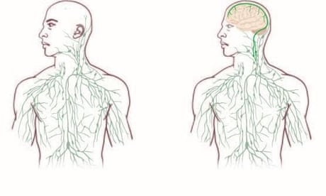
    Last month, researchers from the University of Virginia reported the identification of lymphatic vessels in the central nervous system. They demonstrated that the lymphatic system extends into the dura mater, the thickest and outer-most of the three meningeal membranes that envelope the brain and spinal cord. These vessels run parallel to the major veins and arteries, and split to send branches deep into the brain’s crevices. The researchers believe that they could be linked to the glymphatic system, and may be the second stage of the disposal mechanism, which would transport waste out of the brain and spinal cord altogether.

    The latest study from Nedergaard’s group, published in the Journal of Neuroscience earlier this month, shows that body posture affects the efficiency of the glymphatic system’s waste clearance. Using fluorescence microscopy and radioactive tracing once again, they showed that drainage of the cerebrospinal fluid worked best in mice lying on their sides compared to those lying on their back or standing up.

    The function of sleep was once deeply mysterious, but there’s plenty of evidence that it is critical for memory consolidation, and it would now seem to be required for the effective removal of waste from the brain, too. Although these studies were performed in mice, preliminary results suggest that lymphatic vessels are also present in the human brain and spinal cord, but further research will be needed to confirm that they actually constitute a working waste disposal system.

    Eventually, the link to sleep could have important implications for the treatment of neurodegenerative diseases such as Alzheimer’s and Parkinson’s, all of which involve the build-up of misfolded proteins within and around nerve cells, because of a defective waste disposal system. Indeed, it is now seems clear that good sleep hygiene has a neuroprotective effect and, in line with this, other research shows that sleep disturbances predict the onset of neurodegeneration.

    Sleeping on the side just happens to be the most popular sleeping posture for both mice and humans, and so this preference may have evolved to optimise the waste disposal system and thus ensure that the metropolis of the brain runs as effectively as possible.

    References

    Lee, H. et al. (2015). The Effect of Body Posture on Brain Glymphatic Transport. J. Neurosci, 35: 11034-44. DOI: 10.1523/JNEUROSCI.1625-15.2015.

    Louveau, A., et al. (2015). Structural and functional features of central nervous system lymphatic vessels. Nature, 523: 337-41. DOI: 10.1038/nature14432.

    Xu, L., et al. (2014). Sleep Drives Metabolite Clearance from the Adult Brain. Science, 342: 373-7. DOI: 10.1126/science.1241224. [Full text]

    Iliff, J., et al. (2013). A Paravascular Pathway Facilitates CSF Flow Through the Brain Parenchyma and the Clearance of Interstitial Solutes, Including Amyloid β. Sci. Trans. Med., 4: 147ra111. DOI: 10.1126/scitranslmed.3003748. [Full text]

    Support $15/month
    Unlock All-access digital benefits:

    Monday, September 23, 2024

    The Blood-Cerebrospinal Fluid Barrier Dysfunction in Brain Disorders and Stroke: Why, How, What For?

     FYI.

    The Blood-Cerebrospinal Fluid Barrier Dysfunction in Brain Disorders and Stroke: Why, How, What For?

    • Review
    • Published:

    Access provided by Landsaðgangur

    NeuroMolecular Medicine Aims and scope Submit manuscript
    The Blood-Cerebrospinal Fluid Barrier Dysfunction in Brain Disorders and Stroke: Why, How, What For?

    Abstract

    Ischemic stroke (IS) results in the interruption of blood flow to the brain, which can cause significant damage. The pathophysiological mechanisms of IS include ionic imbalances, oxidative stress, neuroinflammation, and impairment of brain barriers. Brain barriers, such as the blood–brain barrier (BBB) and the blood-cerebrospinal fluid (CSF) barrier (B-CSF), protect the brain from harmful substances by regulating the neurochemical environment. Although the BBB is widely recognized for its crucial role in protecting the brain and its involvement in conditions such as stroke, the B-CSF requires further study. The B-CSF plays a fundamental role in regulating the CSF environment and maintaining the homeostasis of the central nervous system (CNS). However, the impact of B-CSF impairment during pathological events such as IS is not yet fully understood. In conditions like IS and other neurological disorders, the B-CSF can become compromised, allowing the entry of inflammatory substances and increasing neuronal damage. Understanding and preserving the integrity of the B-CSF are crucial for mitigating damage and facilitating recovery after ischemic stroke, highlighting its fundamental role in regulating the CNS during adverse neurological conditions.

    Friday, July 26, 2024

    The anatomy of brainwashing

     How is your doctor making sure this is working correctly to flush out the toxic wastes and prevent dementia? Oh, your doctor doesn't know anything about the problem and has done nothing? Why the fuck are you seeing them?


    .


    Send me hate mail on this: oc1dean@gmail.com. I'll print your complete statement with your name and my response in my blog. Or are you afraid to engage with my stroke-addled mind? I need an explanation of your incompetence on stroke research and why you're not solving stroke.


    The anatomy of brainwashing


  • Lymphatic drainage removes metabolic waste and toxins from tissues, which is crucial for maintaining tissue health. In the central nervous system (CNS), lymphatic drainage relies on meningeal lymphatic vessels located in the dura mater and on the glymphatic system, a recently elucidated network that is responsible for cerebrospinal fluid (CSF) circulation and waste clearance. The interaction between glymphatic flow and meningeal lymphatics also ensures vigilant immune monitoring without perturbing the neuronal environment. How CSF travels through complex vascular and perivascular pathways, and the interactions between CSF flow dynamics and brain metabolic demands, are important for understanding brain health and could lead to the development of therapeutic approaches that might transform the treatment of a variety of neurological diseases.
    Traditionally, the CNS was considered “immune-privileged,” meaning that it was thought to be separated from the immune system, lack proper immune surveillance, and devoid of classical lymphatic drainage. Indeed, healthy brain parenchyma contains no lymphatic vessels. The brain is encapsulated by the meninges, a three-layered membranous cover comprising the dura or dura mater (the outermost layer, closest to the skull), the pia or pia mater (the layer attached to the brain), and the arachnoid that separates them from one another while also forming a subarachnoid space through which CSF flows. About a decade ago, a network of meningeal lymphatic vessels was (re)discovered to be housed in the dura (1); meningeal lymphatic vessels were originally described over 200 years ago but they were ignored by the scientific community. Although meningeal lymphatics are not located within the brain parenchyma, they nevertheless perform the vital function of brain lymphatic drainage (1).
    The presence of meningeal lymphatic vessels in the dura perfectly positions this to be the site at which immune surveillance of the brain occurs because antigens from the brain reach the dura before lymphatic drainage. Indeed, the dura mater, especially at the sites surrounding the dural sinuses, is highly populated by various immune cells, including antigen-presenting cells. Dural antigen-presenting cells take up antigens from the CSF for presentation to patrolling T cells, which could enter the dura relatively easily through dural sinuses. Migration of T cells across the dural sinuses is facilitated by the relatively slow flow of blood, the high expression of adhesion molecules on sinus endothelial cells, and the expression of chemokines and retention molecules by dural fibroblasts located in close proximity to the dural sinuses (1). Performing immune surveillance in the dura allows monitoring of the brain for threats and diseases, without the need for direct entry of immune cells into the brain parenchyma, hence avoiding disturbance of the neurons.
    Arguably, the brain “immune code” [i.e., peptides presented on major histocompatibility complex class I (MHCI) and MHCII molecules] represented on dural antigen-presenting cells would change before diseases ensue. Thus, detecting changes in this code could possibly serve as an early diagnostic tool. Dural presentation of brain antigens could also lead to abnormal immune activation due to viral mimicry, for example, and thus result in detrimental inflammatory responses [virus-specific lymphocytes found in the CSF of patients with neurodegenerative and inflammatory diseases (2, 3) support this hypothesis], eventually leading to parenchymal inflammation. Unraveling the immune code of brain tissue and being able to alter it in the dura mater (for example, through the addition of missing peptides, altering antigen-presenting cells, or interfering with protein processing and presentation) could lead to the development of new therapeutic approaches for neuroinflammatory and neurodegenerative disorders such as Alzheimer’s disease in which adaptive immune cells seem to play a role.
    Although the advances in understanding meningeal immunity and its relationship to brain immune surveillance have provided important answers, several questions remain. For example, how do antigens from the brain reach the dura? CSF was believed to mainly provide the brain with buoyancy and to assist with the removal of waste products. Beyond these basic functions, however, recent research reveals the physiological complexity and importance of the CSF. After its production in the choroid plexus, clean CSF travels through the ventricular network and the subarachnoid space. At the level of the large cortical arteries entering the brain, the CSF encounters perivascular structures called the Virchow-Robin spaces, which are extensions of the subarachnoid space and accompany vessels entering the brain parenchyma. As the arteries penetrate deeper into the brain, these spaces become narrower, but they continue throughout the brain’s blood vessels (excluding capillaries). Arterial pulsation enables CSF from the perivascular spaces to propel along the arteries and also to enter the brain parenchyma across astrocytic endfeet (4). Aquaporin 4 (AQP4) water channels are expressed by astrocytes and polarized to their endfeet, which in part facilitates the transfer of fluid from perivascular spaces into the parenchyma, and vice versa (5). Once inside the brain, this fluid is thought to create a convective flow through the dense brain parenchyma until it reaches the perivenular spaces . This passage of the CSF along the arteries, through the brain, and then out along the veins constitutes the glymphatic system or glymphatic flow, where “g” stands for the role of glial cells (astrocytes) in the process that resembles “lymphatic” flow.
    Glymphatic-lymphatic anatomical connections
    The meningeal layers surrounding the brain comprise a rich dural immune environment, meningeal lymphatic vessels, and channels that allow cerebrospinal fluid (CSF) in the dura to access the skull bone marrow. Two anatomical structures–the arachnoid granulation and the arachnoid cuff exit (ACE) point–allow immune monitoring and toxic waste removal from the brain parenchyma through the CSF.
    GRAPHIC: A. FISHER/SCIENCE
    The glymphatic system and meningeal lymphatic vessels are connected because once the CSF leaves the brain, it drains into the dura, absorbed by the meningeal lymphatic vessels, and from there into the brain-draining cervical lymph nodes. To fully understand the glymphatic-lymphatic connection, some points are to be clarified: for example, how CSF flows along the arteries, what forces facilitate the convective flow within the brain parenchyma, and how CSF reaches the meningeal lymphatics located in the dura .
    Cerebral arterial pulsation is the force driving CSF along the arteries in mice and in humans, where magnetic resonance imaging (MRI) demonstrated a strong correlation between CSF flow and arterial pulsatility. Moreover, perivascular and leptomeningeal macrophages (together referred to as parenchymal border macrophages, or PBMs) constantly degrade the extracellular matrix within the perivascular space, allowing CSF passage (6). Elimination or dysfunction of PBMs results in a build-up of extracellular matrix, which physically clogs the perivascular space and interferes with CSF flow.
    To identify and understand the forces that drive CSF flow within the brain parenchyma, it is necessary to consider how densely populated the parenchyma is. Because there is very little interstitial space, some force is essential to drive the fluid across the parenchyma. Additionally, diffusion alone is not sufficient to explain the rates of CSF perfusion through the brain tissue. An elegant study demonstrated the coupling of hemodynamics and electrophysiological activity with CSF flow using MRI of the fourth ventricle in humans, suggesting that CSF dynamics become intertwined with neural and hemodynamic rhythmicity (7). Neural activity has also been shown to correlate with CSF flow in mice and humans (8, 9). However, direct evidence (in mouse models) that neural activity drives CSF flow through the brain parenchyma was only recently described (10). Inhibiting neural activity in a specific brain region disrupted fluid flow through that area, whereas enhancing neural activity led to increased perfusion. Although the effects on fluid flow were confined to the areas where neural activity was altered (10), the intriguing possibility remains that there may be a central circuitry that regulates fluid flow throughout the brain.
    A conundrum encountered with the above mechanism derives from the empirical finding that during sleep, when arguably fewer neurons are active, fluid flow through the brain tissue is enhanced relative to its flow during wakefulness. One possible explanation is the synchronized neural activity that occurs during sleep (11). Cortical encephalography recordings reveal that different phases of sleep are associated with different wavelengths of neural activity. The slowest waves with high amplitude (delta waves, ranging from 0.5 to 4 Hz) are detected during deep sleep (the most restful phase). The synchronized neural activity that generates delta waves could produce sufficient force and directionality to drive interstitial fluid through the brain tissue (10). This mechanism overcomes discrepancies relating to the production (12) and removal of waste during the sleep?wake cycle. Thus, fewer neurons are active during sleep (and less waste is produced), yet their activity is synchronized, and the waves they produce have enough potential energy to propel the flow of CSF through the parenchyma. Although this hypothesis and its preliminary evidence are promising, further experimental work and new tools that could directly measure movement of water molecules in the tissues are needed to confirm this mechanism.
    How does the CSF reach the dura mater? Venous blood from the brain is delivered through bridging veins to the dural sinuses. These veins pierce the arachnoid to reach the dural sinuses. As the bridging veins penetrate the arachnoid, they carry a sleeve of arachnoid with them (13). Upon entering the dura, the arachnoid sleeve along the bridging veins ends in cuff-like structures called “arachnoid cuff exit” (ACE) points. ACE points are complex structures composed of arachnoid and dural fibroblasts, as well as a variety of immune cells. These are critical sites where the phenotype of endothelial cells changes from that of the blood–brain barrier (for example, with specialized tight junctions) to that of peripheral blood vessels as they continue into the dura. Not only fluid and suspended molecules but also immune cells can traffic through ACE points, making the regulation of these sites crucial for brain health. In neuroinflammatory diseases, the initial invasion of brain parenchyma might happen through ACE points (13) and in diseases of debris accumulation, these sites may be clogged, limiting removal of toxic products (such as amyloid-β in Alzheimer’s disease). The identification of ACE points provides a plausible anatomy of how brain-derived molecules can reach the dura mater on their way to meningeal lymphatics, and how dural immune-derived molecules (cytokines) can reach the brain and affect brain function (1).
    Because mice, like many other small animals with lissencephalic brains, do not have arachnoid granulations, it could be argued that ACE points simply represent primitive, arachnoid granulation-like structures. However, ACE points also exist in humans, facilitating molecular exchange between the dura and the brain parenchyma (13). This raises questions about the roles of ACE points and arachnoid granulations in CSF drainage. Traditionally, it was believed that CSF exits the brain through arachnoid granulations protruding into the venous sinuses, thereby spilling directly into the blood circulation. However, if granulations protrude directly into the sinus, it is unclear how the area of penetration is sealed to prevent blood leakage. Moreover, such a system would imply that CSF, carrying brain antigens and metabolites, drains directly into the blood rather than into the lymphatic circulation, thereby escaping immune surveillance. Recent studies in mice and humans have demonstrated that CSF is drained into the dura before reaching the blood vasculature (13, 14) and that arachnoid granulations, although closely associated with the sinuses, do not protrude into them (while a minor portion of granulations are found inside the sinus, they are separated from the blood by sinus endothelia) and are densely populated by immune cells (15). This suggests that arachnoid granulations likely function as an interface between the CSF and meningeal immunity. It seems plausible that, as the brain evolved and increased in size, so did the need for efficient surveillance of its immune code, and the evolution of arachnoid granulations might have served this purpose.
    The recent discoveries of CSF flow routes, anatomical structures allowing CSF exit, and forces moving CSF through the brain parenchyma provide a new conceptual framework for brain cleansing and immune surveillance (see the figure). Understanding this complex process—comprising numerous functional compartments, each influencing fluid flow—can be expected to facilitate the development of new classes of therapeutic interventions for enhanced brain cleansing. Such interventions—for example, targeting macrophages residing along the vasculature or astrocytic expression and function of AQP4—and inducing synchronized neural activity, could affect neurological disorders in which the accumulation of debris or immune dysfunction is a factor. There is already a precedent for therapeutic intervention using neural stimulation to increase CSF flow to eliminate pathogenic amyloid-β from the brains of people with Alzheimer’s disease (NCT05637801). A better understanding of the anatomy of brainwashing would promote further development of efficient ways not only to enhance brain cleansing but also to improve immune surveillance and effectively engage the immune system in brain diseases, including brain tumors, where immune assistance is likely a powerful solution.

    Acknowledgments

    Thanks to S. Smith for editing of the manuscript and A. Impagliazzo who generated the figure. J.K. holds patents and provisional applications related to topics discussed here.


    Wednesday, February 21, 2024

    Alzheimer's Biomarkers Show Specific Changes 20 Years Before Diagnosis


    Your competent? doctor should use this test on you post stroke to determine the extent of your upcoming dementia and then provide EXACT DEMENTIA PREVENTION PROTOCOLS. 

    Your chances of getting dementia.

    1. A documented 33% dementia chance post-stroke from an Australian study?   May 2012.

    2. Then this study came out and seems to have a range from 17-66%. December 2013.`    

    3. A 20% chance in this research.   July 2013.

    4. Dementia Risk Doubled in Patients Following Stroke September 2018

    The latest here:

    Alzheimer's Biomarkers Show Specific Changes 20 Years Before Diagnosis

    CSF and imaging markers changed in a temporal sequence

    A computer rendering of nerve cells affected by Alzheimer’s disease.

    Biomarkers evolved in a temporal sequence over 20 years in people who developed Alzheimer's disease, a nested case-control study in China showed.

    Over a median follow-up of 19.9 years, cerebrospinal fluid (CSF) and imaging biomarkers changed in a specific order, with differences seen in people who subsequently were diagnosed with sporadic Alzheimer's disease and people who remained cognitively normal, reported Jianping Jia, MD, PhD, of Capital Medical University in Beijing, and co-authors in the New England Journal of Medicineopens in a new tab or window.

    Among nearly 1,300 adults ages 45 to 65, temporal trajectories showed:

    • At 18 years before Alzheimer's diagnosis, CSF amyloid-beta 42 levels deviated between groups
    • At 14 years, the ratio of CSF amyloid-beta 42 to amyloid-beta 40 diverged
    • At 11 years, phosphorylated tau 181 CSF measures in the Alzheimer's group climbed
    • At 10 years, CSF total tau rose
    • At 9 years, CSF neurofilament light chain (NfL), a marker of axonal injury, increased
    • At 8 years, hippocampal atrophy on MRI differed between groups
    • At 6 years, cognitive decline was apparent in the Alzheimer's group based on Clinical Dementia Rating-Sum of Boxes (CDR-SB) assessments

    As cognitive impairment progressed in the Alzheimer's disease group, CSF biomarker changes accelerated at first, and then slowed.

    Biomarker changes in sporadic Alzheimer's disease were "similar in most respects to the temporal sequence of the appearance of differences of biomarkers in studies of autosomal dominant Alzheimer's disease, although the alterations in amyloid-beta 42 concentration became evident nearly a decade later in our study," Jia and colleagues noted.

    The importance of this study "cannot be overstated," wrote Richard Mayeux, MD, of Columbia University in New York City, in an accompanying editorialopens in a new tab or window. "Knowledge of the timing of these physiological events is critical to provide clinicians with useful starting points for prevention and therapeutic strategies," he observed.

    The accuracy of a clinical diagnosis of Alzheimer's disease has been controversial, Mayeux noted.

    "The National Alzheimer's Coordinating Center reported data from the National Institute on Aging Alzheimer's Disease Centers that indicated that the sensitivity of clinical diagnosis ranged from 70.9 to 87.3% and specificity varied from 44.3 to 70.8%, as compared with the reference standard of pathological diagnosis at autopsy," he pointed out. Biomarkers can provide "an opportunity to improve diagnostic accuracy in Alzheimer's disease and to establish objective diagnostic criteria."

    Jia and co-authors used data from a nested study in the nationwide prospective China Cognition and Aging Study (COASTopens in a new tab or window). Participants had CSF tests, cognitive assessments, and brain imaging at 2- to 3-year intervals. All were Han Chinese and were observed for more than 15 years, but not more than 20 years.

    After propensity-score matching on age, sex, and education, 648 participants with Alzheimer's were successfully matched 1:1 with participants who remained cognitively normal at the last follow-up.

    At baseline, participants had a mean age of about 61, and 50.6% were men. People eventually diagnosed with Alzheimer's were more likely than controls to carry an APOE4 allele (37.2% vs 20.4%).

    Cognitive status was assessed at baseline and at each follow-up with three tests. Participants were considered to have no cognitive impairment if they scored 27 or higher on the Mini-Mental State Examination (MMSE, which ranges from 0 to 30, with higher scores representing better performance).

    Scores of 12 or higher on the Logical Memory Test (LMT, which ranges from 0 to 25, with higher scores reflecting better memory) indicated normal baseline cognition. The third scale was the CDR-SB, which ranges from 0 to 18, with higher scores indicating greater impairment.

    Baseline MMSE scores were about 29.5 in each group; LMT scores were 16.8, and CDR-SB scores were 0. At follow-up, cognitively normal was defined as consistently maintaining a CDR-SB score of 0.

    In the Alzheimer's group, the progression of CSF markers appeared to accelerate initially, then peaked at an MMSE score of approximately 25 and an LMT score of approximately 11.

    Because all participants were Han Chinese, the findings may not be generalizable to other populations, Jia and co-authors acknowledged. In addition, people with a familial history of Alzheimer's disease were excluded.

    The study sample might not represent many older adults, the researchers noted. "The exclusion of participants with shorter follow-ups might have yielded a group resembling 'super-agers' -- persons endowed with higher education status, superior health status, and greater health awareness than persons not included in this study," they wrote.

    • Judy George covers neurology and neuroscience news for MedPage Today, writing about brain aging, Alzheimer’s, dementia, MS, rare diseases, epilepsy, autism, headache, stroke, Parkinson’s, ALS, concussion, CTE, sleep, pain, and more. Follow

    Disclosures

    This study was funded by the Key Project of the National Natural Science Foundation of China and others.

    Jia reported relationships with Beijing municipal funding sources, the Beijing Natural Science Foundation, the Ministry of Science and Technology, and the National Natural Science Foundation of China. Co-authors reported no conflicts of interest.

    Mayeux reported no conflicts of interest.

    Primary Source

    New England Journal of Medicine

    Source Reference: opens in a new tab or windowJia J, et al "Biomarker changes during 20 years preceding Alzheimer's disease" N Engl J Med 2024; DOI: 10.1056/NEJMoa2310168.

    Secondary Source

    New England Journal of Medicine

    Source Reference: opens in a new tab or windowMayeux R "Alzheimer's disease biomarkers -- timing is everything" N Engl J Med 2024; DOI: 10.1056/NEJMe2400102.