Changing stroke rehab and research worldwide now.Time is Brain! trillions and trillions of neurons that DIE each day because there are NO effective hyperacute therapies besides tPA(only 12% effective). I have 523 posts on hyperacute therapy, enough for researchers to spend decades proving them out. These are my personal ideas and blog on stroke rehabilitation and stroke research. Do not attempt any of these without checking with your medical provider. Unless you join me in agitating, when you need these therapies they won't be there.

What this blog is for:

My blog is not to help survivors recover, it is to have the 10 million yearly stroke survivors light fires underneath their doctors, stroke hospitals and stroke researchers to get stroke solved. 100% recovery. The stroke medical world is completely failing at that goal, they don't even have it as a goal. Shortly after getting out of the hospital and getting NO information on the process or protocols of stroke rehabilitation and recovery I started searching on the internet and found that no other survivor received useful information. This is an attempt to cover all stroke rehabilitation information that should be readily available to survivors so they can talk with informed knowledge to their medical staff. It lays out what needs to be done to get stroke survivors closer to 100% recovery. It's quite disgusting that this information is not available from every stroke association and doctors group.

Showing posts with label 'pie in the sky'. Show all posts

Sunday, June 5, 2022

Function electrical stimulation mediated by iterative learning control and 3D robotics reduces motor impairment in chronic stroke

 I can't see any use for this. By the time you're chronic insurance has stopped paying and I can't see this being affordable for anyone.  And if it only reduces impairment rather than gets to 100% recovery it really is pie in the sky.

Function electrical stimulation mediated by iterative learning control and 3D robotics reduces motor impairment in chronic stroke

Abstract

Background

Novel stroke rehabilitation techniques that employ electrical stimulation (ES) and robotic technologies are effective in reducing upper limb impairments. ES is most effective when it is applied to support the patients’ voluntary effort; however, current systems fail to fully exploit this connection. This study builds on previous work using advanced ES controllers, and aims to investigate the feasibility of Stimulation Assistance through Iterative Learning (SAIL), a novel upper limb stroke rehabilitation system which utilises robotic support, ES, and voluntary effort.

Methods

Five hemiparetic, chronic stroke participants with impaired upper limb function attended 18, 1 hour intervention sessions. Participants completed virtual reality tracking tasks whereby they moved their impaired arm to follow a slowly moving sphere along a specified trajectory. To do this, the participants’ arm was supported by a robot. ES, mediated by advanced iterative learning control (ILC) algorithms, was applied to the triceps and anterior deltoid muscles. Each movement was repeated 6 times and ILC adjusted the amount of stimulation applied on each trial to improve accuracy and maximise voluntary effort. Participants completed clinical assessments (Fugl-Meyer, Action Research Arm Test) at baseline and post-intervention, as well as unassisted tracking tasks at the beginning and end of each intervention session. Data were analysed using t-tests and linear regression.

Results

From baseline to post-intervention, Fugl-Meyer scores improved, assisted and unassisted tracking performance improved, and the amount of ES required to assist tracking reduced.

Conclusions

The concept of minimising support from ES using ILC algorithms was demonstrated. The positive results are promising with respect to reducing upper limb impairments following stroke, however, a larger study is required to confirm this.

Background

Stroke is a leading cause of death and disability in the UK, and about 50% of people who survive a stroke require some form of rehabilitation to reduce impairment and assist with activities of daily living [1–3]. Upper limb function is particularly important in regaining independence following stroke; impairments impact on daily living and well-being [4, 5].

Research has consistently identified treatment intensity and goal oriented strategies as critical elements for successful therapeutic outcomes [6–10]. To further maximise rehabilitation effects, novel therapeutic and cost-effective rehabilitation interventions need to be developed and may combine different methodological techniques. For example, the combined use of electrical stimulation (ES), robot-aided therapy and virtual reality (VR) environments has been suggested to be particularly promising with respect to upper limb rehabilitation in chronic stroke [10, 11].

Following stroke, robot and ES therapies have been demonstrated to reduce upper limb motor impairments [6, 7, 10, 12–14]. Furthermore, these techniques have been highlighted as a way to facilitate the intensity of the training received [10], and allow training despite muscle weakness and without the aid of a therapist. In addition, when used with a real-time system which displays the participants’ arm and hand movements in a VR environment, the practiced movements can be very task-specific [11, 15]. These types of technologies may be more easily transferred into patients’ homes, increasing the intensity and task specificity of the training and reducing the time and expense constraints on therapists [16].

The therapeutic effect of ES in rehabilitation is known to increase when associated with a person’s voluntary effort [12]. However, a disadvantage of many ES approaches is that they fail to encourage voluntary contribution. In addition, the vast majority of upper limb stroke patient trials using ES employ open-loop or triggered controllers [12, 17], which can lead to imprecise control of movement. In the few cases that closed-loop control has been employed, a simplistic structure and lack of a model means accurate performance is still rarely achieved [18]. Employed mainly with spinal cord injury patients, one of the few advanced control methodologies used comprises artificial neural networks [19, 20]. However such model-free approaches have limited ability to adapt to changing physiological conditions, must be re-trained for use with different movements, and being of a “black-box” structure, do not permit stability and performance analysis.

The study reported in this paper investigates the feasibility and effectiveness of a novel 3D rehabilitation platform which combines robotic support, ES and VR. The system allows patients to receive the benefits of muscle-specific targeted ES within a tightly controlled, safe and motivating environment. In this platform, ES is mediated by iterative learning control (ILC), a technology transferred from industrial robotics which is applicable to systems which repeatedly perform a finite duration tracking operation [21]. After each repetition, ILC uses data gathered on previous executions of the task, often in combination with a model of the underlying system, to update the ES signal that will be applied on the subsequent trial. Hence ILC learns from previous experience the stimulation which maximises performance, and can effectively respond to changes in the model. ILC calculates the required control action in an optimal setting, allowing strict regulation of the amount of ES, its trial-to-trial variation, and the resulting movement error. Through use of appropriate weighting parameters a precise balance can be placed between encouraging voluntary effort and ensuring accurate movement [22, 23].

ILC is one of very few model-based upper limb ES control methodologies that has previously been used in a clinical study [24–26]. During this study, stroke participants attended 18 intervention sessions of 1 hour duration in which they practiced planar reaching tasks, tracking a moving spot of light. These movements were assisted by ILC mediated ES applied to the triceps of the impaired arm. Unassisted tracking performance (i.e., movements without the aid of ES) improved over the course of the intervention and changes in muscle activation patterns towards those of unimpaired participants were also observed [24, 25]. Whilst establishing the feasibility of advanced upper limb ES control approaches in the clinical domain, this planar system did not assist shoulder movement and by providing full mechanical support to the forearm, allowed very limited shoulder elevation.

To address these limitations and increase the potential of this novel approach to stroke rehabilitation, a new system has been developed to assist participants in performing more functional, 3D reaching tasks with ES applied to triceps and anterior deltoid muscles [22, 23]. Termed SAIL: Stimulation Assistance through Iterative Learning, this system comprises a commercial robotic arm support interfaced with custom-designed ES hardware and real-time ES control environment, together with a custom-made VR task display system (see Figure 1).
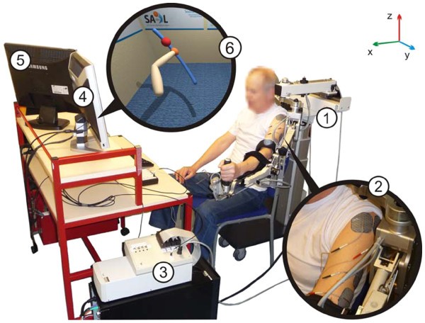
Figure 1
figure 1

SAIL system components: 1) Hocoma ArmeoSpring® support, 2) surface electrodes on triceps brachii and anterior deltoid muscles, 3) realtime processor and interface module, 4) monitor displaying VR task, and 5) monitor displaying therapist user interface. 6) shows an example of a reaching task displayed to a stroke participant with left hemipshere damage. An image of their own arm is shown and they are encouraged to follow a sphere which moves along a reference path (the trajectory); in this case from bottom right to top left.

The commercial exoskeleton robot is a purely passive ‘un-weighing’ system which supports the patient’s arm against gravity via two springs incorporated into the mechanism. Each of its joints contains a resolver which records its angular position and this information is used by both the ES control system, and the VR task display. Whilst building on previous work, the ES controller incorporates substantial developments in terms of biomechanical modelling, identification, and control complexity compared with the planar system previously reported. In particular, a five degree-of-freedom biomechanical model of the combined human and robotic arm system was developed, along with identification procedures using kinetic, kinematic and ES input data which are suitable for patients [23, 27]. Then parallel feedback and feedforward controllers were derived using techniques from nonlinear optimisation to achieve robust tracking whilst maintaining strict trial-to-trial bounds on the change in input, and the patients’ arm dynamics occurring along each trial [22, 23, 28, 29]. Moreover, the muscle structures used in the model, identification procedure and controller have been specifically developed for application to stroke patients [27].

Preliminary tests to assess whether the ILC algorithms were accurately mediating the ES took place with unimpaired participants. Results confirmed that SAIL was effective in moving the arm to produce precise reaching movements, and that tracking performance improved over a series of trials see [22, 28, 29]. The aim of the study reported in this article was to assess the technological feasibility and rehabilitation effectiveness of the SAIL system with chronic stroke participants.

More at link.


Saturday, July 4, 2020

IV tPA is associated with increase in rates of intracerebral hemorrhage and length of stay in patients with acute stroke treated with endovascular treatment within 4.5 hours: should we bypass IV tPA in large vessel occlusion?

You really don't want your doctor questioning what to do when you present at the ER with stroke symptoms. You want EXACT DIAGNOSIS PROTOCOLS LEADING TO EXACT TREATMENT PROTOCOLS AND THEN FIRST WEEK INTERVENTION PROTOCOLS THAT STOP THE NEURONAL CASCADE OF DEATH. FOLLOWED BY EXACT REHAB PROTOCOLS LEADING TO 100% RECOVERY. Yes, this is pie in the sky but until this occurs all persons with stroke are screwed.  Looking forward to being told where my analysis is wrong without excuses about brains being hard to treat and the research is not there yet, or the tyranny of low expectations.  Leaders solve problems, they don't make excuses. Are you a leader or not? If you don't soften up the clot with tPA does endovascular treatment still work? Maybe reduce the size of the bolus substantially by directing tPA there in magnetic nanoparticles.

Or maybe this solution from March, 2015

Magnetic nanoparticles could stop blood clot-caused strokes

The objection to this here:

 Potentially Toxic Magnetic Nanoparticle Pollution Found in Human Brains


Or this from  May, 2012

Future of med devices: Nanorobots in your blood stream

Use them to deliver tPA or drill thru the clot.


The fucking answers are out there, they just need to be researched and implemented. 

The latest here:

IV tPA is associated with increase in rates of intracerebral hemorrhage and length of stay in patients with acute stroke treated with endovascular treatment within 4.5 hours: should we bypass IV tPA in large vessel occlusion?

  1. Ameer E Hassan1,2,3,
  2. Victor M Ringheanu2,
  3. Laurie Preston2,3,
  4. Wondwossen Tekle1,3,
  5. Adnan I Qureshi4,5

Author affiliations


Abstract

Background Endovascular treatment (EVT) is a widely proved method to treat patients diagnosed with intracranial large vessel occlusions (LVOs); however, there has been controversy about the safety and efficacy of incorporating intravenous tissue plasminogen activator (IV tPA) as pretreatment for EVT.
Objective To compare the outcomes of all patients with LVO treated with IV tPA +EVT versus EVT alone within 4.5 hours of stroke onset.
Methods A prospectively collected endovascular database at a comprehensive stroke center between 2012 and 2019 was used to examine variables such as demographics, comorbid conditions, symptomatic/asymptomatic intracerebral hemorrhage (ICH), mortality rate, and good/poor outcomes as shown by the modified Thrombolysis in Cerebral Infarction score and modified Rankin Scale (mRS) assessment at discharge. The outcomes between patients receiving IV tPA+EVT on admission and patients who underwent EVT alone were compared.
Results Of 588 patients with acute ischemic stroke treated with EVT, a total of 189 met the criteria for the study (average age 70.44±12.90 years, 42.9% women). Analysis of 109 patients from the group receiving EVT+IV tPA (average age 68.17±14.28 years, 41.3% women), and 80 patients from the EVT alone group was performed (average age 73.54±9.84 years, 45.0% women). Four patients (5.0%) in the EVT alone group experienced symptomatic ICH versus 15 patients (13.8%) in the IV tPA+EVT group (p=0.0478); significant increases were also noted in the length of stay for patients treated with IV tPA (8.2 days vs 11.0 days; p=0.0056).
Conclusion IV tPA in addition to EVT was associated with an increase in the rate of ICH in patients with LVO treated within 4.5 hours and in patients’ length of stay. Further research is required to determine whether EVT treatment alone in patients with LVO treated within 4.5 hours is a more effective option.
View Full Text

Friday, February 14, 2020

Watch: Hollywood motion capture technology finds a new role in hospital rehabWatch: Hollywood motion capture technology finds a new role in hospital rehab

What a novel idea, get an objective damage report, then your doctor would be required to have protocols that address those EXACT disabilities. I know, 'pie in the sky', maybe 250 years from now. 

Watch: Hollywood motion capture technology finds a new role in hospital rehab

A technology most famous for its use in Hollywood movies is now a rehabilitation tool for those who have experienced a stroke or traumatic brain injury.
Motion capture technology is a staple of blockbuster films. You may have seen A-listers like Tom Hanks or Jim Carrey in behind-the-scenes bonus features dressed in what looks like spandex suits covered in ping-pong balls. Those small spheres are actually reflective markers, which are tracked by infrared cameras during an actor’s performance. The data from those cameras is then used by Hollywood visual effects artists to give computer-generated characters realistic movement.
That very same technology is being used by hospitals to analyze the movements of patients with mobility-limiting conditions such as Parkinson’s disease. Physical therapists can use the data from the motion capture system to make treatment recommendations.

Tuesday, January 8, 2019

Rescue of Transgenic Alzheimer’s Pathophysiology by Polymeric Cellular Prion Protein Antagonists

You'll have to ask your doctor to decipher this into layperson terms and where to get these cocktails, Yes, these are in mice but unless YOU light a fire under your doctor nothing will be done for when you need this.  

Rescue of Transgenic Alzheimer’s Pathophysiology by Polymeric Cellular Prion Protein Antagonists


Open AccessPublished: January 2, 2019DOI:https://doi.org/10.1016/j.celrep.2018.12.021

Highlights

  • •
    Screen for antagonist of PrPC binding to Aβo identifies polymeric antibiotic degradant
  • •
    Class of polymeric nM-potent PrPC antagonists rescue Aβo-induced phenotypes in vitro
  • •
    PrPC antagonists also clear neuroblastoma cells of PrPSc replication
  • •
    Oral PrPC antagonist rescues transgenic AD mouse synapse loss and memory deficits

Summary

Cellular prion protein (PrPC) binds the scrapie conformation of PrP (PrPSc) and oligomeric β-amyloid peptide (Aβo) to mediate transmissible spongiform encephalopathy (TSE) and Alzheimer’s disease (AD), respectively. We conducted cellular and biochemical screens for compounds blocking PrPC interaction with Aβo. A polymeric degradant of an antibiotic targets Aβo binding sites on PrPC with low nanomolar affinity and prevents Aβo-induced pathophysiology. We then identified a range of negatively charged polymers with specific PrPC affinity in the low to sub-nanomolar range, from both biological (melanin) and synthetic (poly [4-styrenesulfonic acid-co-maleic acid], PSCMA) origin. Association of PSCMA with PrPC prevents Aβo/PrPC-hydrogel formation, blocks Aβo binding to neurons, and abrogates PrPSc production by ScN2a cells. We show that oral PSCMA yields effective brain concentrations and rescues APPswe/PS1ΔE9 transgenic mice from AD-related synapse loss and memory deficits. Thus, an orally active PrPC-directed polymeric agent provides a potential therapeutic approach to address neurodegeneration in AD and TSE.

Graphical Abstract

Keywords

Introduction
Extensive evidence points to the oligomeric form of β-amyloid peptide (Aβo) as the trigger to initiate Alzheimer’s pathology (
,
,
,
), but clinical measures to reduce brain Aβ burden have been therapeutically ineffective (
), inspiring exploration for alternate strategies. Discovery that cellular prion protein (PrPC) acts as a high-affinity neuronal receptor required for toxic Aβo signaling (
,
,
,
) has led to the identification of several effectors downstream of Aβo/PrPC interaction, such as mGluR5 (
,
,
,
), Fyn kinase (
,
,
), and Pyk2 kinase (
,
), that can be targeted pharmacologically to rescue the murine brain from AD model pathology. Abrogation of Aβo/PrPC interaction itself in vivo, genetically (
,
) or with antibodies directed against the Aβo-binding domains on PrPC (
,
,
), also reverses synaptic degeneration and restores behavioral performance to impaired AD model mice, even as Aβ load is unaltered. These data indicate the possibility of disease intervention independently of Aβo clearance and identify Aβo/PrPC interaction as an opportune nexus for pharmacological intervention after Aβ accumulation occurs.
Cell-surface PrP is a conformationally diverse protein, originally identified as effecting transmissible spongiform encephalopathy (TSE) via a template-induced proteinase K-resistant form (
). Conversion of PrPC to infectious scrapie (PrPSc) refolds the C-terminal segment, while the toxic action of PrPSc also requires natively unfolded N terminus (
). Recently, we showed that cell surface PrPC engages in phase state changes between soluble, liquid, and hydrogel (
). The mobile liquid state exists at endogenous PrPC levels present in lipid rafts. The relatively immobile hydrogel phase is induced upon association with multivalent Aβo. Lateral mobility of PrPC in the membrane is restricted upon Aβo association, and mGluR5 is trapped in the hydrogel. Upon Aβo association, the unstructured N-terminal PrP domain adopts an α-helical structure, which coincides with the engagement of mGluR5 and consequent synaptotoxic signaling though Fyn and Pyk2 kinases.
Transgenic APPswe/PS1ΔE9 mice (APP/PS1) expressing the human mutant amyloid precursor protein (APP) and presenilin 1 (PS1) proteins that cause early onset AD exhibit certain pathological characteristics (
). Age-dependent accumulation of Aβo and abundant amyloid plaques, synaptic degeneration, dendritic spine loss, Fyn dysregulation, microglial and astrocytic activation, and multiple memory deficits are among APP/PS1 histopathologies and functional deficits (
). Because later-stage AD symptoms such as tau tangle accumulation and cell loss are not evident in APP/PS1 and similar strains (
), these models may reflect an early AD stage, at which Aβo-directed intervention might have the greatest impact.
Here, we describe competitive antagonists of Aβo/PrPC interaction. These compounds target PrPC Aβo-binding domains, thereby preventing Aβo association with PrPC, Aβo action in vitro, and APP/PS1 phenotypes in vivo. Additionally, these N terminus-directed ligands potently inhibit PrPSc propagation in culture, suggesting efficacy across PrPC-mediated neurodegenerative diseases.

Results

 Ceftazidime Degradation Yields a Potent Polymeric Aβo/PrPC Inhibitor Termed Compound “Z”

To search for inhibitors of Aβo/PrPC interaction we engaged in a high throughput cell-based screen using stably PrPC-transfected CV-1 cells. Aβo prepared from biotinylated synthetic Aβ42 peptide associates with these cells in a PrPC-dependent fashion that can be blocked by an antibody (6D11) directed against the Aβo-binding domain at PrPC 90-111 (Figures 1A and 1B). From a screen of 2,560 known drug and 10,130 diverse small molecules, the cephalosporin antibiotic cefixime sample was found to be highly inhibitory. Upon attempted validation, neither fresh cefixime nor a range of cephalosporins was found to possess inhibitory activity, suggesting an impurity or degradation product of cefixime was responsible (compound “X”). To investigate this possibility, five different cephalosporins were allowed to stand in DMSO at 23C for 6 days before re-testing. In addition to cefixime, ceftazidime exhibited activity resulting from prolonged incubation (compound “Z”), while other cephalosporins (cefdinir, cefotaxime, and ceftriaxone) exhibited zero activity either freshly diluted or after 6 days in DMSO (Figure 1C). Inhibitory activity developed progressively from ceftazidime incubated in sodium carbonate at 23°C over 9 days (Figure S1).

Figure thumbnail gr1
Figure 1Cefixime or Ceftazidime Degradation Produces HMW Inhibitor of Aβo/PrPC Interaction

Wednesday, August 8, 2018

Human super intelligence: still science fiction or close to reality?

As stroke survivors most of us probably need some brain augmentation. Probably pie in the sky, but leaders tackle BHAGs(Big Hairy Audacious Goals) of 100% recovery for all survivors! 
Do you really think your doctor is going to read any of the 149 papers?

Human super intelligence: still science fiction or close to reality?


New research published in Frontiers in Neuroscience discusses the facts, fiction and controversy surrounding brain augmentation. 
Creating super intelligence or enhancing the brains of patients with neurological disorders through brain augmentation is currently a hot topic in both scientific literature and the media. This is a remarkable development because just a decade or two ago the idea of brain augmentation was reserved for science fiction. But with the rapid development of neuroscience and related technological and medical fields, many of the past decade’s science fiction themes – such as reading out brain content, sending information to the brain, inter-connecting different brains and adding artificial parts to the brain – are becoming real.
Broadly speaking, the themes of brain augmentation can be divided into three categories:  firstly, approaches that involve recording and decoding brain activity, secondly, approaches that include various ways to stimulate the brain, and finally, futuristic and philosophical considerations around the topic.
So how close are we to brain augmentation approaches that may help in the treatment of neurological and mental conditions, such as paralysis, sensory, motor and cognitive disabilities, or Parkinson’s disease, or to creating super intelligence and enhancing productivity in healthy people who want to excel in their performance?
Recently, more than 600 authors contributed almost 150 research articles investigating brain augmentation – on everything from brain-machine interfaces, neuro-stimulators, the application of neuro-pharmacology and ethical and philosophical considerations around brain enhancement that may seem relatively unimportant today, but given the rapid development of this field, they will become very real and practical in the near future.
The editors of this Research Topic—The augmentation of brain function: facts, fiction and controversy published in Frontiers in Systems Neuroscience—Dr Mikhail Lebedev, from Duke University in the United States, Dr Ioan Opris at the University of Miami School of Medicine and Dr Manuel Casanova at the University of Louisville, also in the U.S. are proud of this comprehensive collection of articles which raise key themes directly related to practical issues around brain enhancement – particularly pertaining to public health.
Indeed, Dr Opris says that the strength of research is in the synergy of collaboration, “Sharing each other’s research is like polishing a diamond and providing new faces to shine”, a sentiment echoed by Dr Casanova, “Research findings need to be communicated in order to be relevant. Personally, communicating our findings forces me to better understand my own results and become critical of them. Sharing my research pushes me to establish a different frame of mind as a writer one that includes other researchers and even lay people.”
One original research paper, Donor/recipient enhancement of memory in rat hippocampus by Dr Sam Deadwyler from the U.S. Wake Forest School of Medicine in Winstom-Salem, New Carolina and his colleagues describes a donor-recipient memory transfer. In this stunning experiment, a donor rat was required to perform a behavioral task requiring memorization. They then decoded the memory content from the hippocampus of the donor rat and, using electrical micro-stimulation, transferred it to the hippocampus of another rat. After the donor rat’s neural activity was processed by a multiple-input multiple-output model, and delivered to the recipient’s brain, the recipient rat successfully reproduced the task behavior.
The authors say that their research provides the basis for utilizing extracted appropriate neural information from one brain to induce, recover, or enhance memory related processing in the brain of another subject and that the results provide important insight for extending donor/recipient procedures to functions performed by other brain regions and other behavioral endpoints, and eventually to similar circumstances involving humans.
In another paper, Transcranial direct current stimulation: five important issues we aren’t discussing (but probably should be), around Dr. Jared Horvath from the University of Melbourne in Australia, discusses several important issues related to the use of transcranial direct current stimulation (tDCS)—constant, low current delivered to the brain area of interest via electrodes on the scalp—as a cognitive enhancement approach. It was originally developed to help patients with brain injuries or psychiatric conditions like a major depressive disorder.
Dr Horvath and his colleagues outline a number of important experimental and technical issues associated with tDCS that they say are simply not being discussed in any meaningful manner. These include the need for an individualized, patient by patient approach to tDCS, the importance of proper controls in tDCS studies, such as sham stimulation and blinding, the interference of motor and cognitive activities with the tDCS effects and changes in electric current related to hair thickness and electrode attachments methods. They argue that if the field of tDCS is to avoid becoming a footnote in the annals of neuro-scientific research it’s time to collectively acknowledge well-known shortcomings and use these issues to guide further research and exploration and well as more comprehensive protocols.
A third paper of the 149 submitted explores something that we all love – sleep! In the paper Sleep for cognitive enhancement, Dr Susanne Diekelmann from the Institute of Medical Psychology and Behavioral Neurobiology at the University of Tubingen in Germany reviews enhancing the potential of sleep for such cognitive functions as attention, language, reasoning, decision making, learning and memory. The article discusses the role of sleep in memory consolidation and the acquisition of new memories after sleep, the role of sleep-specific brain oscillations in these processes and neurotransmitters involved.
Dr Diekelmann suggests that memory processing during sleep can be augmented by cueing memory reactivation with olfactory and auditory cues, electrically inducing sleep-specific brain oscillations, and modulating specific neurotransmitter systems pharmacologically.
At the end of the day, Dr Lebedev says this Research Topic would not have been possible elsewhere, “It was only because of Frontiers publishing model that we were able to compile this collection of articles from the best experts in several disciplines. As far as I can tell, all of our 629 authors enjoyed working with Frontiers, and everybody is looking forward to this research topic published as three eBook volumes: one devoted to decoding of brain signals, the other to neurostimulation approaches to augmentation of brain function, and the third one on futuristic ideas and ethical issues. Judging from the number of page views (more than 700,000 at the moment), this research topic evoked significant interest in our open-access readership, in both the scientific community and the general public. The number of citations is constantly growing for these articles, so hopefully they will be of great educational and scientific value for students, researchers, health care practitioners, and people interested in studies of the brain”.

Lebedev and his fellow Topic co-Editors are finalists of the Frontiers Spotlight Award, where the winners are granted with US$100,000 to host their own conference themed around their Research Topic.

Saturday, February 24, 2018

Wearable tech aids stroke patients

Maybe with this your doctor and therapists could finally get exact diagnosis of your movement disabilities. And use that to correlate exact stroke rehab protocols that fix those disabilities. Pie in the sky I know but someone has to put goals out there. Your stroke medical professionals are doing nothing about goals to 100% recovery.  You do want 100% recovery, don't you? And you've asked your doctor for protocols to get there? Haven't you? Did your doctor ask if you were crazy wanting 100% recovery? Or crazy just for asking?

Wearable tech aids stroke patients


Media captionWearable tech could help stroke patients

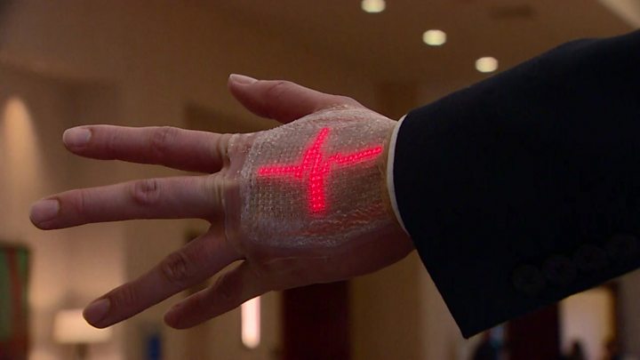
Scientists in the US are developing wearable sensors to speed up the recovery of stroke patients.
The sensors are able to send information to doctors continuously.
The team developing the system says it could allow therapists to more closely monitor the effectiveness of their care.
Details of the study were released at the recent annual meeting of the American Association for the Advancement of Science in Texas.
Lizzy McAninch had a stroke two years ago. She could not move or speak or swallow for several weeks.
Lizzy is testing out wearable sensors that might speed her recovery.
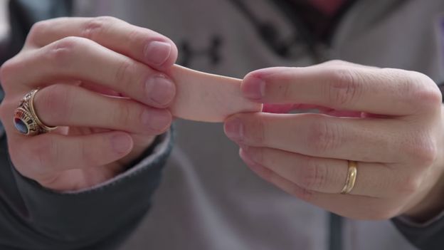
They look like small white sticking plasters, but they send information wirelessly to her medical team.
She is a doctor herself and can see how they could help her.


Sensor Image copyright Shirley Ryan AbilityLab
Image caption They look like small sticking plasters

"This technology to put sensors on the body to assess which muscle groups work or not can really pinpoint the areas affected by the stroke and can target therapies to specifically improve those issues," she told BBC News.
The sensors continue to send back readings even after she has finished her exercises. This means that her therapist Kristen Hohl, from the Shirley Ryan AbilityLab in Chicago, can monitor her progress at home.
"As a therapist, I think about what my patients are doing at home. Are they able to carry through the recommendations I'm giving them as a therapist to do more? Do we see that they are walking more or do we see them engaging in conversations?
"Those are the types of things that I can get feedback from the sensors where currently I have to rely on what they tell me they have done."


Tablet Image copyright Shirley Ryan AbilityLab
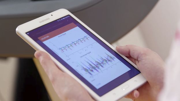
Image caption The team is gathering large amounts of data

The challenge for the scientists was to pack a lot of electronics on to a small flexible material and still make it comfortable for the patient to wear for a long time.
"It is almost mechanically imperceptible to the patient who is wearing the device," according to John Rogers, of Northwestern University in Chicago, who developed the sensors.
"And you can embed all sorts of advanced sensor functionality, microprocessor computing capability, power supplies and WiFi into this very unusual platform, and that is the uniqueness of what we do."
By the end of this year, the research team will have more information than ever before on stroke recovery. The scientists believe that their study could transform the way patients are treated in the future.