Changing stroke rehab and research worldwide now.Time is Brain! trillions and trillions of neurons that DIE each day because there are NO effective hyperacute therapies besides tPA(only 12% effective). I have 523 posts on hyperacute therapy, enough for researchers to spend decades proving them out. These are my personal ideas and blog on stroke rehabilitation and stroke research. Do not attempt any of these without checking with your medical provider. Unless you join me in agitating, when you need these therapies they won't be there.

What this blog is for:

My blog is not to help survivors recover, it is to have the 10 million yearly stroke survivors light fires underneath their doctors, stroke hospitals and stroke researchers to get stroke solved. 100% recovery. The stroke medical world is completely failing at that goal, they don't even have it as a goal. Shortly after getting out of the hospital and getting NO information on the process or protocols of stroke rehabilitation and recovery I started searching on the internet and found that no other survivor received useful information. This is an attempt to cover all stroke rehabilitation information that should be readily available to survivors so they can talk with informed knowledge to their medical staff. It lays out what needs to be done to get stroke survivors closer to 100% recovery. It's quite disgusting that this information is not available from every stroke association and doctors group.

Monday, November 30, 2020

Determinants of Antidepressant Treatment and Outpatient Rehabilitation Within the First Year After Stroke

 You do realize that if you were to have EXACT STROKE PROTOCOLS LEADING TO 100% RECOVERY stroke depression wouldn't exist. Survivors would be too busy counting repetitions to think about anything else, knowing that the end result of their hard work would be recovery.

Determinants of Antidepressant Treatment and Outpatient Rehabilitation Within the First Year After Stroke

First Published November 25, 2020 Research Article 

This study aims to identify individual determinants of antidepressant treatment and outpatient rehabilitation after stroke. People with ischemic stroke (N = 303) recruited at 2 inpatient rehabilitation clinics were included into a prospective longitudinal study with follow-up telephone interviews 6 and 12 months later. Participants reported on their use of antidepressant medication and psychotherapy as well as physical, occupational, speech, and neuropsychological therapy. The use of antidepressants at discharge (n = 65, 23.8%) was predicted by the severity of depressive symptoms, severity of stroke, history of depression, and use of antidepressants at admission (all p < .05, R 2= .55). The number of outpatient rehabilitation services used at follow-ups was predicted by higher functional and cognitive impairment, higher education, younger age, severity of depressive symptoms, and lower self-efficacy (all p < .05; R 2 6M = .24, R 2 12M = .49). The relevance of identified determinants for the improvement of treatment rates after stroke is discussed.

Access Options


Movement Quality: A Novel Biomarker Based on Principles of Neuroscience

This is so simple; One question; 'Are you 100% recovered?', if not, then you provide the protocols to get there

Movement Quality: A Novel Biomarker Based on Principles of Neuroscience

First Published November 13, 2020 Research Article Find in PubMed 

A major problem in neurorehabilitation is the lack of objective outcomes to measure movement quality. Movement quality features, such as coordination and stability, are essential for everyday motor actions. These features allow reacting to continuously changing environment or to resist external perturbations. Neurological disorders affect movement quality, leading to functionally impaired movements. Recent findings suggest that the central nervous system organizes motor elements (eg, muscles, joints, fingers) into task-specific ensembles to stabilize motor tasks performance. A method to quantify this feature has been previously developed based on the uncontrolled manifold (UCM) hypothesis. UCM quantifies movement quality in a spatial-temporal domain using intertrial analysis of covariation between motor elements. In this point-of-view article, we first describe major obstacles (eg, the need for group analysis) that interfere with UCM application in clinical settings. Then, we propose a process of quantifying movement quality for a single individual with a novel use of bootstrapping simulations and UCM analysis. Finally, we reanalyze previously published data from individuals with neurological disorders performing a wide range of motor tasks, that is, multi-digit pressing and postural balance tasks. Our method allows one to assess motor quality impairments in a single individual and to detect clinically important motor behavior changes. Our solution may be incorporated into a clinical setting to assess sensorimotor impairments, evaluate the effects of specific neurological treatments, or track movement quality recovery over time. We also recommended the proposed solution to be used jointly with a typical statistical analysis of UCM parameters in cohort studies.


Blowing up Neural Repair for Stroke Recovery

 It is what I've been screaming about for years. STROKE HAS NO STRATEGY AND NO LEADERSHIP. Articles like this will do nothing.  Our fucking failures of stroke associations  have done nothing for decades. Did you somehow miss the 'Stroke is Treatable' meme from World Stroke Day a few years ago? Obviously stroke research is no longer needed. We can repurpose all those researchers into personal care attendants for stroke survivors that somehow didn't obey the meme and fully recover.

What a lying piece of shit

Blowing up Neural Repair for Stroke Recovery

Preclinical and Clinical Trial Considerations
Originally publishedhttps://doi.org/10.1161/STROKEAHA.120.030486Stroke. 2020;51:3169–3173

The repair and recovery of the brain after stroke is a field that is emerging in its preclinical science and clinical trials. However, recent large, multicenter clinical trials have been negative, and conflicting results emerge on biological targets in preclinical studies. The coalescence of negative clinical translation and confusion in preclinical studies raises the suggestion that perhaps the field of stroke recovery faces a fate similar to stroke neuroprotection, with interesting science ultimately proving difficult to translate to the clinic. This review highlights improvements in 4 areas of the stroke neural repair field that should reorient the field toward successful clinical translation: improvements in rodent genetic models of stroke recovery, consideration of the biological target in stroke recovery, stratification in clinical trials, and the use of appropriate clinical trial end points.


Study: Lower Door-to-Needle Times Linked to Lower All-Cause Mortality

 But that is the wrong measurement isn't it? 100% recovery is the only measurement you should be doing.

Study: Lower Door-to-Needle Times Linked to Lower All-Cause Mortality

A new study shows that every 15-minute decrease in door-to-needle time is linked to lower all-cause mortality. How can we work to reduce door-to-needle times?

When a neurologist utters the phrase "time is brain," the implication is that every minute counts in treating each stroke patient. A recent study is further confirming what stroke teams have known since 1993: efficient treatment is everything to better outcomes in stroke care. 

A New Study Further Confirms: Time Is Brain.

This study, published by the Journal of the American Medical Association, shows a definitive correlation between door-to-needle times and outcomes in stroke cases for patients treated with tPA. According to researchers, “each 15-minute increase in door-to-needle times was significantly linked to higher all-cause mortality within 90 minutes after arriving to the hospital.”

audio-clip-stroke-patient-1180x700That wasn’t the only connection researchers found. Each 15 minute increase in treatment time also equaled higher all-cause readmission. One of the researchers involved in the study, Greg Fonarow, MD, of the University of California Los Angeles, summarized his takeaway from the findings: “Faster treatment translates into better long-term outcomes in patients with stroke. For every 15 minutes improvement in treatment time, improvements in 1-year outcomes were observed."

In 1993, neurologist Camilo Gomez, MD, coined the phrase “time is brain” to illustrate the principle that time is of the essence when it comes to treating strokes. For every minute a stroke goes untreated, the average patient loses 1.9 million neurons. Every minute counts, which is why door-to-needle times have become a mark of quality stroke treatment.

This new study reinforces that shorter door-to-needle times do indeed improve long-term outcomes in ischemic stroke. Christopher C. Muth, MD, assistant professor in the department of neurological sciences at Rush University Medical Center and senior editor of JAMA, explained its significance: “This study fills an important gap in the literature by convincingly documenting the association between faster treatment with intravenous [tissue plasminogen activator] and better long-term outcomes, including 1-year mortality. The findings are yet another reason for clinicians and health systems to design stroke services that can treat patients with acute ischemic stroke with thrombolytic therapy in a rapid fashion.”


This research is important because strokes are the leading cause of cardiovascular disease-related deaths in the United States. Every year, 13.7 million people worldwide suffer a stroke. Of these, over 5 million will die, and another 5 million are permanently disabled. Stroke is the leading cause of adult disability in Canada, and the number of Australians living with the negative effects of a stroke is predicted to more than double to one million within the next thirty years. 

Rapid treatment is vital to giving each patient the best possible chance at not only surviving a stroke, but also maintaining their quality of life once they recover. 

How Are Hospitals Working to Reduce Door-to-Needle Times? 

Given the gravity of these statistics, hospitals around the world are looking for new and innovative ways to reduce their door-to-needle times and improve patients' chances of a good outcome. Most of these approaches revolve around one concept: efficiency. 

Recently, a research team in Germany conducted a study using interactive clocks to improve D2N times by incentivizing physicians to be as efficient as possible. During the study, the clocks displayed the amount of time that had elapsed since the patient’s arrival, and would sound a buzzer at intervals to let physicians know whether they were hitting the target time for each stage of treatment. Physicians could avoid the buzzer by completing each stage of treatment within the allotted time and hitting a button to register their progress.

The researchers concluded that the use of the clock did, in fact, help accelerate door-to-needle times: “This study showed that the use of a stroke clock demanding active feedback significantly improves acute stroke-management metrics and, thus, represents a potential low-cost strategy for streamlining time-sensitive stroke treatment.” An emphasis on efficiency and an awareness of the elapsed time makes a significant difference in efficient—and effective—stroke treatment.

Hospitals are also finding that poor or inefficient communication is often a culprit behind inflated door-to-needle times. Coordinating care for a stroke patient takes quite a bit of time and effort. Many stroke coordinators have to make upwards of ten phone calls to coordinate care for a single patient. Non-interoperable communication methods like pagers, fax machines, and phones require information to be repeated multiple times, wasting precious treatment time for patients.

staff-device-gloves-stroke-1200x630In order to make treatment more efficient, some hospitals have turned to mobile technology to streamline communication. Communication platforms that have been specifically designed for healthcare teams and fit naturally into existing workflows allow clinicians to save time that they would normally spend on care coordination. When clinicians can instantly build care teams around a patient's needs and communicate with one another in real time, door-to-needle times have been shown to decrease by significant percentages. 

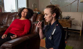
One mobile platform that enables this type of communication is Pulsara, a healthcare communication and telehealth app designed to simplify care coordination for caregivers. Pulsara allows clinicians to communicate with one another and share vital information on the same HIPAA compliant patient channel. This year, several hospitals have released data showing how streamlining their communication with Pulsara has helped greatly reduce time wasted in trying to get patients in for treatment.

Last month, Wendy Barrilleaux, the Neuroscience Service Line Administrator for St. Dominic Hospital in Jackson, Mississippi, shared that St. Dominic’s saw an 18% improvement in their door-to-needle times within six months of implementing mobile technology for communication between team members. “With Pulsara, we now have a secure HIPAA compliant video network that is accessible from the device that all clinicians carry right in their pocket,” said Barrilleaux. “With mobile telemedicine, we not only have a network of providers connected for telemedicine, but these consultations can take place within a patient's channel, which is primed for real-time communication.” St. Dominic’s is continuing to implement new technology to improve stroke care in every way possible. They recently announced that they’ve become the fifth hospital in the world to integrate two leading healthcare technologies, Pulsara and RapidAI, resulting in faster, more efficient stroke care. 

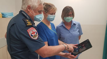
CHRISTUS Good Shepherd Medical Center - Longview in Longview, Texas struggled with synchronizing their stroke team on one universal clock. “One challenge we found was that everybody providing care was looking at their own clocks, which weren’t necessarily in sync,” said Jennifer Reeves, RN, MSN, ASC-BC, the stroke program coordinator at CHRISTUS Good Shepherd. “I may be looking at the time on my watch and the charge nurse could be looking at the one on the computer.” In an effort to make sure all members of the care team were watching the same clock, CHRISTUS Good Shepherd implemented Pulsara. This year, they reported a 46-minute average door-to-needle for patients receiving tPA, a 59% decrease from their previous average time of 110 minutes. They now have 100% of their door-to-needle completed in under 60 minutes—and 58% in under 30 minutes. By improving communication and a fostering greater awareness of the passage of time between the members of their stroke team, Christus Good Shepherd has greatly increased their stroke patients' chances for positive outcomes.

latrobe-mark-janet-carolyn-2-900x501In 2019, Latrobe Regional Hospital, near Melbourne, Australia, was already doing incredibly well with stroke care, averaging 22 minutes on their door-to-CT times. After dealing with frustrating hindrances in communication between Ambulance Victoria and the hospital, they implemented Pulsara near the beginning of 2020. “Now with Pulsara, we are able to pre-register the patients, so stroke patients can go directly to CT on arrival,” said Janet May, Stroke and Pulsara Coordinator at Latrobe. Comparing the data from March through July of 2019 to the same period in 2020, Latrobe Regional Hospital improved door-to-CT times from 22 minutes down to just 7 minutes on average—a 68% reduction in treatment time. Eliminating the frustrating runaround of missed phone calls and pages has helped Latrobe take their treatment times from great to excellent. 

The research is clear: in stroke cases, faster treatment equals better outcomes. If we can reduce door-to-needle times for stroke patients, we can help make sure patients are given the best shot at recovery and the best possible outcomes. Efficiency awareness and mobile technology are helping hospitals around the world to improve treatment times for their patients. What are you doing to reduce D2N times at your facility?

pulsara-logo-only

Want to look into improving your treatment times, but aren't sure where to start, or even what questions to ask? Check out our blog post: To Improve Treatment Times, It Pays to Become Obsessed With the Process.


Mobile Interventional Stroke Team Model Improves Early Outcomes in Large Vessel Occlusion Stroke


You don't know what you are doing do you? You have no clue how fast tPA needs to be delivered to get 100% recovery.  You are not even measuring 100% recovery you blithering idiots, you are measuring recanalization. 

Mobile Interventional Stroke Team Model Improves Early Outcomes in Large Vessel Occlusion Stroke

The NYC MIST Trial
and on behalf of the Mount Sinai Stroke Investigators*
Originally publishedhttps://doi.org/10.1161/STROKEAHA.120.030248Stroke. 2020;51:3495–3503

Background and Purpose:

Triage of patients with emergent large vessel occlusion stroke to primary stroke centers followed by transfer to comprehensive stroke centers leads to increased time to endovascular therapy. A Mobile Interventional Stroke Team (MIST) provides an alternative model by transferring a MIST to a Thrombectomy Capable Stroke Center (TSC) to perform endovascular therapy. Our aim is to determine whether the MIST model is more time-efficient and leads to improved clinical outcomes compared with standard drip-and-ship (DS) and mothership models.

Methods:

This is a prospective observational cohort study with 3-month follow-up between June 2016 and December 2018 at a multicenter health system, consisting of one comprehensive stroke center, 4 TSCs, and several primary stroke centers. A total of 228 of 373 patients received endovascular therapy via 1 of 4 models: mothership with patient presentation to a comprehensive stroke center, DS with patient transfer from primary stroke center or TSC to comprehensive stroke center, MIST with patient presentation to TSC and MIST transfer, or a combination of DS with patient transfer from primary stroke center to TSC and MIST. The prespecified primary end point was initial door-to-recanalization time and secondary end points measured additional time intervals and clinical outcomes at discharge and 3 months.

Results:

MIST had a faster mean initial door-to-recanalization time than DS by 83 minutes (P<0.01). MIST and mothership had similar median door-to-recanalization times of 192 minutes and 179 minutes, respectively (P=0.83). A greater proportion had a complete recovery(How many?) (National Institutes of Health Stroke Scale of 0 or 1) at discharge in MIST compared with DS (37.9% versus 16.7%; P<0.01). MIST had 52.8% of patients with modified Rankin Scale of ≤2 at 3 months compared with 38.9% in DS (P=0.10).

Conclusions:

MIST led to significantly faster initial door-to-recanalization times compared with DS, which was comparable to mothership. This decrease in time has translated into improved short-term outcomes and a trend towards improved long-term outcomes.

Registration:

URL: https://www.clinicaltrials.gov. Unique identifier: NCT03048292.


Sunday, November 29, 2020

Clinical Features, Risk Factors, and Early Prognosis for Wallerian Degeneration in the Descending Pyramidal Tract after Acute Cerebral Infarction

Useless, NOTHING ON HOW TO PREVENT THIS.

Clinical Features, Risk Factors, and Early Prognosis for Wallerian Degeneration in the Descending Pyramidal Tract after Acute Cerebral Infarction

Published:November 27, 2020DOI:https://doi.org/10.1016/j.jstrokecerebrovasdis.2020.105480

Abstract

Background

Wallerian degeneration(WD) occurs in the descending pyramidal tract(DPT) after cerebral infarction commonly, but studies of its degree evaluation, influencing factors and effects on nervous function are still limited.

Objectives

The purpose of this study was to describe these findings and estimate their clinical significance.

Methods

In total, 133 patients confirmed acute cerebral infarction and restricted diffusion in the DPT of the cerebral peduncle by MRI scans. These cases were retrospectively reviewed. We describe their clinical characteristics and analyze influence factors of WD, including the timespan from symptom onset to MRI and TOAST classification. Their NIHSS scores at admission and first 7 days NIHSS improvement rate after admission were also analyzed.

Results

These patients were divided into three groups by timespan ≤7 days( n = 45),7–14 days( n = 70) and >14 days( n = 18). The mean WD degree (%)of these three groups was 44.41 ± 22.51,52.35 ± 22.61and 44.31 ± 19.35,respectively( p = 0.122).According to the TOAST classification, the mean WD degree(%) of the cardioembolism group( n = 28, 62.80 ± 25.12) was significantly different from both the large-artery atherosclerosis group( n = 73,45.08 ± 20.03, p = 0.000) and the small-vessel occlusion group( n = 23,39.68 ± 16.95, p = 0.000). The mean NIHSS score upon admission of the WD degree≤50% group( n = 82,8.17 ± 5.87) was different from that of the >50% group( n = 51,11.31 ± 7.00)( p = 0.006). However, the mean 7 days NIHSS improvement rate(%) of the WD degree≤50% group( n = 79,11.83 ± 23.76)and >50% group( n = 50,13.40 ± 27.88) was not significantly different( p = 0.733).

Conclusions

Early WD in ischemic stroke patients has a correlation with serious baseline functional defects. Therefore, we should give close attention to imaging change, especially in those with cardioembolism .
 

Muscle Temperature Sensing and Control with a Wearable Device for Hand Rehabilitation of People After Stroke

But you only did partial work, you didn't follow thru to specify the protocols needed to recover the hand. All teachers would give you a failing grade on this. But just maybe you could repurpose this to deliver warm and cold alternatively instead of buckets of water.

Facilitation of Sensory and Motor Recovery by Thermal Intervention for the Hemiplegic Upper Limb in Acute Stroke Patients
Basically 15 seconds warm 30 seconds cool.

The latest here:

Muscle Temperature Sensing and Control with a Wearable Device for Hand Rehabilitation of People After Stroke


Abstract:
Muscle spasm affects the hand rehabilitation of the person after stroke. This paper presents the muscle temperature sensing and control with a wearable device. The device mainly consists of three layers, i.e. a Graphite heat dissipation film, a Peltier array, tailor-made radiation fins, from bottom to top. Multiple temperature sensors PT1000 installed between the film and the Peltier pieces are employed for distributed detecting the surface temperature of the muscle which drives the motion of fingers. The Peltier array is used to control the temperature of the muscle with a PID controller for regulating the voltage supplied for the Peltier array. The direction of the current through the Peltier array can be adjusted for cold and heat stimulation of the muscle. By precisely controlling the temperature of the muscle, this device could alleviate the muscle spasm and reduce the edema of hand for better rehabilitation treatment after stroke. The device can also be used for investigating other symptoms alleviation needing cold stimulation or thermotherapy.
Date of Conference: 15-17 Oct. 2020
 

Towards a robotic knee exoskeleton control based on human motion intention through EEG and sEMG signals

I looked but didn't find anything that suggested that my problem of knee snapping was addressed by any of these. I'm assuming that this is because my pre-motor cortex is mostly dead. My doctor explained absolutely nothing about why my deficits were occurring, he was completely useless.

 Towards a robotic knee exoskeleton control based on human motion intention through EEG and sEMG signals

 A.C.Villa-Parra a,b,
D.Delisle-Rodríguez a,c, 
A. López-Delis c, 
T. Bastos-Filho a,*,
R. Sagaró d, 
A. Frizera-Neto a
a Post-Graduate Program in Electrical Engineering, Universidade Federal do Espírito Santo, Vitória,Brazil
b GIIB, Universidad Politécnica Salesiana, Cuenca, Ecuador
c Center of Medical Biophysics, Universidad de Oriente, Santiago,Cuba
d  Mechanical and Design Engineering Department, Universidad de Oriente, Santiago,Cuba

Abstract

The integration of lower limb exoskeletons with robotic walkers allows obtaining a system to improve mobility and security duringgait rehabilitation. In this work, the evaluation of human motion intention (HMI) based on electroencephalogram (EEG) and surface electromyography (sEMG) signals are analyzed for a knee exoskeleton control as a preliminary study for gait neuro-rehabilitation with a hybrid robotic system. This system consists of the knee exoskeleton H2 and the UFES’s Smart Walker, which are used to restore the neuromotor control function of subjects with neural injuries. An experimental protocol was developed to identify patterns to control the exoskeleton in accordance with the HMI-based on EEG/sEMG. The EEG and sEMG signals are recorded during the following activities: stand-up/sit-down and knee flexion/extension. HMI is analyzed through  both event-related desynchronization/synchronization (ERD/ERS) and slow cortical potential, as well as the myoelectric  pattern classification related to lower limb. The feature extraction from sEMG signals is based on vector combinations in time and frequency domain which are used for a pattern classification stage trough an artificial neural network with Levenberg Marquadt training algorithm and support vector machine. Preliminary results shown that a combination of EEG/sEMG signals can be used to define a control strategy for the robotic system.©2015The Authors.Published by Elsevier

Friday, November 27, 2020

Taking Charge after Stroke: A randomized controlled trial of a person-centered, self-directed rehabilitation intervention

Once again you fucking blithering idiots dropping all responsibility for recovering on the survivor. RESPONSIBILITY FOR RECOVERY BELONGS TO THE DOCTOR IN CHARGE, THEY ARE BEING PAID TO RECOVER STROKE SURVIVORS. DEMAND THEY DO THEIR JOB!  

Taking Charge after Stroke: A randomized controlled trial of a person-centered, self-directed rehabilitation intervention

First Published April 15, 2020 Research Article 

“Take Charge” is a novel, community-based self-directed rehabilitation intervention which helps a person with stroke take charge of their own recovery. In a previous randomized controlled trial, a single Take Charge session improved independence and health-related quality of life 12 months following stroke in Māori and Pacific New Zealanders. We tested the same intervention in three doses (zero, one, or two sessions) in a larger study and in a broader non-Māori and non-Pacific population with stroke. We aimed to confirm whether the Take Charge intervention improved quality of life at 12 months after stroke in a different population and whether two sessions were more effective than one.

We randomized 400 people within 16 weeks of acute stroke who had been discharged to institution-free community living at seven centers in New Zealand to a single Take Charge session (TC1, n = 132), two Take Charge sessions six weeks apart (TC2, n = 138), or a control intervention (n = 130). Take Charge is a “talking therapy” that encourages a sense of purpose, autonomy, mastery, and connectedness with others. The primary outcome was the Physical Component Summary score of the Short Form 36 at 12 months following stroke comparing any Take Charge intervention to control.

Of the 400 people randomized (mean age 72.2 years, 58.5% male), 10 died and two withdrew from the study. The remaining 388 (97%) people were followed up at 12 months after stroke. Twelve months following stroke, participants in either of the TC groups (i.e. TC1 + TC2) scored 2.9 (95% confidence intervals (CI) 0.95 to 4.9, p = 0.004) points higher (better) than control on the Short Form 36 Physical Component Summary. This difference remained significant when adjusted for pre-specified baseline variables. There was a dose effect with Short Form 36 Physical Component Summary scores increasing by 1.9 points (95% CI 0.8 to 3.1, p < 0.001) for each extra Take Charge session received. Exposure to the Take Charge intervention was associated with reduced odds of being dependent (modified Rankin Scale 3 to 5) at 12 months (TC1 + TC2 12% versus control 19.5%, odds ratio 0.55, 95% CI 0.31 to 0.99, p = 0.045).

Confirming the previous randomized controlled trial outcome, Take Charge—a low-cost, person-centered, self-directed rehabilitation intervention after stroke—improved health-related quality of life and independence.

http://www.anzctr.org.au. Unique identifier: ACTRN12615001163594


Carotid plaque vulnerability on magnetic resonance imaging and risk of future ischemic events: a systematic review and meta-analysis

The only thing that would have caught mine is if my Dad's doctor had told him to have any children tested for carotid blockage after he was diagnosed with 80% blockage. But he didn't so as a result I had a stroke. For non-symptomatic people like me there will never be any MRI scans. So you'll have to come up with something easier that then requires a MRI scan


Carotid plaque vulnerability on magnetic resonance imaging and risk of future ischemic events: a systematic review and meta-analysis</

>
Affiliations

Abstract

Introduction: Magnetic resonance imaging (MRI) can characterize carotid plaque features, including intraplaque hemorrhage (IPH), lipid-rich necrotic core (LRNC), and thin/ruptured fibrous cap (TRFC), that have increased tendency to cause future cerebrovascular ischemic events. We performed a systematic review and meta-analysis of studies evaluating association of MRI-identified high-risk plaque features, including IPH, LRNC, and TRFC, with risks of subsequent ischemic events of stroke, transient ischemic attack (TIA), or amaurosis fugax (AF) over follow-up duration of ≥3 months.

Evidence acquisition: Multiple databases were searched for relevant publications between January 2000 and March 2020. Studies reporting outcomes of future ischemic events of stroke, TIA, or AF for individual MRI-identified high-risk carotid plaque features over follow-up duration of ≥3 months were included. Random effects meta-analysis was performed to estimate odds ratios (OR) and 95% confidence intervals (CI) comparing outcomes between MRI-positive and MRI-negative groups.

Evidence synthesis: Fifteen studies including 2350 patients were included. The annual rate of future ischemic events was 11.9% for MRI-positive IPH, 5.4% for LRNC, and 5.7% for TRFC. IPH, LRNC, and TRFC were associated with increased risk of future ischemic events (OR 6.37; 95% CI, 3.96 to 10.24), (OR 4.34; 95% CI, 1.65 to 11.42), and (OR 10.60, 95% CI 3.56 to 31.58), respectively.

Conclusions: The current study findings strengthen the assertion that MRI-positive "high-risk" or "vulnerable" plaque features, including IPH, LRNC, and/or TRFC can predict risks of future ischemic events of stroke, TIA, or AF.


Pupil measurements added to American Heart Association guidance for brain injury prognosis


You better figure out how to beat this test because if you don't the plug gets pulled on you and you die.

Pupil measurements added to American Heart Association guidance for brain injury prognosis

Updated American Heart Association Guidelines for CPR and Emergency Cardiovascular Care included the Neurological Pupil Index and automated pupillometry as an objective measure of brain injury prognosis after cardiac arrest.

Health care providers can assess pupillary light reflex with automated infrared pupillometry, according to a press release on the updated guidelines. The NPi-200 Pupillometer from NeurOptics, a medical technology company, measures pupil size and reactivity and calculates a patient’s neurological pupil index (NPi) value. NPi scores range from 0 to 4.9; scores under 3 are considered abnormal, according to the release.

NeurOptics NPI-200 Pupillometer

NeurOptics' NPi-200 Pupillometer is a handheld device that provides accurate, reliable and objective pupil size and reactivity data.

(Photo/NeurOptics Inc.)

“The NPi scale removes subjectivity from the neurological evaluation, providing clinicians with more accurate, objective and reliable pupil data that can be trended over time and allows earlier detection of changes for timelier patient treatment,” William Worthen, president and CEO of NeurOptics, said in the release.

According to the new guidelines from the American Heart Association, most deaths attributed to brain injury after cardiac arrest result from removal of life-sustaining treatment because of predictions for a poor neurological outcome. Automated evaluation of pupillary reactivity, as is done with the NPi scale, offers a standard way to examine pupil size and reactivity to inform a prognosis of a poor neurological outcome in patients who remain comatose 72 hours after cardiac arrest.


Big Surprise: The Brain Can Recover Many Years After a Stroke

 This is great, but HOW DO YOU MAKE THIS EXACTLY REPEATABLE? If I were to try swimming now I would drown, so this person is definitely much higher functioning than me in regards to arm use.

Big Surprise: The Brain Can Recover Many Years After a Stroke

Abstract

Most doctors think that after a patient has a stroke, recovery only happens for 6 months to a year; after that there is no point in continuing with rehabilitation therapy. We described a patient who had a severe stroke at age 15 and was left with a completely useless left hand. Then 23 years later, after he started swimming regularly to lose weight, he had some movement in the fingers of his left hand. He began intensive therapy with exercises using a special glove, and now, 37 years after the stroke, he is still improving. The way his brain “rewired” itself all over both sides of the brain is shown with a special imaging method called functional magnetic resonance imaging. This means that intensive physiotherapy and maybe new approaches to brain recovery including stem cell therapy, need to be tried much longer after the stroke than we used to think.

Introduction

What most doctors believe is that after a stroke

, the patient can only recover for a short time, such as 6 months or a year. Young children who have strokes recover much better than people who have strokes as adults.

This is a story of a man who had a stroke at age 15 and had no use of his left hand until he began to recover 23 years later, after taking up swimming to try to lose weight [1]. He is still improving 37 years after the stroke. His story gives hope for recovery much later than we thought possible, and suggests that intensive physiotherapy, and maybe new approaches to brain recovery such as stem cell

therapy might improve recovery long after a stroke [25].


DWI cerebellar infarct volume as predictor of outcomes after endovascular treatment of acute basilar artery occlusion

In what universe do you live where predicting failure is more important than doing the research that will prevent poor outcomes?

DWI cerebellar infarct volume as predictor of outcomes after endovascular treatment of acute basilar artery occlusion

  1. Isabelle Mourand1,
  2. Mehdi Mahmoudi2,
  3. Cyril Dargazanli2,
  4. Frederique Pavillard3,
  5. Caroline Arquizan1,
  6. Julien Labreuche4,
  7. Imad Derraz2,
  8. Nicolas Gaillard1,
  9. Genevieve Blanchet-Fourcade5,
  10. Pierre Henri Lefevre2,
  11. Yassine Boukriche6,
  12. Gregory Gascou2,
  13. Lucas Corti1,
  14. Vincent Costalat2,
  15. Emmanuelle Le bars2,
  16. Federico Cagnazzo2

Author affiliations

Abstract

Background Preprocedural predictors of outcome in patients with acute basilar artery occlusion (ABAO) who have undergone endovascular treatment (EVT) remain controversial. Our aim was to determine if pre-EVT diffusion-weighted imaging cerebellar infarct volume (CIV) is a predictor of 90-day outcomes.

Methods We analyzed consecutive MRI-selected endovascularly treated patients with ABAO within the first 24 hours after symptom onset. Successful reperfusion was defined as a modified Thrombolysis in Cerebral Infarction score of 2b–3. Using the initial MRI, baseline CIV was calculated in mL on an apparent diffusion coefficient map reconstruction (Olea Sphere software). CIV was analyzed in univariate and multivariable models as a predictor of 90-day functional independence (modified Rankin Scale (mRS) 0–2) and mortality. According to receiver operating characteristic (ROC) analysis, the optimal cut-off was determined by maximizing the Youden index to evaluate the prognostic value of CIV.

Results Of the 110 MRI-selected patients with ABAO, 64 (58.18%) had a cerebellar infarct. The median CIV was 9.6 mL (IQR 2.7–31.4). Successful reperfusion was achieved in 81.8% of the cases. At 90 days the proportion of patients with mRS ≤2 was 31.8% and the overall mortality rate was 40.9%. Baseline CIV was significantly associated with 90-day mRS 0–2 (p=0.008) in the univariate analysis and was an independent predictor of 90-day mortality (adjusted OR 1.79, 95% CI 1.25 to 2.54, p=0.001). The ROC analysis showed that a CIV ≥4.7 mL at the initial MRI was the optimal cut-off to discriminate patients with a higher risk of death at 90 days (area under the ROC curve (AUC)=0.74, 95% CI 0.61 to 0.87, sensitivity and specificity of 87.9% and 58.1%, respectively).

Conclusions In our series of MRI-selected patients with ABAO, pre-EVT CIV was an independent predictor of 90-day mortality. The risk of death was increased for baseline CIV ≥4.7 mL.

View Full Text

Effects of Treatment Intensity in Upper Limb Robot-Assisted Therapy for Chronic Stroke: A Pilot Randomized Controlled Trial

 Your stroke hospital has had 9 years, have they done ONE DAMN THING with this? Or do YOU get to stay disabled because of their incompetency? I'd suggest firing the board of directors for not setting correct goals.

Effects of Treatment Intensity in Upper Limb Robot-Assisted Therapy for Chronic Stroke: A Pilot Randomized Controlled Trial

2011, Neurorehabilitation and Neural Repair
 Yu-wei Hsieh, MS 1, 
Ching-yi Wu, ScD 2, 
Wan-wen Liao, MS 1, 
Keh-chung Lin, ScD 1 3,
Kuen-yuh Wu, PhD 1, 
and Chia-yi Lee, MS 4

Abstract

Background and Objectives.
Robot-assisted therapy (RT) is a current promising intervention in stroke rehabilitation, but more research is warranted for examining its efficacy and the dose–benefit relation. The authors investigated the effects of higher intensity versus lower intensity RT on movements of forearm pronation–supination and wrist flexion–extension relative to conventional rehabilitation (CR) in patients poststroke for a mean of 21 months.
 Methods.
In this pilot study, 18 patients with initial mean Fugl-Meyer Assessment (FMA) of 37 to 44 for the upper extremity were randomized to higher intensity RT, lower intensity RT, or CR intervention for 4 weeks. The dose of the higher intensity RT was twice the number of repetitions in the lower intensity RT. Outcome measures at pretreatment and posttreatment were administered to patients to evaluate beneficial and adverse effects of interventions. Primary outcomes were the FMA and Medical Research Council scale.
Results.
There were significant differences in motor function (P = .04) and daily performance (P = .03) among the 3 groups. The higher intensity RT group showed better improvement in motor function, muscle strength, performance of daily activities, and bimanual ability than the other 2 groups. The intensive RT intervention did not induce higher levels of an oxidative DNA biomarker.
Conclusions.
Higher intensity of RT that assists forearm and wrist movements may lead to greater improvement in motor ability and functional performance in stroke patients. A sample size of only 20 to 25 in each arm of a larger randomized controlled trial is needed to confirm the findings for similar subjects.

Stroke Ready Very Brief Intervention Improves Immediate Postintervention Stroke Preparedness

I'll just focus on a couple of points in the letter:

1.  'Stroke is Treatable' : Bullshit, your definition of treatable is vastly different than survivors. Treatable is 100% RECOVERY, nothing less.

2. Everything here is about patient responsibility, NOTHING on what doctors should be doing to ensure 100% recovery.

Stroke Ready Very Brief Intervention Improves Immediate Postintervention Stroke Preparedness

First page image

Examining Hospital Variation on Multiple Indicators of Stroke Quality of Care

 You can tell from the title that this was not correctly examined.  'Care' NOT RESULTS! Survivors don't give a crap about care,they want 100% recovery. Nothing here will get them there.

There is no quality here if you don't measure the right things.

  1. tPA full recovery? Better than 12%?
  2. 30 day deaths? Better than competitors?
  3. rehab full recovery? Better than 10%?


Examining Hospital Variation on Multiple Indicators of Stroke Quality of Care

Originally publishedhttps://doi.org/10.1161/CIRCOUTCOMES.120.006968Circulation: Cardiovascular Quality and Outcomes. ;0

Background

Provider profiling involves comparing the performance of hospitals on indicators of quality of care. Typically, provider profiling examines the performance of hospitals on each quality indicator in isolation. Consequently, one cannot formally examine whether hospitals that have poor performance on one indicator also have poor performance on a second indicator.

Methods

We used Bayesian multivariate response random effects logistic regression model to simultaneously examine variation and covariation in multiple binary indicators across hospitals. We considered 7 binary patient-level indicators of quality of care for patients presenting to hospital with a diagnosis of acute stroke. We examined between-hospital variation in these 7 indicators across 86 hospitals in Ontario, Canada.

Results

The number of patients eligible for each indicator ranged from 1321 to 14 079. There were 7 pairs of indicators for which there was a strong correlation between a hospital’s performance on each of the 2 indicators. Twenty-nine of the 86 of hospitals had a probability higher than 0.90 of having worse performance than average on at least 4 of the 7 indicators. Seven of the 86 of hospitals had a probability higher than 0.90 of having worse performance than average on at least 5 indicators. Fourteen of the 86 of hospitals had a probability higher than 0.50 of having worse performance than average on at least 6 indicators. No hospitals had a probability higher than 0.50 of having worse performance than average on all 7 indicators.

Conclusions

These findings suggest that there are a small number of hospitals that perform poorly on at least half of the quality indicators, and that certain indicators tend to cluster together. The described methods allow for targeting quality improvement initiatives at these hospitals.

Footnotes

Peter C. Austin, PhD, ICES, G106, 2075 Bayview Ave, Toronto, ON M4N 3M5, Canada. Email