Changing stroke rehab and research worldwide now.Time is Brain! trillions and trillions of neurons that DIE each day because there are NO effective hyperacute therapies besides tPA(only 12% effective). I have 523 posts on hyperacute therapy, enough for researchers to spend decades proving them out. These are my personal ideas and blog on stroke rehabilitation and stroke research. Do not attempt any of these without checking with your medical provider. Unless you join me in agitating, when you need these therapies they won't be there.

What this blog is for:

My blog is not to help survivors recover, it is to have the 10 million yearly stroke survivors light fires underneath their doctors, stroke hospitals and stroke researchers to get stroke solved. 100% recovery. The stroke medical world is completely failing at that goal, they don't even have it as a goal. Shortly after getting out of the hospital and getting NO information on the process or protocols of stroke rehabilitation and recovery I started searching on the internet and found that no other survivor received useful information. This is an attempt to cover all stroke rehabilitation information that should be readily available to survivors so they can talk with informed knowledge to their medical staff. It lays out what needs to be done to get stroke survivors closer to 100% recovery. It's quite disgusting that this information is not available from every stroke association and doctors group.

Friday, July 31, 2020

Brain Health: A Concept Analysis

Sounds useful IF it could be applied to stroke.  

Brain Health: A Concept Analysis


The concept of brain health has been inconsistently used across disciplines. This concept analysis sought to clarify brain health and construct a unified definition that may lead to consistent use of this concept. The analysis used Walker and Avant's framework to identify scholarly reports on the concept of brain health from various electronic databases. Building on the identified data sources, brain health can be understood as the brain's ability to optimally adapt to internal and external human conditions through cognitive and emotional responses across one's lifespan, which result in sustainable positive changes in brain structures and functional features. This analysis emphasized that maintaining brain health has positive implications on an individual's lifelong quality of health, independence, and delaying cognitive decline. By clarifying uses and definitions of the concept of brain health, this concept analysis may enable researchers and clinicians to evaluate and interpret the concept related data consistently.

Evidence-based ways to prevent cognitive decline

But this has nothing specific.  So start guessing.  

You lost 5 cognitive years from the stroke,  on your own, because your doctor knows nothing and will do nothing. Guidelines don't count, they are practically useless.


Evidence-based ways to prevent cognitive decline

Cognitive decline impairs quality of life, threatens independence, and places more burden on an already-strained healthcare system. Fortunately, there are interventions that help keep the brain sharp and nimble.
how to prevent cognitive decline
Evidence-based lifestyle changes may help improve cognition and delay cognitive decline.
“Overall research findings support positive effects of cognitive and physical activity, social engagement, and therapeutic nutrition in optimizing cognitive aging,” wrote the authors of a literature review published in the Journal of Psychosocial Nursing and Mental Health Services.
The following are eight evidence-based lifestyle interventions that not only stave off cognitive decline but also boost cognitive faculties.

Eat healthy fats

Proper nutrition—in particular, lipid intake—impacts brain function and health, which affects emotions, cognitive functions, neuroendocrine function, behavior, and synaptic plasticity, as well as neuroprotection or detriment.

In a meta-analysis spanning 12 randomized-controlled trials, researchers found that low levels of omega-3 supplementation (<1.73 g/day) significantly reduced cognitive decline compared with placebo. Intriguingly, the same effect was not observed with higher levels of supplementation, so there’s probably no need to go overboard with omega-3 intake.
From earlier research this came out: So ask your doctor to reconcile
--------------------------------------------------------------------------------------------------------

Omega-3s

Although taking omega-3 fatty acid supplements doesn’t seem to protect against Alzheimer’s disease or slow cognitive decline in older adults, omega-3s found in fish and other foods may offer some benefit for adults with rheumatoid arthritis. While omega-3s have shown mixed results in patients with cardiovascular disease (CVD), eating seafood high in omega-3s has been linked to healthier and increased longevity in older adults.

--------------------------------------------------------------------------------------------------------------
“The subanalysis suggests that low-dose but not higher-dose omega-3 supplementation has positive effects on cognitive function. This variation may in part explain the contrasting reports observed with clinical trials of omega-3 and cognitive function. However, what is important to note is the positive impact of low-dose omega-3 on memory,” the researchers wrote.
“The mechanism(s) of effect remains to be elucidated, but may include reducing the production of amyloid beta-protein, thus reducing the plaque burden on neuronal cells and ultimately preventing neuronal cell death. This in turn would prevent memory loss and/or reduce the speed of cognitive deterioration,” they concluded.

Solve puzzles (I do Ken-ken and sudoku)

Some activities that stimulate cognition can be quite fun.
“Chess and bridge are leisure activities that demand working memory and reasoning skills,” wrote authors of the aforementioned literature review. “Older adults who play bridge score higher on working memory and reasoning measures compared to nonplayers and working crossword puzzles has also been associated with maintained cognition in older adults.”
The authors pointed out that although most correlational studies indicated benefits from cognitively demanding activities, more research needs to be done to suss out whether such activities truly contribute to better cognition.

Cultivate strong relationships(Yep, without marriage)

One familiar adage is “happy wife, happy life.” Unfortunately, there’s no similar saying for “happy husband.” (What rhymes with husband?) Nevertheless, married people seem to be healthier in certain contexts, including cognitive health.

In a systematic review and high-powered meta-analysis published in the Journal of Neurology, Neurosurgery & Psychiatry, researchers compared dementia risk in married people with those who are single, widowed, or divorced. They found that lifelong single people had a 42% increased risk of developing dementia and widowed people had a 20% higher risk compared with married people. No associations were observed in divorced people. Notably, decreased risk of dementia in married people remained after considering covariates.
The researchers had some ideas as to why married people have a lower risk of dementia. “Being married may change individuals’ exposure to other protective and risk factors throughout their subsequent lifespan; this is supported by our identification of confounding factors affecting this risk and evidence showing married people to be more likely to have a healthy lifestyle,” they wrote.
They also suggested the following: “Developing dementia could be related to other underlying cognitive or personality traits meaning that in societies where marriage was the social norm, people with difficulties in flexibility of thought or communication and consequent smaller lifelong cognitive reserve (therefore more likely to develop dementia) may be less likely to marry.”

Get out and garden(Nope, just hour walks in the woods near me.)

A growing corpus of research indicates that physical activity helps prevent cognitive decline and Alzheimer disease. Conversely, sedentary behavior heightens the risk of age-associated cognitive decline.  In fact, experts estimate that a 25% reduction in sedentary behavior could prevent more than 1 million cases of Alzheimer disease globally.
In a longitudinal study published in the Journal of Alzheimer’s Disease, other researchers evaluated the effect of caloric expenditure from gardening, mowing, and raking, as well as various types of exercise, on gray matter volumes in the elderly people with and without cognitive impairment. “In assessing physical activity, caloric expenditure is a proxy marker reflecting the sum total of multiple physical activity types conducted by an individual,” they noted. 
Using MRI, the researchers found that increased gray-matter volumes in the frontal, parietal, and temporal lobes, as well as the hippocampus, thalamus, and basal ganglia were linked to higher caloric expenditure. Moreover, higher levels of calorie expenditure mitigated neurodegenerative volume loss in the precuneus, posterior cingulate, and cerebellar vermis. In short, the more activity that older people do, the bigger their brains. 

Reduce stress(Retired, I have none.)

According to the authors of a study published in Science, stress affects brain function. “Chronic stress, mainly through the release of corticosteroids, affects executive behavior through sequential structural modulation of brain networks. Stress-induced deficits in spatial reference, working memory, and behavioral flexibility are associated with synaptic and dendritic reorganization in both the hippocampus and the medial prefrontal cortex,” they wrote.
Although there’s likely no surefire way to decrease stress, investigators publishing in the journal Frontiers in Behavioral Neuroscience found that writing about previous failures before taking a challenging 5-minute test attenuates stress responses.
“In a real-world setting, this information may be valuable to clinicians, as well as educators hoping to improve attentional performance,” they wrote. “Since writing about test anxieties has already been shown to protect against the negative effects of stress on performance on a high-stakes exam in a classroom setting, this writing manipulation may be especially valuable to populations who exhibit high levels of performance anxiety.”

Meditate(Nope, can't succeed at that, although I do think a lot on my walks.)

In recent years, mindfulness has received a lot of attention. Mindful meditation practices (MMP) entail giving full attention to internal and external present-moment experiences and accepting emotional states in a nonjudgmental manner. Sounds pretty zen, right?
In the long term, MMP facilitates executive function and attention span. But even brief interventions seem to help. In a study published in Consciousness and Cognition, just a few sessions of MMP improved cognition when compared with a control group.
“After four sessions of either meditation training or listening to a recorded book, participants with no prior meditation experience were assessed with measures of mood, verbal fluency, visual coding, and working memory. Both interventions were effective at improving mood but only brief meditation training reduced fatigue, anxiety, and increased mindfulness,” researchers wrote.

“Moreover, brief mindfulness training significantly improved visuo-spatial processing, working memory, and executive functioning. Our findings suggest that 4 days of meditation training can enhance the ability to sustain attention; benefits that have previously been reported with long-term meditators,” they concluded.

Cut back on sugar(No clue how much I get, whatever is in the foods I eat, no juices, no  sodas.)

If the brain were a motor, then sugar could be considered its optimal fuel. But it’s easy to overdo it with sugar, especially with its abundance in Western diets. Excessive sugar intake is linked to both impaired memory and dementia risk. The WHO recommends that “free sugar” intake be limited to less than 10% of total caloric energy intake, with cutting back below 5% linked to even more health benefits.
Per the WHO: “Free sugars refer to monosaccharides (such as glucose, fructose) and disaccharides (such as sucrose or table sugar) added to foods and drinks by the manufacturer, cook or consumer, and sugars naturally present in honey, syrups, fruit juices and fruit juice concentrates.”
However, this definition “does not refer to the sugars in fresh fruits and vegetables, and sugars naturally present in milk, because there is no reported evidence of adverse effects of consuming these sugars.”
The WHO stresses that sugars are often “hidden” in processed foods, so beware! For instance, 1 tablespoon of ketchup contains 4 g of free sugars, equivalent to about 1 teaspoon. Additionally, one can of sugar-sweetened soda may contain 40 g of sugar, a whopping 10 teaspoons!

Combine physical and cognitive exercises(Both)

In a society that seems to focus on physical training—including cardio and strength training—it’s easy to forget that other forms of exercise exist. There’s also motor training, which focuses on coordination, balance, and flexibility. 

While physical training activities are repetitive and automatic, thus demanding high energy output but low neuromuscular input, motor training is often the opposite (think Tai Chi). Nevertheless, there are exceptions like tennis, which burns a lot of calories and requires thought and coordination.
In a review article published in Frontiers in Medicine, the author recommends a mix of both physical and motor training to boost cognition in different ways.
“In the physical training category, it is the intensity of training that enhances neuroplasticity and consequently improves cognition, while in the motor activities it is the task complexity that increases neuroplasticity, which improves cognition. Dual-task training, which includes cognitive demands in addition to physical or motor activity, has proven more effective in improving cognitive functioning than a single task,” the author wrote. 
“The implications are that if all training components traditionally recommended by official bodies—physical as well as motor training—are efficient in enhancing cognition, then we merely have to emphasize the inclusion of all exercise modes in our routine exercise regimen for physical as well as cognitive health in advanced age,” she concluded.

Parkland(Dallas, Texas) receives Get With The Guidelines-Stroke Gold Plus Quality Achievement Award

Big fucking whoopee.


 But you tell us NOTHING ABOUT RESULTS. They remind us they 'care' about us 3 times but never tell us how many 100% recovered.

Three measurements will tell me if the stroke hospital is possibly not completely incompetent; DO YOU MEASURE ANYTHING?

  1. tPA full recovery? Better than 12%?
  2. 30 day deaths? Better than competitors?
  3. rehab full recovery? Better than 10%?


You'll want to know results so call that hospital president(Whoever that is)
and demand to know what the RESULTS are; tPA efficacy, 30 day deaths, 100% recovery. Because there is no point in going to that hospital if they are not willing to publish results.

 The latest chest thumping here:


Parkland receives Get With The Guidelines-Stroke Gold Plus Quality Achievement Award

Parkland Health & Hospital System has received the American Heart Association/American Stroke Association’s Get With The Guidelines®-Stroke Gold Plus Quality Achievement Award. The award recognizes the hospital’s commitment to ensuring stroke patients receive the most appropriate treatment according to nationally recognized, research-based guidelines based on the latest scientific evidence.
Parkland earned the award by meeting specific quality achievement measures for the diagnosis and treatment of stroke patients at a set level for a designated period. These measures include evaluation of the proper use of medications and other stroke treatments aligned with the most up-to-date, evidence-based guidelines with the goal of speeding recovery and reducing death and disability for stroke patients. Before discharge, patients should also receive education on managing their health, scheduling a follow-up visit and other care transition interventions.
“A stroke patient loses 1.9 million neurons each minute stroke treatment is delayed. Parkland is dedicated to continuously improving the quality of care for our stroke patients by implementing the American Heart Association’s Get With The Guidelines-Stroke initiative. The tools and resources provided help us track and measure our success in meeting evidenced-based clinical guidelines developed to improve patient outcomes,” said Jennifer Cross, APRN, ACNS-BC, Parkland Stroke Program Coordinator.
This marks the fifth consecutive year that Parkland has achieved a Gold Plus or higher Get With The Guidelines Award from the American Heart Association/American Stroke Association.
Parkland has also met specific scientific guidelines as a Comprehensive Stroke Center, featuring a comprehensive system for rapid diagnosis and treatment of stroke patients admitted to the emergency department.
“We are pleased to recognize Parkland Health & Hospital System for their commitment to stroke care,” said Lee H. Schwamm, MD, national chairperson of the Quality Oversight Committee and Executive Vice Chair of Neurology, Director of Acute Stroke Services, Massachusetts General Hospital, Boston, Massachusetts. “Research has shown that hospitals adhering to clinical measures through the Get With The Guidelines quality improvement initiative can often see fewer readmissions and lower mortality rates.”
According to the American Heart Association/American Stroke Association, stroke is the No. 5 cause of death and a leading cause of adult disability in the United States. On average, someone in the U.S. suffers a stroke every 40 seconds and nearly 795,000 people suffer a new or recurrent stroke each year.
To learn more about Parkland services, visit www.parklandhospital.com.


NMMC(North Mississippi Medical Center) Gilmore-Amory received Stroke Gold Plus Quality Achievement Award

Big fucking whoopee.


 But you tell us NOTHING ABOUT RESULTS. They remind us they 'care' about us 3 times but never tell us how many 100% recovered.

Three measurements will tell me if the stroke hospital is possibly not completely incompetent; DO YOU MEASURE ANYTHING?

  1. tPA full recovery? Better than 12%?
  2. 30 day deaths? Better than competitors?
  3. rehab full recovery? Better than 10%?


You'll want to know results so call that hospital president(Whoever that is)
and demand to know what the RESULTS are; tPA efficacy, 30 day deaths, 100% recovery. Because there is no point in going to that hospital if they are not willing to publish results.

 The latest chest thumping here:

NMMC Gilmore-Amory received Stroke Gold Plus Quality Achievement Award


AMORY – North Mississippi Medical Center Gilmore-Amory has received the American Heart Association/American Stroke Association’s Get With The Guidelines®-Stroke Gold Plus Quality Achievement Award. The award recognizes the hospital’s commitment to ensuring stroke patients receive the most appropriate treatment according to nationally recognized, research-based guidelines based on the latest scientific evidence.
NMMC Gilmore-Amory earned the award by meeting specific quality achievement measures for the diagnosis and treatment of stroke patients at a set level for a designated period. These measures include evaluation of the proper use of medications and other stroke treatments aligned with the most up-to-date, evidence-based guidelines with the goal of speeding recovery and reducing death and disability for stroke patients. Before discharge, patients should also receive education on managing their health, get a follow-up visit scheduled, as well as other care transition interventions
“NMMC Gilmore-Amory is dedicated to improving the quality of care for our stroke patients by implementing the American Heart Association’s Get With The Guidelines-Stroke initiative,” said Cathy Mitchell, chief nursing officer at NMMC Gilmore-Amory. “The tools and resources provided help us track and measure our success in meeting evidence-based clinical guidelines developed to improve patient outcomes.”
“We are pleased to recognize NMMC Gilmore-Amory for their commitment to stroke care,” said Lee H. Schwamm, M.D., national chairperson of the Quality Oversight Committee and executive vice chair of neurology, director of acute stroke services, Massachusetts General Hospital in Boston. “Research has shown that hospitals adhering to clinical measures through the Get With The Guidelines quality improvement initiative can often see fewer readmissions and lower mortality rates.”
According to the American Heart Association/American Stroke Association, stroke is the No. 5 cause of death and a leading cause of adult disability in the United States. On average, someone in the U.S. suffers a stroke every 40 seconds and nearly 795,000 people suffer a new or recurrent stroke each year.

Passive-elastic knee-ankle exoskeleton reduces the metabolic cost of walking

Useless for us, tested in healthy subjects.  And since we have NO STROKE LEADERSHIP,  we have no one to ask to get this tested in stroke subjects. 

Passive-elastic knee-ankle exoskeleton reduces the metabolic cost of walking


Abstract

Background

Previous studies have shown that passive-elastic exoskeletons with springs in parallel with the ankle can reduce the metabolic cost of walking. We developed and tested the use of an unpowered passive-elastic exoskeleton for walking that stores elastic energy in a spring from knee extension at the end of the leg swing phase, and then releases this energy to assist ankle plantarflexion at the end of the stance phase prior to toe-off. The exoskeleton uses a system of ratchets and pawls to store and return elastic energy through compression and release of metal springs that act in parallel with the knee and ankle, respectively. We hypothesized that, due to the assistance provided by the exoskeleton, net metabolic power would be reduced compared to walking without using an exoskeleton.

Methods

We compared the net metabolic power required to walk when the exoskeleton only acts at the knee to resist extension at the end of the leg swing phase, to that required to walk when the stored elastic energy from knee extension is released to assist ankle plantarflexion at the end of the stance phase prior to toe-off. Eight (4 M, 4F) subjects walked at 1.25 m/s on a force-measuring treadmill with and without using the exoskeleton while we measured their metabolic rates, ground reaction forces, and center of pressure.

Results

We found that when subjects used the exoskeleton with energy stored from knee extension and released for ankle plantarflexion, average net metabolic power was 11% lower than when subjects walked while wearing the exoskeleton with the springs disengaged (p = 0.007), but was 23% higher compared to walking without the exoskeleton (p < 0.0001).

Conclusion

The use of a novel passive-elastic exoskeleton that stores and returns energy in parallel with the knee and ankle, respectively, has the potential to improve the metabolic cost of walking. Future studies are needed to optimize the design and elucidate the underlying biomechanical and physiological effects of using an exoskeleton that acts in parallel with the knee and ankle. Moreover, addressing and improving the exoskeletal design by reducing and closely aligning the mass of the exoskeleton could further improve the metabolic cost of walking.

Introduction

Reducing the metabolic cost of walking through use of assistive devices such as exoskeletons would allow humans to walk further with less effort and fatigue and could allow those with physical disabilities to be able to walk. The idea of creating mechanical devices to assist human movement and reduce metabolic cost has been around since the year 1890 [1]. During the twentieth century, many scientists have focused their efforts on creating mechanical devices that reduce the metabolic cost of human movement [2], and during the last decade, use of an unpowered passive-elastic exoskeleton has reduced the metabolic cost of walking compared to walking without an exoskeleton by improving efficiency (quotient of mechanical and metabolic power) [3, 4].
Distinct biomechanical tasks needed to walk, such as supporting body weight and redirecting/accelerating the center of mass, require leg muscle force and work, and thus incur a metabolic cost. The single limb support phase of walking has been modelled as an inverted pendulum [5,6,7]. In this model, the body’s mass is represented by a point mass and the stance leg by a rigid massless strut [6, 7]. During the single support phase, mechanical energy is conserved through the phasic exchange of kinetic and gravitational energy. However, the muscles of the leg must produce force to support body weight during single support and thus require metabolic energy [8]. The muscles of the leg must also generate mechanical work to transition body mass from step to step during the double support phase and this incurs a greater metabolic cost than body weight support [8,9,10]. Redirecting the center of mass during the step-to-step transition requires approximately 45% of the overall net metabolic power; whereas supporting body weight requires approximately 28% of the overall net metabolic power needed for steady-speed level-ground walking [8]. To facilitate walking, the muscles surrounding the ankle, knee, and hip joints dissipate and generate mechanical work; these changes in negative and positive energy could be exploited by a passive-elastic exoskeleton to reduce metabolic cost.
The muscles surrounding the ankle joint are primarily responsible for absorbing/producing power to facilitate the redirection of the center of mass during the step-to-step transition [11]. Over a stride, the muscles surrounding the knee joint dissipate or absorb/store net negative mechanical power and work, whereas the muscles surrounding the ankle and hip joints generate net positive mechanical power and work [12]. 
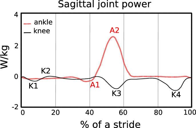
Negative and positive peaks in joint power indicate when mechanical energy is absorbed and generated, respectively, during a stride (Fig. 1). Negative peak power indicates eccentric contraction of the ankle plantar-flexor muscles from heel-strike through tibial progression, knee extensor muscles during heel-strike, rectus femoris during late stance, and biceps femoris during late stance (Fig. 1). Positive power regions primarily correspond to concentric contraction of the ankle plantar-flexor muscles during late stance, knee extensor muscles during early stance and hip flexor muscles during early swing phase. All these muscle contractions incur a metabolic cost. Thus, an exoskeleton that stores energy corresponding with the eccentric contraction of the knee extensor muscles and returns this energy during the concentric contraction of the ankle plantar-flexor muscles could decrease the metabolic cost of walking.

Fig. 1
figure1
Average sagittal plane ankle (A), and knee (K) joint mechanical power during level ground walking at 1.10 m/s over a stride for one leg, starting at heel strike. Data are from a previous study [13]. Negative peak power regions for the ankle and knee joints are denoted as A1 and K1, K3, and K4, respectively. Mechanical power and thus energy, is dissipated/absorbed during negative ankle (A1) and knee (K4) joint minimums [11]. At ~ 35–40% of the stride, the ankle plantar-flexors contract eccentrically to control ankle joint dorsiflexion. During terminal swing (K4), the hamstrings contract eccentrically to slow the speed of the swinging leg and avoid knee hyperextension just prior to the subsequent heel-strike (~ 90% of the stride). Positive mechanical power regions are labelled as A2 and K2 and correspond to the concentric contraction of the ankle plantar-flexors during late stance and the knee extensors during early stance, respectively
In order to reduce the metabolic cost of walking, use of an exoskeleton should not alter kinematic gait parameters such as stride length and step width, or kinetic parameters such as ground reaction forces. Previous studies have shown that when people walk with stride lengths and stride frequencies different from preferred, the metabolic cost of walking increases [14,15,16]. Walking speed is the product of stride length and stride frequency. At a fixed walking speed, the relationship between stride frequency and metabolic cost is represented by a U–shaped curve with the minimum metabolic cost corresponding to the preferred stride frequency [17]. Similarly, previous studies show that when humans walk with wider or narrower step widths compared to preferred, their metabolic cost increases [18,19,20]. Step width indicates the lateral distance between the midlines of the feet [21]. At a fixed walking speed, metabolic cost increases with the square of step width [19]. Thus, use of an exoskeleton that results in changes to stride length, stride frequency and step width compared to preferred could increase the metabolic cost of walking.
The development of wearable devices such as exoskeletons has been motivated by the challenge to reduce the metabolic cost of walking. In 1890, Nicholas Yagn conceptualized and received a patent for the first exoskeleton for assisting walking, running, and jumping using pneumatically powered gas bags [1]. Since then, many investigators have developed electrically powered or battery-powered lower limb exoskeletons for medical applications, neurorehabilitation therapy, augmentation, and military use [2, 22,23,24,25]. Most of the recent powered exoskeletons use actuators to provide assistance at the ankle joint during powered plantarflexion at the end of the stance phase of walking [25,26,27,28,29,30]. Specifically, use of powered exoskeletons has reduced the muscle activity and lower limb joint work needed by the user during level-ground walking compared to wearing the exoskeleton with the power turned off. Together with the timing of the assistance, the weight of these devices (12 kg to 38 kg [22]) may be one of the reasons why use of a powered exoskeleton does not decrease metabolic cost compared to normal walking without any wearable system [24].
With new methodological innovations, current research shows that exoskeletons can improve the metabolic cost of walking. Malcolm et al. [30] used optimal actuation timing predicted by a mathematical model combined with a tethered electrically-powered exoskeleton that assists ankle plantarflexion, and found that use of the exoskeleton reduced the metabolic cost of walking at 1.38 m/s by 6.0 ± 2.0% (mean ± SD) compared to walking without the exoskeleton. Mooney et al. [25] developed a battery-powered exoskeleton that utilizes a mathematical model to control the magnitude of positive mechanical power provided by the exoskeleton during ankle powered plantarflexion, and reduced metabolic cost by 8 ± 3% compared to walking without an exoskeleton at 1.5 m/s. Thus, through the optimal timing and magnitude of applied power, use of a powered exoskeleton can reduce the metabolic cost of walking.
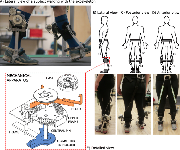
The way that an exoskeleton is attached to a person can affect the assistance provided to the person and thus the metabolic cost of walking. Panizzolo et al. [31] have investigated the use of an exosuit equipped with compliant textiles that provide assistance instead of rigid structures, such as those used in other powered exoskeletons. This approach aimed to improve the interface between the exoskeleton and the body [32] and reduce the mass on distal body segments to have less of an effect on metabolic cost [33, 34]. In particular, the exosuit was designed to provide assistance during both ankle joint plantarflexion at the end of the stance phase and hip joint flexion during the early swing phase. Using this exosuit, net metabolic power in the powered condition was 14.2 ± 6.1% lower than in the unpowered condition, but net metabolic power was not reduced with respect to normal walking at 1.5 m/s.
Use of passive-elastic exoskeletons has reduced the metabolic cost of walking by enhancing the mechanism of elastic energy storage and return at the ankle joint, with springs in parallel to the Achilles tendon [3, 25]. Recent studies demonstrate that storing and returning elastic energy during the phases of ankle joint negative and positive mechanical power can significantly reduce metabolic cost [3, 35]. Collins et al. [3] has shown that use of a passive-elastic exoskeleton in parallel with the ankle joint that stores and returns energy during the negative and positive phases of ankle joint power (Fig. 1) reduced metabolic cost by 7.2 ± 2.6% (mean ± SD) compared to walking without an exoskeleton at 1.25 m/s. Many others have investigated how use of a passive-elastic exoskeleton, which does not require an external power supply and is not equipped with sensors or actuators, affects walking [3, 36,37,38,39]. Panizzolo et al. [3] demonstrated that it is possible to reduce the metabolic cost of walking by more than 3% with a passive device that assists the hip joint compared to normal walking. Rome et al. [38] found that use of rubber bands in parallel with the hip joint can reduce the metabolic costs of carrying loads during walking, whereas Dean et al. [39] has shown that the use of a two-joint passive-elastic exoskeleton that works in parallel with the hip and knee joints can reduce the activity of lower limb muscles compared to normal walking [39].
We aimed to determine if a passive-elastic exoskeleton in parallel with the knee and ankle joints could reduce the metabolic cost of walking. We built an exoskeleton that stores energy from knee extension during the late leg swing phase, which corresponds to negative peak knee power (Fig. 1, K4) since it represents the greatest magnitude of energy absorption/storage during the stride [12]. Then, we designed the exoskeleton to release the energy stored from knee extension to assist ankle powered plantarflexion, which corresponds to positive peak ankle power during late stance (Fig. 1, A2). We designed our experiments to test three hypotheses. First, we hypothesized that the use of a passive-elastic exoskeleton that resists knee extension during the late leg swing phase would reduce metabolic power during level-ground walking compared to walking without an exoskeleton. Second, we hypothesized that the use of a passive-elastic exoskeleton that stores energy from knee extension during the late leg swing phase and returns energy for ankle powered plantarflexion during late stance would reduce metabolic power during level-ground walking compared to walking without an exoskeleton. Third, we hypothesized that the use of a passive-elastic exoskeleton would not change stride length, ground contact time, peak ground reaction forces, and step width during level ground walking compared to walking without an exoskeleton.

Material and methods

Participants

Eight healthy subjects [4 M and 4 F, mean ± SD age: 25 ± 3 years, mass: 73 ± 15 kg, height: 174 ± 10 cm, standing leg length: 83 ± 5 cm] participated in the study. We measured their leg lengths from the greater trochanter to the medial malleolus and averaged the right and left leg lengths. All subjects gave informed written consent before participating according to the University of Colorado Boulder Institutional Review Board.
We measured metabolic rates, ground reaction forces, and center of pressure while subjects walked on a dual-belt force measuring treadmill with the exoskeleton springs disengaged (no springs), engaged in parallel with the knee only, engaged in parallel with the knee and ankle, engaged in parallel with the knee only but with a longer engagement rope length, and engaged in parallel with the knee and ankle but with a longer engagement rope length, and without the exoskeleton.

Description of the exoskeleton

We custom-made the passive-elastic exoskeleton, which consists of a lightweight aluminum frame secured to the lower leg with a modified knee brace (Ottobock HealthCare LP, Austin, US). The exoskeleton is equipped with a mechanical apparatus comprised of six parts (Fig. 2, panel e). The primary frame has a central pin that is fixed on the external side of a modified knee brace, and all the other parts are attached to this frame. An asymmetric pin holder includes two small pins and rotates around the central pin, and an upper frame is fixed to the primary frame and holds the system of springs and a pawl. A block, with three arms and a ratchet wheel rotates about the central pin and compresses the system of springs on the upper frame. A case, which contains a spiral spring is attached distally on the longest arm of the block. In addition, the exoskeleton includes two inextensible ropes (r1, r2) that are fixed proximally on the anterior and posterior side of a belt worn by the subject positioned just above the iliac spines and glutei (Fig. 2), and distally to the exoskeletal mechanical apparatus on the pawl (r1) and the asymmetric pin holder (r2). Specifically, r2 is comprised of two parts (Fig. 2b). The superior part consists of a 4 cm wide piece of nylon webbing that lies posteriorly over the middle of the gluteus, whereas the lower part is a nylon rope that is attached to the mechanical apparatus. 4 cm wide pieces of nylon webbing were used to increase the surface area on the skin and prevent potential discomfort related to the pressure exerted by the rope on the skin. A third inextensible rope (r3) that originates from the exoskeletal case, is attached to the ankle frame surrounding the subject’s heel (Fig. 2a). The total mass of the exoskeleton is ~ 1.4 kg per leg.

Fig. 2
figure2
a Lateral view of a subject walking on the treadmill while using the exoskeleton just as the rope (r3) provides plantarflexion assistance to the trailing leg at the end of the stance phase. b Lateral, c Posterior, and d Anterior views of the exoskeleton attached to the body and corresponding pictures of a subject wearing the exoskeleton with ropes attached from the anterior belt to the apparatus (r1), posterior belt to the apparatus (r2) and apparatus to the ankle frame (r3). E) An exploded view of the mechanical apparatus. We designed the upper frame so that it could engage up to three linear springs. We used two springs during the experimental sessions in order to attain an angular spring stiffness of 17.45 Nm/rad. The mechanical apparatus is comprised of a frame that anchors the upper frame to the braces on the shank. That frame provides a central pin for the rotation of the asymmetric pin holder and block. The case rotates around a pin (not shown), fixed on the longer arm of the block
We 3D printed plastic ratchets and a pawl that were attached to the lateral portion of the brace and allowed energy from the movement of the knee to be stored through compression of the metal springs. The exoskeleton is attached to each leg, but we describe the effects of the exoskeleton for the right leg (Fig. 3). At the end of the swing phase, after hip extension is maximal, r2 is tensioned and rotates the asymmetric pin holder counter-clockwise (Fig. 3a), which pushes the block against the system of springs and compresses them (Fig. 3b). At the same time, the 3D-printed pawl engages the ratchet wheel to keep the spring compressed and prevent the clockwise rotation of the block (Fig. 3b, c, d). As r2 slackens, the asymmetric pin holder returns to its original position and the knee can freely flex from the loading response through mid-stance without any interaction with the engaged mechanical apparatus. Prior to the beginning of the experimental trial, we set the length of r2 to ensure it only tensioned in late swing, at maximum hip flexion. As the shank moves forward, due to its inertia, the tension in rope r2 provides a force that compresses the springs and allows the exoskeleton to decelerate the shank prior to heel strike. Also, the biological ankle’s motion is not affected by the exoskeleton due to a spiral spring inside the case. This spring allows the case to rotate from mid-swing through mid-stance (Fig. 3a, b, c, d), while r3 remains slack and does not interfere with the motion of the ankle. r3 only undergoes tension during late stance, when the hip extends, and the case rotation is locked. During this phase, just prior to powered plantarflexion, r1 is extended along the anterior side of the upper leg, disengages the pawl, and releases the compressed springs (Fig. 3e). The pawl disengagement allows the system of springs to release the stored energy; the block rotates clockwise lifting the case attached to its longer arm, which pulls up on r3 and assists the ankle joint during the push–off phase (Fig. 3f). The posterior case, attached to the longer arm of the block, rotates freely during a stride except for at the end of the stance phase, when the central ratchet is released. Ankle joint plantarflexion is therefore assisted by the elastic energy return of the exoskeleton. The exoskeleton does not constrain the biological range of motion of the ankle joint, and it is positioned at the same height and just lateral to the ankle, which connects the brace and heel frame of the exoskeleton. The ankle frame does not include a bearing but is designed to have low friction and the lever arm of the ankle frame is approximately 0.125 m.

Fig. 3
figure3
Engagement and disengagement of the exoskeleton mechanical apparatus during different phases of a walking stride. The upper portion of the figure shows the action of the exoskeleton during a stride and the lower portion provides a detailed view of the knee mechanism. Red arrows indicate the direction that ropes r1, r2 and r3 are moving during a specific phase (in panel E, red arrows are also used to indicate the extension of the linear springs). a During Mid-Swing, r2 begins to stretch due to knee extension and pulls the central pin. This knee extension movement continues until (b) Terminal Swing, and results in the rotation of the block which, in turn, compresses the springs. r3 also undergoes tension and causes the counterclockwise rotation of the case during Terminal Swing (considering the lateral side of the right leg). c During the Loading Response, both r2 and r3 become slack as the hip extends, which causes clockwise rotation of the central pin and case, respectively. d During Mid-Stance, r3 is stretched again as the hip rotates over the ankle and the case is locked so that there is no additional counterclockwise rotation. e During Terminal Stance, r1 is stretched due to hip extension, which pulls the pawl out of the ratchet, and allows the linear springs to extend and release their stored elastic energy. In this way, the block rotates clockwise and pulls on r3, which provides assistance during ankle powered plantarflexion. f At the subsequent Initial Swing only r1 is still stretched, while all of the other components of the device are disengaged


Cardiac MRI Reveals Post-Recovery Heart Damage in 78 Percent of COVID-19 Patients

This is not good. Be careful out there. 

Cardiac MRI Reveals Post-Recovery Heart Damage in 78 Percent of COVID-19 Patients

July 28, 2020
Scans show cardiovascular impact lingers after the acute phase of viral infection and into recovery.

MRI scans reveal that heart damage caused by COVID-19 remains present in 78 percent of patients who test positive for -- and recover from -- the virus, a new study has revealed.
In an article published July 27 in JAMA Cardiology, investigators from Germany revealed that the majority of patients who recover from the viral infection sustain cardiovascular damage regardless of any pre-existing conditions or their severity of disease.
“The results of our study provide important insights into the prevalence of cardiovascular involvement in the early convalescent stage,” wrote the team led by Valentina Puntmann, M.D., Ph.D., from the Institute for Experimental and Translational Cardiovascular Imaging at the University Hospital Frankfurt. “Our findings demonstrate that participants with a relative paucity of pre-existing cardiovascular condition and with mostly home-based recovery had frequent cardiac inflammatory involvement, which was similar to the hospitalized sub-group with regards to severity and extent.”

High-sensitivity troponin T level on the day of cardiac magnetic resonance imaging was 17.8 pg/mL. The patient recovered at home from COVID-19 illness with minimal symptoms, which included loss of smell and taste and only mildly increased temperature lasting 2 days. There were no known previous conditions or regular medication use. Histology revealed intracellular edema as enlarged cardiomyocytes with no evidence of interstitial or replacement fibrosis. A and B, Immunohistochemical staining revealed acute lymphocytic infiltration (lymphocyte function–associated antigen 1 and activated lymphocyte T antigen CD45R0), as well as activated intercellular adhesion molecule 1. C and D, Cardiac magnetic resonance imaging revealed enlarged volumes in myocardial mapping acquisitions, including significantly raised native T1 and native T2. E and F, Pericardial effusion and enhancement (yellow arrowheads) and epicardial and intramyocardial enhancement (white arrowheads) were seen on late gadolinium enhancement (LGE) acquisition. Courtesy: JAMA Cardiology

In addition, the study analysis revealed that the cardiovascular damage lingers even after the acute phase of the disease has passed and patients have begun their recovery. The team identified the presence of ongoing inflammation in the heart muscle and the heart lining in most patients, pointing to two potentially dangerous heart conditions – myocarditis and pericarditis.
Consequently, they said, there is a need for more research to determine the full extent of the long-term impacts of COVID-19 on the heart.
To investigate how the virus affects the heart, Puntmann’s team evaluated 3T MRI scans from an unselected cohort of 100 patients who had recently recovered from COVID-19. To their knowledge, this was the first such study. Most patients (53 percent) were male, the average age was 46, and the majority (67 percent) recovered at home. The severity of their disease course varied from asymptomic to severe enough to require hospitalization. Pre-existing conditions present among all patients included hypertension, diabetes, previously identified coronary artery disease, asthma, and chronic obstructive pulmonary disease. No patient had prior heart failure or cardiomyopathy.
Additionally, compared to healthy patients, individuals who recovered from COVID-19 experienced left ventricular ejection fraction, higher left ventricle volumes, higher left ventricle mass, and raised native T1 and T2, indicating active inflammatory processes. Among the specific results, 78 percent of patients had abnormal cardiac MRI findings, including raised myocardial native T1 in 73 percent of patients and increased myocardial native T2 in 60 percent of patients, as well as detectable high-sensitivity troponin in 76 percent of patients. Thirty-two percent of patients experienced myocardial late gadolinium enhancement, and 22 percent saw pericardial enhancement.
“Our findings may provide an indication of potentially considerable burden of inflammatory disease in large and growing parts of the population and urgently require confirmation in a larger cohort,” the team said. “Although the long-term health effects of these findings cannot yet be determined, several of the abnormalities described have been previously related to worse outcome in inflammatory cardiomyopathies.”
Most of the imaging findings, they said, indicate ongoing perimyocarditis after the acute phase of COVID-19 infection.
There were limitations to the study, however, the team said. The findings have not yet been validated with the pediatric patient population, nor do the results represent patients who have current acute COVID-19 infection.
Even with these limitations, though, the results from this study are informative, said Clyde Yancey, M.D., chief of cardiology at Northwestern University Feinberg School of Medicine in an accompanying editorial. Not only do they point to the presence of residual left ventricular dysfunction and sustained inflammation, but they also highlight other important clinical patho-physiologies, such as clinical syndromes consistent with acute myocarditis, immunologic responses, and microvascular clot formation.
“When added to the postmortem pathological findings [from other studies,] we see that plot thickening, and we are inclined to raise a new and very evident concern that cardiomyopathy and heart failure related to COVID-19 may potentially evolve as the natural history of this infection becomes clearer,” said Yancey, who is also the deputy editor of JAMA Cardiology. “We wish not to generate additional anxiety, but rather to incite other investigators to carefully examine existing and prospectively collect new data in other populations to confirm or refute these findings.”
Further investigation of these practical concerns, he said, could continue to shed light on additional aspects of the COVID-19 crisis.