Use the labels in the right column to find what you want. Or you can go thru them one by one, there are only 33,991 posts. Searching is done in the search box in upper left corner. I blog on anything to do with stroke. DO NOT DO ANYTHING SUGGESTED HERE AS I AM NOT MEDICALLY TRAINED, YOUR DOCTOR IS, LISTEN TO THEM. BUT I BET THEY DON'T KNOW HOW TO GET YOU 100% RECOVERED. I DON'T EITHER BUT HAVE PLENTY OF QUESTIONS FOR YOUR DOCTOR TO ANSWER.
Changing stroke rehab and research worldwide now.Time is Brain!trillions and trillions of neuronsthatDIEeach day because there areNOeffective hyperacute therapies besides tPA(only 12% effective). I have 523 posts on hyperacute therapy, enough for researchers to spend decades proving them out. These are my personal ideas and blog on stroke rehabilitation and stroke research. Do not attempt any of these without checking with your medical provider. Unless you join me in agitating, when you need these therapies they won't be there.
What this blog is for:
My blog is not to help survivors recover, it is to have the 10 million yearly stroke survivors light fires underneath their doctors, stroke hospitals and stroke researchers to get stroke solved. 100% recovery. The stroke medical world is completely failing at that goal, they don't even have it as a goal. Shortly after getting out of the hospital and getting NO information on the process or protocols of stroke rehabilitation and recovery I started searching on the internet and found that no other survivor received useful information. This is an attempt to cover all stroke rehabilitation information that should be readily available to survivors so they can talk with informed knowledge to their medical staff. It lays out what needs to be done to get stroke survivors closer to 100% recovery. It's quite disgusting that this information is not available from every stroke association and doctors group.
Brains
are like puzzles, requiring many nested and codependent pieces to
function well. The brain is divided into areas, each containing many
millions of neurons connected across thousands of synapses. These
synapses, which enable communication between neurons, depend on even
smaller structures: message-sending boutons (swollen bulbs at the
branch-like tips of neurons), message-receiving dendrites (complementary
branch-like structures for receiving bouton messages), and
power-generating mitochondria. To create a cohesive brain, all these
pieces must be accounted for.
However, in the aging brain, these pieces can get lost or altered and no longer fit in the greater brain puzzle.
"Fifty percent of people experience loss of working memory with old
age, meaning their ability to hold and manipulate information in the
short-term decreases," says co-first author Courtney Glavis-Bloom, a
senior staff scientist in Salk Institute Professor John Reynolds' lab.
"We set out to understand why some individuals maintain healthy working
memory as they age, while others do not. In the process, we discovered a
novel mechanism for the synaptic basis of cognitive impairment."
Prior studies had found that brains lose synapses as they age, and
the researchers saw this pattern in their non-human primate model, too.
But when they looked at the synapses that remained, they found evidence
of a breakdown in coordination between the size of boutons and the
mitochondria they contained. A fundamental neuroscientific principle,
the ultrastructural size principle, explains that whenever one part of
the synaptic complex changes in size, so too must all the other
parts. The synapse, the mitochondria, the boutons-;all these parts must
scale in accordance with one another. Before the Salk team's study,
published in Frontiers in Aging Neuroscience on April 12, 2023, nobody had asked whether this principle could be violated with age or disease.
"To
examine this, we turned to electron microscopy," says co-first author
Casey Vanderlip, a former research assistant in Reynolds' lab. "This
enabled us to visualize these components across many synapses. We found
that synaptic loss occurred with healthy and impaired aging, but what
differed was the breakdown in the correlation between the sizes of
boutons and their mitochondria."
"It is a ripple effect, with unfathomably small synaptic structures
altering networks of neurons, brain function, and behavior," says
Glavis-Bloom. "Investigating these microscopic dysfunctions is uncharted
territory that could revolutionize our understanding of aging and its
impact on cognition."
The team found that adherence to the ultrastructural size principle
was essential for avoiding working memory impairment with age. By
viewing violation of the ultrastructural size principle and
mitochondria-related failures as the key to age-related cognitive
impairment, the study ushers in a new era for aging research.
The images we have captured of synapses are snapshots of a dynamic
process. With these snapshots in hand, we can begin to think first about
the mechanisms that coordinate the expansion and contraction of the
various parts of the synaptic complex, then ask how disruption of these
mechanisms can explain age-related cognitive decline. This opens an
entirely new way of thinking about cognitive decline that could lead to
new targets for future therapeutics."
John Reynolds, Salk Institute Professor, Holder of the Fiona and Sanjay Jha Chair in Neuroscience
Other authors include Sammy Weiser Novak and Uri Manor of the Salk
Institute; and Masaaki Kuwajima, Lyndsey Kirk, and Kristen M. Harris of
the University of Texas at Austin.
Glavis-Bloom, C., et al.
(2023) Violation of the ultrastructural size principle in the
dorsolateral prefrontal cortex underlies working memory impairment in
the aged common marmoset (Callithrix jacchus). Frontiers in Aging Neuroscience.doi.org/10.3389/fnagi.2023.1146245.
Your doctor has had 4 years to figure out how to use this capacity to get you recovered. Now she just needs to figure out how to speedily access all that information.
Data from the Salk Institute shows brain’s memory capacity is in the petabyte range, as much as entire Web
January 20, 2016
LA JOLLA—Salk researchers and collaborators have achieved critical
insight into the size of neural connections, putting the memory capacity
of the brain far higher than common estimates. The new work also
answers a longstanding question as to how the brain is so energy
efficient and could help engineers build computers that are incredibly
powerful but also conserve energy.
“This is a real bombshell in the field of neuroscience,” says Terry Sejnowski, Salk professor and co-senior author of the paper, which was published in eLife.
“We discovered the key to unlocking the design principle for how
hippocampal neurons function with low energy but high computation power.
Our new measurements of the brain’s memory capacity increase
conservative estimates by a factor of 10 to at least a petabyte, in the
same ballpark as the World Wide Web.”
Our memories and thoughts
are the result of patterns of electrical and chemical activity in the
brain. A key part of the activity happens when branches of neurons, much
like electrical wire, interact at certain junctions, known as synapses.
An output ‘wire’ (an axon) from one neuron connects to an input ‘wire’
(a dendrite) of a second neuron. Signals travel across the synapse as
chemicals called neurotransmitters to tell the receiving neuron whether
to convey an electrical signal to other neurons. Each neuron can have
thousands of these synapses with thousands of other neurons.
“When
we first reconstructed every dendrite, axon, glial process, and synapse
from a volume of hippocampus the size of a single red blood cell, we
were somewhat bewildered by the complexity and diversity amongst the
synapses,” says Kristen Harris, co-senior author of the work and
professor of neuroscience at the University of Texas, Austin.
“While I had hoped to learn fundamental principles about how the brain
is organized from these detailed reconstructions, I have been truly
amazed at the precision obtained in the analyses of this report.”
Synapses are still a mystery, though their dysfunction can cause a range of neurological diseases.
Larger synapses—with more surface area and vesicles of
neurotransmitters—are stronger, making them more likely to activate
their surrounding neurons than medium or small synapses.
The Salk
team, while building a 3D reconstruction of rat hippocampus tissue (the
memory center of the brain), noticed something unusual. In some cases, a
single axon from one neuron formed two synapses reaching out to a
single dendrite of a second neuron, signifying that the first neuron
seemed to be sending a duplicate message to the receiving neuron.
At
first, the researchers didn’t think much of this duplicity, which
occurs about 10 percent of the time in the hippocampus. But Tom Bartol, a
Salk staff scientist, had an idea: if they could measure the difference
between two very similar synapses such as these, they might glean
insight into synaptic sizes, which so far had only been classified in
the field as small, medium and large.
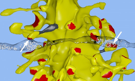
In
a computational reconstruction of brain tissue in the hippocampus, Salk
scientists and UT-Austin scientists found the unusual occurrence of two
synapses from the axon of one neuron (translucent black strip) forming
onto two spines on the same dendrite of a second neuron (yellow).
Separate terminals from one neuron’s axon are shown in synaptic contact
with two spines (arrows) on the same dendrite of a second neuron in the
hippocampus. The spine head volumes, synaptic contact areas (red), neck
diameters (gray) and number of presynaptic vesicles (white spheres) of
these two synapses are almost identical.
To
do this, researchers used advanced microscopy and computational
algorithms they had developed to image rat brains and reconstruct the
connectivity, shapes, volumes and surface area of the brain tissue down
to a nanomolecular level.
The scientists expected the synapses
would be roughly similar in size, but were surprised to discover the
synapses were nearly identical.
“We were amazed to find that the
difference in the sizes of the pairs of synapses were very small, on
average, only about eight percent different in size. No one thought it
would be such a small difference. This was a curveball from nature,”
says Bartol.
Because the memory capacity of neurons is dependent
upon synapse size, this eight percent difference turned out to be a key
number the team could then plug into their algorithmic models of the
brain to measure how much information could potentially be stored in
synaptic connections.
It was known before that the range in sizes
between the smallest and largest synapses was a factor of 60 and that
most are small.
But armed with the knowledge that synapses of all
sizes could vary in increments as little as eight percent between sizes
within a factor of 60, the team determined there could be about 26
categories of sizes of synapses, rather than just a few.
“Our data
suggests there are 10 times more discrete sizes of synapses than
previously thought,” says Bartol. In computer terms, 26 sizes of
synapses correspond to about 4.7 “bits” of information. Previously, it
was thought that the brain was capable of just one to two bits for short
and long memory storage in the hippocampus.
“This is roughly an order of magnitude of precision more than anyone has ever imagined,” says Sejnowski.
What
makes this precision puzzling is that hippocampal synapses are
notoriously unreliable. When a signal travels from one neuron to
another, it typically activates that second neuron only 10 to 20 percent
of the time.
“We had often wondered how the remarkable precision
of the brain can come out of such unreliable synapses,” says Bartol. One
answer, it seems, is in the constant adjustment of synapses, averaging
out their success and failure rates over time. The team used their new
data and a statistical model to find out how many signals it would take a
pair of synapses to get to that eight percent difference.
The
researchers calculated that for the smallest synapses, about 1,500
events cause a change in their size/ability (20 minutes) and for the
largest synapses, only a couple hundred signaling events (1 to 2
minutes) cause a change.
“This means that every 2 or 20 minutes,
your synapses are going up or down to the next size. The synapses are
adjusting themselves according to the signals they receive,” says
Bartol.
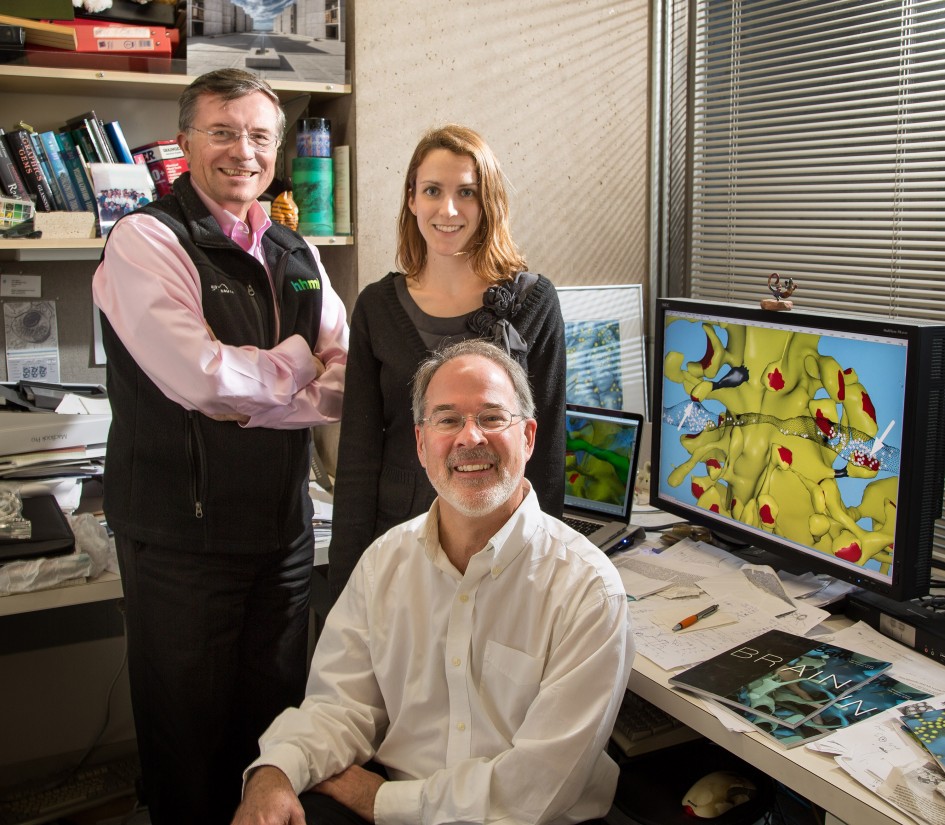
From left: Terry Sejnowski, Cailey Bromer and Tom Bartol
“Our
prior work had hinted at the possibility that spines and axons that
synapse together would be similar in size, but the reality of the
precision is truly remarkable and lays the foundation for whole new ways
to think about brains and computers,” says Harris. “The work resulting
from this collaboration has opened a new chapter in the search for
learning and memory mechanisms.” Harris adds that the findings suggest
more questions to explore, for example, if similar rules apply for
synapses in other regions of the brain and how those rules differ during
development and as synapses change during the initial stages of
learning.
“The implications of what we found are far-reaching,”
adds Sejnowski. “Hidden under the apparent chaos and messiness of the
brain is an underlying precision to the size and shapes of synapses that
was hidden from us.”
The findings also offer a valuable
explanation for the brain’s surprising efficiency. The waking adult
brain generates only about 20 watts of continuous power—as much as a
very dim light bulb. The Salk discovery could help computer scientists
build ultraprecise, but energy-efficient, computers, particularly ones
that employ “deep learning” and artificial neural nets—techniques
capable of sophisticated learning and analysis, such as speech, object
recognition and translation.
“This trick of the brain absolutely
points to a way to design better computers,” says Sejnowski. “Using
probabilistic transmission turns out to be as accurate and require much
less energy for both computers and brains.”
Other authors on the paper were Cailey Bromer of the Salk Institute; Justin Kinney of the McGovern Institute for Brain Research; and Michael A. Chirillo and Jennifer N. Bourne of the University of Texas, Austin.
Sedentary mice given the drug ran longer without training.
FULL STORY
Salk scientists move one step closer to
developing 'exercise-in-a-pill.' Partial view of a mouse calf muscle
stained for different types of muscle fibers: oxidative slow-twitch
(blue), oxidative fast-twitch (green), glycolytic fast-twitch (red).
Credit: Salk Institute/Waitt Center
Every week, there seems to be another story
about the health benefits of running. That's great -- but what if you
can't run? For the elderly, obese or otherwise mobility-limited, the
rewards of aerobic exercise have long been out of reach.
Salk Institute scientists, building on earlier work that identified a
gene pathway triggered by running, have discovered how to fully
activate that pathway in sedentary mice with a chemical compound,
mimicking the beneficial effects of exercise, including increased fat
burning and stamina. The study, which appears in Cell Metabolism
on May 2, 2017, not only deepens our understanding of aerobic
endurance, but also offers people with heart conditions, pulmonary
disease, type 2 diabetes or other health limitations the hope of
achieving those benefits pharmacologically.
"It's well known that people can improve their aerobic endurance
through training," says senior author Ronald Evans, Howard Hughes
Medical Institute investigator and holder of Salk's March of Dimes Chair
in Molecular and Developmental Biology. "The question for us was: how
does endurance work? And if we really understand the science, can we
replace training with a drug?"
Developing endurance means being able to sustain an aerobic activity
for longer periods of time. As people become more fit, their muscles
shift from burning carbohydrates (glucose) to burning fat. So
researchers assumed that endurance is a function of the body's
increasing ability to burn fat, though details of the process have been
murky. Previous work by the Evans lab into a gene called PPAR delta
(PPARD) offered intriguing clues: mice genetically engineered to have
permanently activated PPARD became long-distance runners who were
resistant to weight gain and highly responsive to insulin -- all
qualities associated with physical fitness. The team found that a
chemical compound called GW1516 (GW) similarly activated PPARD,
replicating the weight control and insulin responsiveness in normal mice
that had been seen in the engineered ones. However, GW did not affect
endurance (how long the mice could run) unless coupled with daily
exercise, which defeated the purpose of using it to replace exercise.
In the current study, the Salk team gave normal mice a higher dose of
GW, for a longer period of time (8 weeks instead of 4). Both the mice
that received the compound and mice that did not were typically
sedentary, but all were subjected to treadmill tests to see how long
they could run until exhausted.
Mice in the control group could run about 160 minutes before
exhaustion. Mice on the drug, however, could run about 270 minutes --
about 70 percent longer. For both groups, exhaustion set in when blood
sugar (glucose) dropped to around 70 mg/dl, suggesting that low glucose
levels (hypoglycemia) are responsible for fatigue.
To understand what was happening at the molecular level, the team
compared gene expression in a major muscle of mice. They found 975 genes
whose expression changed in response to the drug, either becoming
suppressed or increased. Genes whose expression increased were ones that
regulate breaking down and burning fat. Surprisingly, genes that were
suppressed were related to breaking down carbohydrates for energy. This
means that the PPARD pathway prevents sugar from being an energy source
in muscle during exercise, possibly to preserve sugar for the brain.
Activating fat-burning takes longer than burning sugar, which is why the
body generally uses glucose unless it has a compelling reason not to --
like maintaining brain function during periods of high energy
expenditure. Although muscles can burn either sugar or fat, the brain
prefers sugar, which explains why runners who "hit the wall" experience
both physical and mental fatigue when they use up their supply of
glucose.
"This study suggests that burning fat is less a driver of endurance
than a compensatory mechanism to conserve glucose," says Michael Downes,
a Salk senior scientist and co-senior author of the paper. "PPARD is
suppressing all the points that are involved in sugar metabolism in the
muscle so glucose can be redirected to the brain, thereby preserving
brain function."
Interestingly, the muscles of mice that took the exercise drug did
not exhibit the kinds of physiological changes that typically accompany
aerobic fitness: additional mitochondria, more blood vessels and a shift
toward the type of muscle fibers that burn fat rather than sugar. This
shows that these changes are not exclusively driving aerobic endurance;
it can also be accomplished by chemically activatin
g a genetic pathway.
In addition to having increased endurance, mice who were given the drug
were also resistant to weight gain and more responsive to insulin than
the mice who were not on the drug.
"Exercise activates PPARD, but we're showing that you can do the same
thing without mechanical training. It means you can improve endurance
to the equivalent level as someone in training, without all of the
physical effort," says Weiwei Fan, a Salk research associate and the
paper's first author.
Although the lab's studies have been in mice, pharmaceutical
companies are interested in using the research to develop clinical
trials for humans. The team can envision a number of therapeutic
applications for a prescription drug based on GW, from increasing fat
burning in people suffering from obesity or type 2 diabetes to improving
patients' fitness before and after surgery.
Story Source: Materials provided by Salk Institute. Note: Content may be edited for style and length.